

Abstract
Aims:
To evaluate the clinical and microbiological effects of local drug delivery of moxifloxacin and ibuprofen gel as an adjunct to conventional periodontal therapy in chronic periodontitis patients.
Subjects and Methods:
Twenty patients with moderate-to-severe chronic generalized periodontitis with probing pocket depth (PPD) of ≥5 mm and <8 mm were randomly assigned to one of the following two treatment modalities: scaling and root planing (SRP) group and moxifloxacin and ibuprofen combination gel as an adjunct to SRP group. Clinical parameters include plaque index (PI), gingival index (GI), probing depths and clinical attachment level (CAL) that were recorded at baseline and 1 and 3 months after the treatment, and microbiologic assessment was done using dark-field microscopy.
Results:
A statistically significant difference in mean PI and GI scores and reduction in PPD and gain in CAL were observed at different study intervals with greater difference in the test group. On microbiological examination, the percentage of cocci increased, while a statistically significant decrease in the mean percentage of bacilli and spirochetes was observed in both groups at given intervals. In-vitro dissolution showed controlled release of both the drugs.
Conclusions:
Among the two treatment modalities, treatment with moxifloxacin and ibuprofen local delivery as an adjunct to SRP gave superior results in clinical and microbiological parameters compared to SRP group.
Keywords: Chronic periodontitis, ibuprofen, microbiology, moxifloxacin, spirochetes
INTRODUCTION
Chronic periodontitis is an infectious bacterial disease resulting in inflammation within the supporting tissues of the teeth, which results in progressive attachment and bone loss and is characterized by pocket formation and/or recession of the gingiva.
Elimination or adequate suppression of periodontopathic microorganisms in the subgingival microbiota is necessary for periodontal healing. Reestablishment of subgingival microflora occurs in 120–140 days despite supra- and subgingival instrumentation.[1] The main objective of systemic antibiotic therapy is to reinforce mechanical periodontal treatment for bacterial elimination and to support the host defense system by destroying subgingival pathogens.[2,3] A local route of drug delivery can attain up to 100-fold higher concentrations of an antimicrobial agent in subgingival sites as compared with a systemic drug regimen. To overcome the high clearance rate of drug in gingival crevicular fluid, Goodson et al.[4] first proposed the concept of controlled drug delivery in the treatment of periodontitis. Various vehicles are used to assure the therapeutic concentrations of antimicrobial agent in periodontal pocket.[5]
Although penicillins were widely accepted drugs as the anti-infective treatment of choice, due to the high incidence of bacterial resistance, quinolones were introduced later.[6] They represent a class of broad-spectrum antibiotics having one or more fluorine substitutions, and are called fluoroquinolones which have advantages of good penetration into tissues and antibacterial activity within the cells.[7]
Goodson et al. noted that PGE2 levels in inflamed gingiva were found to be 10-fold higher than that in healthy gingiva.[8] The major side effect of systemically administered nonsteroidal anti-inflammatory drugs (NSAIDs) is gastrointestinal tract irritation and to overcome this effect, the local delivery of these agents was proposed in periodontal treatment with successful outcome and minimal side effects. In general, topical application of NSAIDs is possible because these agents are lipophilic and are well absorbed into the gingival tissues.[9]
In periodontal diseases, both antibacterial and NSAIDs are used simultaneously to treat infection and inflammation. A single delivery system which can deliver both drugs locally is expected to be more convenient and beneficial and improve patient compliance.[10] Hence, the present study is conducted to evaluate the clinical and microbiological effects of local drug delivery (LDD) of moxifloxacin and ibuprofen gel as an adjunct to conventional periodontal therapy in chronic periodontitis patients.
Aims and objectives
The present study aimed to evaluate the clinical and microbiological effects of scaling and root planing (SRP) alone and LDD of moxifloxacin and ibuprofen gel as an adjunct to SRP in chronic periodontitis patients.
SUBJECTS AND METHODS
Study design
The study was a single-center, randomized, split-mouth, controlled clinical study.
Clearance from the institutional ethical committee was obtained for the study. Twenty patients were selected from the outpatient section.
Inclusion criteria
Patients above 18 years of age and diagnosed with moderate-to-severe chronic generalized periodontitis with probing pocket depth (PPD) >5 mm and <8 mm were included in the study.
Exclusion criteria
Patients with systemic disease which could influence the outcome of therapy, pregnant women and lactating mothers, those who underwent active periodontal treatment within the last 6 months, patients allergic to moxifloxacin, teeth with furcation involvement and smokers were excluded from the study.
A detailed case history was recorded in a specially prepared pro forma which included information regarding the patient's overall medical health and oral health, and an informed consent was obtained.
Randomization
After baseline examination, simple randomization by coin method was done to assign patients in a split-mouth design to one of the following two treatment modalities: SRP group (only SRP) and moxifloxacin and ibuprofen as an adjunct to SRP group (SRP + LDD).
Clinical parameters
Plaque index (PI), gingival index (GI), probing depths (PD) and clinical attachment level (CAL) were recorded at baseline and 1 and 3 months after the treatment. A custom-made acrylic stent and a Williams graduated periodontal probe were used to standardize the measurement of PD and CAL [Figure 1a and b]. Dark-field microscopic examination to assess the presence of morphologically different microorganisms was done. Subgingival plaque sample was collected from tooth with pocket depth of ≥5 mm with a sterile curette at baseline and at 3 months [Figure 2].
Figure 1.

(a) Probing pocket depth at baseline in scaling and root planing group; (b) probing pocket depth in scaling and root planing + local drug delivery group
Figure 2.

Collection of plaque sample
Methods
After baseline examination and recording of all clinical parameters, all patients received routine oral hygiene instructions and full-mouth SRP. For patients in SRP group, no additional treatment was provided. In SRP + LDD group, along with SRP, LDD of 0.1 ml of moxifloxacin and ibuprofen gel was injected into two sites having deep periodontal pocket using a syringe with a blunt cannula [Figure 3]. After that, the periodontal pack was placed on that area for better retention of the local drug.
Figure 3.

Local drug delivery of moxifloxacin and ibuprofen gel in scaling and root planing + local drug delivery group
Preparation of moxifloxacin and ibuprofen gel
It was prepared at the Department of Pharmaceutics, Vikas College of Pharmaceutical Sciences, Suryapet, Nalgonda district. It was prepared by using 50 mg of moxifloxacin and 50 mg of ibuprofen in 750 mg of polycaprolactone gel (biodegradable) by using 188.67 μl of dichloromethane as solvent [Figure 4].
Figure 4.
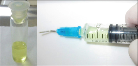
Moxifloxacin and ibuprofen gel
Sol–gel transition
Sol–gel transition occurs by two phenomena. One is due to change in temperature, and the other is due to change in pH. For this drug formulation, pH-based sol–gel transition was used, i.e., at 6.8 pH, the prepared formulation was transformed to gel form.
Statistical analysis
The data were analyzed using the SPSS software 19.00 program (SPSS Inc., Chicago, IL, USA). Comparison of plaque and GI at baseline and at 1 and 3 months was done by Wilcoxon matched-pairs test. The intragroup comparison of clinical parameters such as PD and CAL was done by using paired t-test at various study intervals, and intergroup comparison between SRP group and SRP + LDD group was done by using unpaired t-test at various study intervals. Intragroup changes in the subgingival microbiota such as percentage of cocci and bacilli and spirochetes scores were compared at various intervals in both groups by using Wilcoxon matched-pairs test, and intergroup comparison of microbial changes between SRP group and SRP + LDD group was done by using Mann–Whitney U-test. Differences were considered statistically significant at P < 0.05*.
RESULTS
The present study was designed to assess the effect of SRP (control group), SRP plus moxifloxacin and ibuprofen gel (active group) on the clinical parameters (PI, GI, PPD and CAL) and changes in subgingival microflora in periodontal pockets at various study intervals of baseline and at 1 and 3 months within and between the two groups.
Plaque index and gingival index
There was a gradual reduction in PI and GI scores over the study period. The mean PI score at baseline was 1.32 ± 0.23, at the end of 1 month was 0.67 ± 0.09 and at the end of 3 months was 0.40 ± 0.09. The mean GI score at baseline was 1.64 ± 0.21, at the end of 1 month was 0.80 ± 0.18 and at the end of 3 months was 0.38 ± 0.08. The mean PI and GI scores were statistically significant when compared from the baseline score to the end of 1 and 3 months [Table 1].
Table 1.
Comparison of baseline, 1 and 3 months with respect to plaque index and gingival index scores
| Parameters | Observation period | Mean value±SD | Comparison | Mean difference | P |
|---|---|---|---|---|---|
| PI | Baseline | 1.32±0.23 | - | - | - |
| 1st month | 0.67±0.09 | Baseline versus 1st month | 0.65 | 0.0001* | |
| 3rd month | 0.40±0.09 | 1st month versus 3rd month | 0.27 | 0.0001* | |
| - | Baseline versus 3rd month | 0.92 | 0.0001* | ||
| GI | Baseline | 1.64±0.21 | - | - | - |
| 1st month | 0.80±0.18 | Baseline versus 1st month | 0.84 | 0.0001* | |
| 3rd month | 0.38±0.08 | Versus 2nd month | 0.42 | 0.0001* | |
| - | Baseline versus 3rd month | 1.26 | 0.0001* |
*P<0.05. SD: Standard deviation, PI: Plaque index, GI: Gingival index
Pocket probing depth
The mean PPD (mm) difference from baseline to 1 month in SRP group was 1.20 ± 0.30 and SRP + LDD group was 1.83 ± 0.34, and the difference from baseline to 3 months in SRP group was 1.83 ± 0.47 and SRP + LDD group was 2.85 ± 0.33. By comparison of SRP group and SRP + LDD group, there was statistically significant difference of reduction in PPD (mm) at different study intervals with greater reduction in the test group [Table 2 and Figure 5a, b].
Table 2.
Intra- and inter-group comparison of scaling and root planing and scaling and root planing + local drug delivery groups at various study intervals with respect to probing pocket depth scores
| Groups | Mean±SD | ||||
|---|---|---|---|---|---|
| Baseline | 1 month | 3 months | Changes from baseline to | ||
| 1 month | 3 months | ||||
| SRP | 6.08±0.57 | 4.88±0.56 | 4.25±0.57 | 1.20±0.30 | 1.83±0.47 |
| SRP + LDD | 6.03±0.62 | 4.20±0.57 | 3.18±0.57 | 1.83±0.34 | 2.85±0.33 |
| Percentage of change in SRP | 19.75# P=0.00001* |
30.04# P=0.00001* |
|||
| Percentage of change in SRP + LDD | 30.29# P=0.00001* |
47.30# P=0.00001* |
|||
| t | 0.2665 | 3.7769 | 5.9540 | −6.2194 | −8.0326 |
| P | 0.7913 | 0.0005* | 0.00001* | 0.00001* | 0.00001* |
*P<0.05, #Applied paired t-test. LDD: Local drug delivery, SRP: Scaling and root planing, SD: Standard deviation
Figure 5.

(a) Probing pocket depth at 3 months in scaling and root planing group; (b) probing pocket depth at 3 months in scaling and root planing + local drug delivery group
Clinical attachment level
The mean gain in CAL (mm) from baseline to 1 month in SRP group was 1.23 ± 0.34 and SRP + LDD group was 1.88 ± 0.28, and the difference from baseline to 3 months in SRP group was 1.95 ± 0.22 and SRP + LDD group was 2.85 ± 0.29. By comparison of both the groups, there was statistically significant gain in CAL (mm) at different study intervals with greater gain in the test group [Table 3].
Table 3.
Intra- and inter-group comparison of scaling and root planing and scaling and root planing + local drug delivery groups at various study intervals with respect to clinical attachment level scores
| Groups | Mean±SD | ||||
|---|---|---|---|---|---|
| Baseline | 1 month | 3 months | Changes from baseline to | ||
| 1 month | 3 months | ||||
| SRP | 4.65±0.65 | 3.43±0.59 | 2.70±0.70 | 1.23±0.34 | 1.95±0.22 |
| SRP + LDD | 4.65±0.65 | 2.78±0.75 | 1.80±0.73 | 1.88±0.28 | 2.85±0.29 |
| Percentage of change in SRP | 26.34# P=0.00001* |
41.94# P=0.00001* |
|||
| Percentage of change in SRP + LDD | 40.32# P=0.00001* |
61.29# P=0.00001* |
|||
| t | 0.0000 | 3.0398 | 3.9832 | −6.6096 | −11.0959 |
| P | 1.0000 | 0.0043* | 0.0003* | 0.00001* | 0.00001* |
*P<0.05, #Applied paired t-test. LDD: Local drug delivery, SRP: Scaling and root planing, SD: Standard deviation
Microbiologic assessment
The Mean cocci increased in both groups compared to baseline, with a greater increase of 67.61% and 68.12% in the test group at 1 and 3 months, respectively. This increase was statistically significant between the two groups at both time intervals. The percentage reduction of mean bacilli from baseline to different study intervals was compared, and statistically significant difference was found from baseline to 1 month and from baseline to 3 months in both the groups, with higher reduction in SRP + LDD group. The spirochetes percentage was greatly reduced in SRP + LDD group at different study intervals rather than compared with SRP group [Table 4 and Figures 6a-c].
Table 4.
Intra- and inter-group comparison of scaling and root planing and scaling and root planing + local drug delivery groups at various study intervals with respect to microbiota
| Groups | Mean±SD | ||||
|---|---|---|---|---|---|
| Baseline | 1 month | 3 months | Changes from baseline to | ||
| 1 month | 3 months | ||||
| SRP | 39.05±7.10 | 55.95±6.93 | 55.75±7.61 | 16.90±5.88 | 16.70±6.95 |
| SRP + LDD | 39.05±7.10 | 65.45±4.88 | 65.65±4.77 | 26.40±6.85 | 26.60±6.95 |
| Percentage of change in SRP | 43.28# P=0.0001* |
42.77# P=0.0001* |
|||
| Percentage of change in SRP + LDD | 67.61# P=0.0001* |
68.12# P=0.0001* |
|||
| t | 0.0000 | −4.0169 | −4.1522 | −3.9628 | −3.7870 |
| P | 1.0000 | 0.0001* | 0.00001* | 0.0001* | 0.0002* |
*P<0.05, #Applied Wilcoxon matched pairs test. SD: Standard deviation, SRP: Scaling and root planing, LDD: Local drug delivery
Figure 6.

(a) Microbiologic assessment at baseline; (b) microbiologic assessment at 3 months in scaling and root planing group; (c) microbiologic assessment at 3 months in scaling and root planing + local drug delivery group
In-vitro dissolution
The in-vitro dissolution results showed that there was release of drug up to 168 h, i.e., 7 days after single application of gel into the dissolution medium. At the end of 168 h, 96.34% of moxifloxacin and 95.45% of ibuprofen were released into the medium. The drug release pattern shows that both the drugs were released slowly for 168 h by following first-order kinetics. This indicates that there was a controlled release of both the drugs in in-vitro studies [Table 5 and Graph 1].
Table 5.
In-vitro release of drug formulation
| Time (h) | F5 | |
|---|---|---|
| Percentage drug release of Moxifloxacin | Percentage drug release of Ibuprofen | |
| 0.083 | 3.57 | 3.24 |
| 0.166 | 9.83 | 15.55 |
| 0.333 | 9.64 | 16.93 |
| 0.5 | 20.34 | 32.55 |
| 1 | 22.36 | 34.21 |
| 1.5 | 32.33 | 47.021 |
| 2 | 40.77 | 57.42 |
| 3 | 56.18 | 75.38 |
| 4 | 70.37 | 65.95 |
| 5 | 58.29 | 68.25 |
| 6 | 62.38 | 70.94 |
| 8 | 72.69 | 67.28 |
| 10 | 83.54 | 76.01 |
| 12 | 82.05 | 75.14 |
| 24 | 86.56 | 72.96 |
| 48 | 76.13 | 74.36 |
| 96 | 92.21 | 85.74 |
| 144 | 93.90 | 89.95 |
| 168 | 96.34 | 95.45 |
Graph 1.

In-vitro drug release of moxifloxacin and ibuprofen (pH 6.8)
DISCUSSION
The primary objective of periodontal therapy for patients with chronic periodontitis is to halt disease progression and to resolve inflammation. It is aimed at reducing etiologic factors below the threshold level capable of producing periodontal tissue breakdown, thereby allowing the repair of the affected region. Very few patients can maintain periodontal health over a lifetime without the benefit of a regular dental care which consists primarily of oral hygiene instructions and nonsurgical periodontal therapy (NSPT).[11] A novel approach is the local application of antimicrobial agents that are effective against periodontopathogens and can reduce pocket depth. This approach consists of the administration of local biodegradable sustained or controlled release antimicrobial agents in periodontal pockets. Several biodegradable local delivery systems containing different antimicrobial agents have been developed and introduced as an adjunctive measure to mechanical therapy.[12,13] The benefits of subgingival application of drugs include:
Better patient compliance
Enhanced or improved pharmacokinetic response
Greater access and ability to position the drug adjacent to the tissue
The ability to deliver a lower total dosage of the drug at a more controlled concentration.
Moxifloxacin alone has been used earlier as LDD in the treatment of chronic periodontitis,[7] whereas ibuprofen has been used earlier systemically in patients with chronic periodontitis.[14] To our knowledge, the present combination of the drugs has not been investigated before, which prompted us to explore further to find their efficacy.
The present study showed significant reduction in the PI and GI scores over the study period after NSPT. Flemmig et al.[7] reported significant reduction in gingival and PI scores following NSPT, which were in accordance with the present study.
The mean reduction of PPD (2.85 mm) and gain in CAL (2.56 mm) observed at the end of the study period in the SRP + LDD group of the present study was greater than that observed in the studies of Azmak et al.[15] and Timmerman et al.,[16] which could be attributed to different antimicrobial agents used as adjunct to SRP in the abovementioned studies, i.e., chlorhexidine (CHX) chips and 2% minocycline gel, respectively. The better reduction in PPD and gain in CAL in the present study could be due to the greater efficacy of moxifloxacin as a local delivery agent and the synergistic effect of ibuprofen which acts as an inhibitor of further alveolar bone loss.
The reduction in PPD (2.85 mm) and gain in CAL (2.56 mm) in the present study were significantly more in comparison with a study using only antimicrobials in different LDD forms (tetracycline fibers, metronidazole gel, minocycline gel, minocycline microspheres, CHX chips and doxycycline polymer) by Greenstein.[17] The reduction in PPD ranged from 0.93 to 2.5 mm, whereas the gain in CAL ranged from 0.76 to 1.5 mm for the abovementioned antimicrobials. The significant results of the present study may be attributed to the addition of NSAID to antimicrobial agent which contributed to the improved clinical outcome of treatment when compared to antimicrobial agents alone.
Both the groups showed statistically significant increase in the number of cocci and decrease in motile rods and spirochetes when compared to baseline, which was statistically significant. The increase in cocci and decrease in motile rods and spirochetes were greater in the SRP + LDD group than in the SRP group at all-time intervals, and the difference was found to be statistically significant (P < 0.05). The results of the present study were in accordance with the studies of Okuda et al.[18] and Pradeep et al.,[19] where they used minocycline and azithromycin as LDD, respectively. There was a statistically significant change in the percentage of microorganisms from baseline to 1 and 3 months in both the groups, but when compared between 1st-month and 3rd-month intervals, the change was not statistically significant. This could be due to “rebound” return, i.e., a rapid reduction followed by a slow return to pretreatment levels, which was seen for the total subgingival cell counts, which is in accordance with the studies of Listgarten and Levin[20] and Slots et al.[21]
According to our knowledge, this is the first indigenously prepared LDD formulation containing a broad-spectrum antimicrobial agent, i.e., moxifloxacin and an anti-inflammatory agent, i.e., ibuprofen that proved to be a better LDD formulation in terms of pharmacokinetic profile and clinical and microbiological effects as compared to other formulations in the treatment of chronic periodontitis patients. The strengths of the present study could be the nature of the vehicle, split-mouth design used, ease of application and better retention of drug in the periodontal pocket due to sol–gel conversion. Apart from the above significant results, this combination of drugs did not result in any adverse events and thus can be used safely as LDD in the treatment of chronic periodontitis.
CONCLUSION
Among the two treatment modalities, treatment with moxifloxacin and ibuprofen local delivery as an adjunct to SRP gave superior results in clinical and microbiological parameters compared to SRP group with statistically significant results. However, long-term studies with greater sample size need to be carried out to confirm the observations seen in the present study.
Financial support and sponsorship
Nil.
Conflicts of interest
There are no conflicts of interest.
REFERENCES
- 1.Magnusson I, Lindhe J, Yoneyama T, Liljenberg B. Recolonization of a subgingival microbiota following scaling in deep pockets. J Clin Periodontol. 1984;11:193–207. doi: 10.1111/j.1600-051x.1984.tb01323.x. [DOI] [PubMed] [Google Scholar]
- 2.Slots J. Subgingival microflora and periodontal disease. J Clin Periodontol. 1979;6:351–82. doi: 10.1111/j.1600-051x.1979.tb01935.x. [DOI] [PubMed] [Google Scholar]
- 3.Ciancio SG. Systemic medications: Clinical significance in periodontics. J Clin Periodontol. 2002;29(Suppl 2):17–21. [PubMed] [Google Scholar]
- 4.Goodson JM, Haffajee A, Socransky SS. Periodontal therapy by local delivery of tetracycline. J Clin Periodontol. 1979;6:83–92. doi: 10.1111/j.1600-051x.1979.tb02186.x. [DOI] [PubMed] [Google Scholar]
- 5.Kornman KS. Controlled-release local delivery antimicrobials in periodontics: Prospects for the future. J Periodontol. 1993;64:782–91. doi: 10.1902/jop.1993.64.8s.782. [DOI] [PubMed] [Google Scholar]
- 6.Al-Nawas B, Walter C, Morbach T, Seitner N, Siegel E, Maeurer M, et al. Clinical and microbiological efficacy of moxifloxacin versus amoxicillin/clavulanic acid in severe odontogenic abscesses: A pilot study. Eur J Clin Microbiol Infect Dis. 2009;28:75–82. doi: 10.1007/s10096-008-0587-2. [DOI] [PubMed] [Google Scholar]
- 7.Flemmig TF, Petersilka G, Völp A, Gravemeier M, Zilly M, Mross D, et al. Efficacy and safety of adjunctive local moxifloxacin delivery in the treatment of periodontitis. J Periodontol. 2011;82:96–105. doi: 10.1902/jop.2010.100124. [DOI] [PubMed] [Google Scholar]
- 8.Page RC. The role of inflammatory mediators in the pathogenesis of periodontal disease. J Periodontal Res. 1991;26:230–42. doi: 10.1111/j.1600-0765.1991.tb01649.x. [DOI] [PubMed] [Google Scholar]
- 9.Oringer RJ. Research, Science, and Therapy Committee of the American Academy of Periodontology. Modulation of the host response in periodontal therapy. J Periodontol. 2002;73:460–70. doi: 10.1902/jop.2002.73.4.460. [DOI] [PubMed] [Google Scholar]
- 10.Karthikeyan K, Durgadevi R, Saravanan K, Shivsankar K, Usha S, Saravanan M. Formulation of bioadhesive carbomer gel incorporating drug-loaded gelatin microspheres for periodontal therapy. Trop J Pharm Res. 2012;11:335–43. [Google Scholar]
- 11.Greenstein G. Nonsurgical periodontal therapy in 2000: A literature review. J Am Dent Assoc. 2000;131:1580–92. doi: 10.14219/jada.archive.2000.0087. [DOI] [PubMed] [Google Scholar]
- 12.Goodson JM, Holborow D, Dunn RL, Hogan P, Dunham S. Monolithic tetracycline-containing fibers for controlled delivery to periodontal pockets. J Periodontol. 1983;54:575–9. doi: 10.1902/jop.1983.54.10.575. [DOI] [PubMed] [Google Scholar]
- 13.Heijl L, Dahlen G, Sundin Y, Wenander A, Goodson JM. A 4-quadrant comparative study of periodontal treatment using tetracycline-containing drug delivery fibers and scaling. J Clin Periodontol. 1991;18:111–6. doi: 10.1111/j.1600-051x.1991.tb01699.x. [DOI] [PubMed] [Google Scholar]
- 14.Taiyeb Ali TB, Waite IM. The effect of systemic ibuprofen on gingival inflammation in humans. J Clin Periodontol. 1993;20:723–8. doi: 10.1111/j.1600-051x.1993.tb00697.x. [DOI] [PubMed] [Google Scholar]
- 15.Azmak N, Atilla G, Luoto H, Sorsa T. The effect of subgingival controlled-release delivery of chlorhexidine chip on clinical parameters and matrix metalloproteinase-8 levels in gingival crevicular fluid. J Periodontol. 2002;73:608–15. doi: 10.1902/jop.2002.73.6.608. [DOI] [PubMed] [Google Scholar]
- 16.Timmerman MF, van der Weijden GA, van Steenbergen TJ, Mantel MS, de Graaff J, van der Velden U, et al. Evaluation of the long-term efficacy and safety of locally-applied minocycline in adult periodontitis patients. J Clin Periodontol. 1996;23:707–16. doi: 10.1111/j.1600-051x.1996.tb00599.x. [DOI] [PubMed] [Google Scholar]
- 17.Greenstein G. Local drug delivery in the treatment of periodontal diseases: Assessing the clinical significance of the results. J Periodontol. 2006;77:565–78. doi: 10.1902/jop.2006.050140. [DOI] [PubMed] [Google Scholar]
- 18.Okuda K, Wolff L, Oliver R, Osborn J, Stoltenberg J, Bereuter J, et al. Minocycline slow-release formulation effect on subgingival bacteria. J Periodontol. 1992;63:73–9. doi: 10.1902/jop.1992.63.2.73. [DOI] [PubMed] [Google Scholar]
- 19.Pradeep AR, Sagar SV, Daisy H. Clinical and microbiologic effects of subgingivally delivered 0.5% azithromycin in the treatment of chronic periodontitis. J Periodontol. 2008;79:2125–35. doi: 10.1902/jop.2008.070589. [DOI] [PubMed] [Google Scholar]
- 20.Listgarten MA, Levin S. Positive correlation between the proportions of subgingival spirochetes and motile bacteria and susceptibility of human subjects to periodontal deterioration. J Clin Periodontol. 1981;8:122–38. doi: 10.1111/j.1600-051x.1981.tb02352.x. [DOI] [PubMed] [Google Scholar]
- 21.Slots J, Mashimo P, Levine MJ, Genco RJ. Periodontal therapy in humans I Microbiological and clinical effects of a single course of periodontal scaling and root planing, and of adjunctive tetracycline therapy. J Periodontol. 1979;50:495–509. doi: 10.1902/jop.1979.50.10.495. [DOI] [PubMed] [Google Scholar]