F1000Research logoLink to F1000Research
. 2020 May 27;8:1923. Originally published 2019 Nov 15. [Version 2] doi: 10.12688/f1000research.20700.2

Cajuputs candy impairs Candida albicans and Streptococcus mutans mixed biofilm formation in vitro

Siska Septiana 1, Boy Muchlis Bachtiar 2, Nancy Dewi Yuliana 1, Christofora Hanny Wijaya 1,a
PMCID: PMC7269370  PMID: 32528653

Version Changes

Revised. Amendments from Version 1

In response to the referee, we have revised the manuscript as suggested: - rephrased aim of the study ( third sentence of the Introduction’s last paragraph) - added the information on the last sentence in microbial strains and MCEO samples section. - rephrased the term “susceptibility” as suggested into “ efficacy” throughout the paper and rephrased the fourth sentence in the first result section according to this correction. - to enhance the readers’ understanding we have revised Figure 1 and the figure legend as well. “Figure 1. … absorbance at 600nm. The letters on histogram represented the significantly different values compared to each other formula within the groups in 0, 3, or 24 hours according to Duncan’s test (p <0.05)…” - removed the term of “significantly” regarding the SEM analysis as suggested since we didn’t conduct any quantitative analysis on this image data - to give a better understanding, we have revised several sentences as suggested by the reviewers : the sixth sentence on mixed biofilm formation paragraph-Method section, the third sentence in Result’s fourth paragraph, the fifth sentence in Discussion’s fourth paragraph, and last sentence in the fifth paragraph of the Discussion section. - added the additional sentence according to the possible bioactive compounds (the sixth sentence on the Discussion’s the last paragraph) - added new relevant references and updated the “unpublished report” since it has just published this year.

Abstract

Background: Cajuputs candy (CC), an Indonesian functional food, utilizes the bioactivity of Melaleuca cajuputi essential oil (MCEO) to maintain oral cavity health. Synergistic interaction between Candida albicans and Streptococcus mutans is a crucial step in the pathogenesis of early childhood caries. Our recent study revealed several alternative MCEOs as the main flavors in CC. The capacity of CC to interfere with the fungus-bacterium relationship remains unknown. This study aimed to evaluate CC efficacy to impair biofilm formation by these dual cariogenic microbes.

Methods: The inhibition capacity of CC against mixed-biofilm comprising C. albicans and S. mutans was assessed by quantitative (crystal violet assay, tetrazolium salt [MTT] assay, colony forming unit/mL counting, biofilm-related gene expression) and qualitative analysis (light microscopy and scanning electron microscopy).

Result: Both biofilm-biomass and viable cells were significantly reduced in the presence of CC. Scanning electron microscopy imaging confirmed this inhibition capacity, demonstrating morphology alteration of C. albicans, along with reduced microcolonies of S. mutans in the biofilm mass. This finding was related to the transcription level of selected biofilm-associated genes, expressed either by C. albicans or S. mutans. Based on qPCR results, CC could interfere with the transition of C. albicans yeast form to the hyphal form, while it suppressed insoluble glucan production by S. mutans. G2 derived from Mojokerto MCEO showed the greatest inhibition activity on the relationship between these cross-kingdom oral microorganisms (p < 0.05).

Conclusion: In general, all CC formulas showed biofilm inhibition capacity. Candy derived from Mojokerto MCEO showed the greatest capacity to maintain the yeast form of C. albicans and to inhibit extracellular polysaccharide production by S. mutans. Therefore, the development of dual-species biofilms can be impaired effectively by the CC tested.

Keywords: Cajuputs candy, essential oil, caries, mixed biofilm, Candida albicans, Streptococcus mutans

Abbreviations

CC, Cajuputs candy; MCEO, Melaleuca cajuputi essential oil; G0, untreated biofilm, control group, without the addition of CC formula; G1, biofilm group treated with Cajuputs candy made with Melaleuca cajuputi essential oil from Pulau Buru; G2, biofilm group treated with Cajuputs candy made with Melaleuca cajuputi essential oil from Mojokerto; G3, biofilm group treated with Cajuputs candy made with Melaleuca cajuputi essential oil from Ponorogo; G4, biofilm group treated with Cajuputs candy made with Melaleuca cajuputi essential oil from Pasuruan; G5, biofilm group treated with Cajuputs candy made with Melaleuca cajuputi essential oil from Kuningan.

Introduction

Candida albicans is the most prevalent fungus in oral microbiota 1. This opportunistic fungus grows as yeast, pseudohyphae, and hyphae based on environmental conditions 2. The hyphal form is relevant for its virulence as it allows penetration and invasion of epithelial cells 3.

Streptococcus mutans is a strong acidogenic and aciduric bacteria, defined as the major cause of dental caries. The critical virulence factor of S. mutans is its capacity to convert dietary sugars to produce an extracellular polysaccharide (EPS) matrix, mainly through glucosyltransferase enzymes (Gtfs) 4. EPS is the main building block of the biofilm. It can provide a binding site for colonization by other microbes and creates an acidic environment 5, 6.

Several studies have reported that C. albicans is frequently found with S. mutans in early childhood caries (ECC) 79. The presence of both microbes indicates cross-kingdom feeding 10. Furthermore, GtfB from S. mutans plays a significant role in mediating this dual-species interaction 11. Their co-species interaction enhanced cell accumulation, biofilm formation, and Gtf gene expression 8, 12. Therefore, targeting the synergism of C. albicans and S. mutans in mixed biofilms has become a promising strategy for oral antimicrobial exploration 1315.

In accordance with the efficacy of essential oils as natural antimicrobial substances, Cajuputs candy (CC) has been developed using Melaleuca cajuputi essential oil (MCEO) as the main flavor ingredient. Previous work in our lab revealed the efficacy of CC in inhibiting biofilm formation by single oral microbes such as S. mutans (unpublished report) and C. albicans 16. This functional candy may have interfered with their synergistic relationship in dual-species biofilm 5, 7, 8, 10. Therefore, this study aimed to evaluate the capacity of CC to impair their symbiotic interaction. This finding will provide novel evidence for CC in interfering with the traits of cariogenic oral microorganisms.

Methods

Microbial strains and MCEO samples

A C. albicans and S. mutans Xc were used for this study. C. albicans was obtained from the Oral Biology Laboratory stock culture previously isolated from the patients with their consent in the dental hospital of Universitas Indonesia 17. S. mutans Xc was kindly provided by Prof. Yoshihisa Yamashita, Department of Preventive Dentistry, Kyushu University, Japan 17. They were maintained as glycerol stocks at -80°C in our laboratory. C. albicans was grown in Sabouraud dextrose broth (SDB) (Oxoid, UK) for 24 hours at 37°C. S. mutans was cultured in brain heart infusion (BHI) (Himedia Laboratories, India) for 24 hours under anaerobic conditions (10% CO 2, 10% H 2, 10% N 2). The cell densities of each culture were quantified using total plate count on an agar medium.

Five essential oils were obtained. MCEO from Mojokerto, Ponorogo, Pasuruan, and Kuningan were provided by Perhutani Indonesia, whereas MCEO from Pulau Buru was obtained from local villages where they produce the MCEO on a small scale by home distilling. For this, approximately 300kg of sun-dried leaves of Melaleuca cajuputi are placed in the boiler of the distilling apparatus and hydrodistillation is performed for six hours. After passing through the condenser, the extracted oil is collected and separated from the residual water. A similar method was also used for the other extracts. The essential oil is stored in a dark bottle.

CC preparation

The candies were prepared by mixing 98 g isomalt (Beneo-Palatinit GmbH, Germany), 0.1 g Acesulfame K (Anhui Jinhe Industries, China) and 0.1 g water 18. The mixed ingredients were heated to 150°C with continuous stirring. As the temperature decreased to 135°C, 820 µL MCEO and 180 µL peppermint oil (Brataco Chemika, Indonesia) was added and the dough was molded. Peppermint oil was used as a secondary flavor in addition to MCEO. To identify the most active MCEO, the MCEOs were varied among the candies. Pulau Buru was used as the targeted reference as it has been utilized from the beginning of our research series 16, 18 and needed to be replaced with other potent MCEOs due to its currently limited amounts. Four MCEOs were selected from our previous work as they had similar sensory characteristics to MCEO Pulau Buru 19. Five kinds of CCs were prepared using MCEO from different origins with Pulau Buru as the reference, and Mojokerto, Ponorogo, Pasuruan, and Kuningan as the alternative MCEOs.

Mixed biofilm formation

A mixed biofilm was prepared on a 96-well plate by inoculating approximately 2 × 10 4 colony forming units per milliliter (CFU/mL) of C. albicans suspended in SDB and 2 × 10 6 CFU/mL of S. mutans in BHI in an equal suspension volume (50 µL). The well was previously coated with fetal bovine serum (FBS) (Biosera, South America) with one-hour incubation at 37°C. Similar with saliva, FBS coating aims to induce phenotype-associated C. albicans biofilm formation 2022. Supernatants were removed after a 90-minute incubation under anaerobic conditions 15. Then, 140µL of tryptic soy broth (Oxoid, UK) supplemented with 1% sucrose was added to each well followed by 60 µL of CC formula (each CC was dissolved in sterile distilled water (1:2 w/v) prior to the analysis). For the untreated control, the formula was replaced by 60 µL sterile phosphate-buffered saline (PBS). The biofilm group treated with CC made from Pulau Buru MCEO (as the reference) was represented as G1. Other treated groups G2, G3, G4, and G5 represented biofilms with the addition of CC made from Mojokerto, Ponorogo, Pasuruan, and Kuningan MCEOs, respectively. The untreated control (G0) was mixed biofilm without addition of the test CC formula. A light microscope equipped with a mobile camera (Primo Vert, Zeiss, Germany) was used to observe biofilm formation.

Mixed biofilm analysis

The plates mentioned previously were incubated for zero, three, and 24 hours at 37°C under anaerobic conditions. The supernatants were aspirated and washed twice with 200 µL PBS. Attached biofilms were stained using 100 µL crystal violet (CV) 0.5% (v/v). Total biomass was extracted using absolute ethanol and absorbance at 600 nm was measured. This CV assay was performed in triplicate from two independent experiments.

Similar to the CV assay, the mixed biofilms on 96-well plates were washed twice with PBS after zero, three, and 24 hours of incubation at 37°C. Next, 50 µL of 5 mg/mL MTT (3-(4,5-Dimethylthiazol-2-yl)-2,5-diphenyltetrazolium bromide) were added for total cell viability analysis. The plates were incubated for three hours followed by tetrazolium salt extraction using 100 µL acidified isopropanol. After re-incubation for two hours at 37°C under anaerobic conditions, absorbance was measured at 600 nm. Three independent experiments were conducted in triplicate.

Total plate count of C. albicans and S. mutans

The 24 hour biofilms on 96-well plates were washed twice with PBS. The biofilms at the bottom of the well were manually scraped and diluted with 300 µL PBS. The solutions obtained from each well underwent serial dilution and were grown for 24 hours at 37°C in separate media in triplicates. Sabouraud dextrose agar was used for C. albicans, whereas brain heart infusion agar was used for S. mutans.

Morphology analysis of dual-species biofilm formation

A 24-well plate supplemented with 8 mm acrylic resin discs inside was used to grow the mixed biofilms. The biofilms were fixed by immersion in 1 mL of 2.5% glutaraldehyde for 1 hour followed by 20 minutes dehydration with each ethanol series (10, 25, 50, 75, and 90%). They were then immersed in 100% alcohol for one hour. The plates were dried at 37°C for 24 h 20. The mixed biofilm on the acrylic resin disc was analyzed using an FEI Quanta 650 Scanning Electron Microscope (SEM) (Thermo Scientific, Chicago).

Mixed biofilm-related gene expression

The biofilm was harvested after 24 hours incubation. RNA was extracted using Trizol reagent (Sangon Biotech, China). cDNA synthesis was performed using ReverTra Ace qPCR RT Master Mix (Cat. No. FSQ-301; Toyobo, Japan) following the manufacturer’s protocol. cDNA concentration was measured using a Qubit RNA HS Assay Kit (Cat. No. Q32852; Thermo Fisher Scientific, USA). The PCR mixture contained 10 µL SensiFAST SYBR Hi-ROX (Cat. No. BIO-92020; Bioline Reagents, UK), 0.8 µL of the forward and reverse primer, nuclease free water, and 50 ng/mL of template-diluted cDNA to achieve a 20 µL final volume. Table 1 shows the list of primers used for C. albicans and S. mutans specific genes based on the literature 13. The PCR program for C. albicans genes was started with five minutes initial denaturation at 95 oC, followed by 40 cycles of 15 seconds at 95°C and 60°C for one minute. For S. mutans, the PCR was run at 95°C for two minutes followed by 40 cycles of 95°C for five seconds and 60–61°C for 30 seconds. qRT-PCR was run on a StepOnePlus Real-Time PCR System (Applied Biosystems, USA). The relative gene expression was calculated as 2 -ΔΔCt and normalized to 18S rRNA and 16S rRNA for C. albicans and S. mutans genes, respectively.

Table 1. Primers used in qRT-PCR analysis of dual-species biofilm.

Primers Sequences *
ALS3 F: CAACTTGGGTTATTGAAACAAAAACA
R: AGAAACAGAAACCCAAGAACAACC
HWP1 F: GCTCCTGCTCCTGAAATGAC
R: CTGGAGCAATTGGTGAGGTT
YWP1 F: GCTACTGCTACTGGTGCTA
R: AACGGTGGTTTCTTGAC
gtfB F: AGCAATGCAGCCAATCTACAAAT
R: ACGAACTTTGCCGTTATTGTCA
gtfD F: ACAGCAGACAGCAGCCAAGA
R: ACTGGGTTTGCTGCGTTTG
16S rRNA F: CCTACGGGAGGCAGCAGTAG
R: CAACAGAGCTTTACGATCCGAAA
18S rRNA F: CACGACGGAGTTTCACAAGA
R: CGATGGAAGTTTGAGGCAAT

*Primer sequences were produced based on a previous study 13.

Statistical analysis

Data analysis was performed using IBM SPSS Statistics 22 (IBM Corp., New York, USA). A one-way analysis of variance (ANOVA) followed by Duncan’s test (p <0.05) were used to analyze total biomass, cell viability, and CFU/mL. The means of gene expression were evaluated by Student’s t-test. All the graphs were produced using GraphPad Prism 8.0.2 (GraphPad Software, San Diego, California)

Results

Effect of CC on dual-species biofilm development

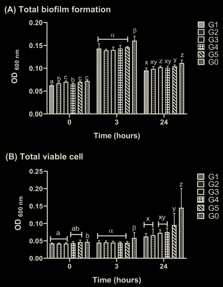
All the CC formulae significantly inhibited biofilm development during the early-prematurity phase (0–3 hours) until the maturity phase (24 hours) ( Figure 1A). Total viable cell analysis showed comparable results 23. CC effectively suppressed both C. albicans and S. mutans viable cells (0–3 hours) ( Figure 1B) . Cell viability (24 hours) was also reduced in the presence of the CC formula, with G2 exhibiting the strongest capacity, similar to the reference group (G1). Figure 2 showed that CC exposure had similar efficacy against both microbes in single biofilm. G3, G4, and G5 did not interfere significantly with the number of C. albicans and S. mutans organisms, whereas G2 exhibited the highest inhibition capacity.

Figure 1.

Figure 1.

Cajuputs candy exposure inhibited C. albicans and S. mutans biofilm development: ( A) total biomass on C. albicans and S. mutans dual-species biofilm, evaluated by crystal violet (CV) assay; ( B) total viable cells on C. albicans and S. mutans dual-species biofilm based on MTT assay. The values were presented as mean and standard deviation of absorbance at 600nm. The letters on histogram represented the significantly different values compared to each other formula within the groups in 0, 3, or 24 hours according to Duncan’s test (p <0.05). G0: untreated control, biofilm group treated with Cajuputs candy made with Melaleuca cajuputi essential oil from different origins denoted by G1: Pulau Buru, G2: Mojokerto, G3: Ponorogo, G4: Pasuruan, and G5: Kuningan.

Figure 2.

Figure 2.

Total plate count (colony forming units/mL) of C. albicans and S. mutans on mixed biofilm ( in vitro): ( A) mean and standard deviation of the C. albicans colonies; ( B) mean and standard deviation of the S. mutans colonies. Different letters represented the significantly different values among the groups according to Duncan’s test (p <0.05). G0: untreated control, biofilm group treated with Cajuputs candy made with Melaleuca cajuputi essential oil from different origins denoted by G1: Pulau Buru, G2: Mojokerto, G3: Ponorogo, G4: Pasuruan, and G5: Kuningan.

Effect of CC on the morphology of dual-species biofilm

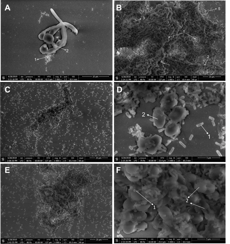
Biofilm development started with the germ-tube formation of C. albicans in the 90 minutes before formula treatments ( Figure 3A). In the maturity stage (24 hours), the hyphal form of C. albicans dominated the biofilm, surrounded by S. mutans accumulation in the untreated control (G0) ( Figure 3B). A corncob-like structure 24 was observed in the mixed biofilm ( Figure 3C).

Figure 3.

Figure 3.

Light microscopy analysis of C. albicans and S. mutans interaction in mixed biofilm: ( A) germ tube formation in the first stage of biofilm development in 90 minutes (40× magnification); ( B) hyphal growth on untreated control of mixed biofilm after 24 hours incubation (20× magnification); ( C) dual-species interaction formed a corn cob-like structure for biofilm treated with Cajuputs candy (40× magnification). Grayscale color adjustment has been performed in order to clarify the figures.

SEM analysis confirmed the germ tube formation in the first 90 minutes in which S. mutans was found close to C. albicans ( Figure 4A). As shown in Figure 4B, hyphal cells grew progressively in the untreated biofilm (G0), enclosed within the self-produced EPS matrix. This co-species population formed a complex structure within the biofilm. Interestingly, the presence of CC altered the architecture of the mixed biofilm. C. albicans tended to be maintained in yeast form, whereas S. mutans adherence to C. albicans was obviously reduced, especially for G2 ( Figure 4C–D). The microcolonies formed were not as many as those in the untreated control. However, exposure to G5 did not affect the interaction and a matrix-rich biofilm was still formed ( Figure 4E–F).

Figure 4.

Figure 4.

In vitro dual-species biofilm formation of C. albicans and S. mutans by scanning electron microscopy: ( A) initial germ-tube formation (3000× magnification); ( B) mixed biofilm of untreated control group (G0) (1000× magnification); ( CD) mixed biofilm under G2 exposure (1000× and 5000× magnification, respectively); ( EF) mixed biofilm under G5 exposure (1000× and 5000× magnification, respectively). The presence of Cajuputs candy reduced the hyphal cells of C. albicans and inhibited matrix production after 24 hours biofilm formation. (1. S. mutans cell; 2. C. albicans yeast and hyphal cells; 3. water channel; 4. extracellular polysaccharides matrix; 5. microcolony).

Effect of CC on the expression of biofilm-related genes

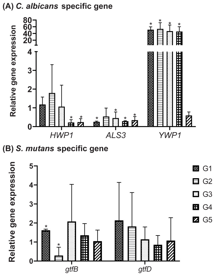
All of the CC groups demonstrated significant downregulation of ALS3, the adhesion-specific gene of C. albicans. HWP1, which is responsible for hyphal filamentation, was still expressed in G1, G2, and G3. However, the expression of YWP1, the yeast-specific gene, was had a higher upregulation in almost all of the CC groups than other specific genes (HWP1 and ALS3) ( Figure 5A). This result confirmed the results of the SEM imaging, that CC exposure tends to maintain the commensal form of C. albicans.

Figure 5.

Figure 5.

qRT-PCR assay of C. albicans and S. mutans biofilm-related genes: ( A) C. albicans-specific genes expression; ( B) S. mutans specific genes expression. An untreated control (G0) was defined as ‘1’. The values were shown as mean and SD. *Significantly regulated than the untreated control (G0) according to Student t-test (p<0.05). G0: untreated control, biofilm group treated with Cajuputs candy made with Melaleuca cajuputi essential oil from different origins denoted by G1: Pulau Buru, G2: Mojokerto, G3: Ponorogo, G4: Pasuruan, and G5: Kuningan.

As for S. mutans gene expression, the greatest downregulation was observed for gtfB in the mixed biofilm exposed to G2, whereas exposure to other formulas still allowed the expression of this insoluble glucan-specific enzyme ( Figure 5B). Regarding gtfD expression (the gene for the soluble glucan enzyme), none of the CC groups had a significant effect on gene regulation compared to the untreated control (G0).

Discussion

CC is a lozenge that has been known as an emerging functional food in Indonesia. Further studies have shown its capability in maintaining oral cavity health due to the antimicrobial capacity of MCEO as its flavor against pathogenic oral microbes 16, 18, 25. In addition to the existing MCEO (PB), we successfully identified four additional MCEOs as potential CC flavors 19. However, the mechanism by which CC interferes with the relationship between the fungus and cariogenic bacteria ( S. mutans) remains unknown. CC consists of isomalt and peppermint oil in addition to MCEO as the main flavor. These ingredients were each added at the same concentration in all of the formulas. Hence, their effect can be assumed as background activity. So far, no studies have been performed to evaluate the efficacy of CC derived from several alternative MCEOs in attenuating the mixed biofilm of S. mutans and C. albicans. Our data show that all the CC groups showed a potent capacity in reducing the biofilm formation composed of these oral microflorae, as well as the viability of biofilm cells, until the biofilm reached its maturation stage. We observed that a higher total biofilm in the early prematurity phase (three hours) dominantly contributed to matrix production since cell viability was maintained at a low level. The colony number confirmed that viability reduction in the mature biofilm was contributed by the reduction in cell numbers of both microbes, with G2 demonstrating the strongest inhibition capacity, similar to our existing MCEO (G1) used as the reference 18.

The interkingdom interaction might begin in the first 90 minutes of biofilm growth, in which a corn-cob-like structure was observed (shown in Figure 3C). This result is in accordance with that of Zijnge et al. 24, who first found that S. mutans cells adhere to the hyphal cells of C. albicans to form this structure. This occurred due to the high affinity of S. mutans cells to the O-mannan group in the C. albicans cell wall 7, 26. Our study showed that G2 exposure intervenes in the C. albicans and S. mutans interaction, indicated by reduction in total biofilm and cell viability (CV and MTT assays, respectively). SEM imaging confirmed these quantitative results. The inhibition effect was related to the morphology alteration of C. albicans into the yeast form, inhibition of S. mutans adherence, and lack of microcolonies compared to the untreated control (G0).

The molecular mechanism underlying the CC inhibition capacity was explained by the expression patterns of selected biofilm-related genes. The adhesion trait of C. albicans was suppressed by ALS3 downregulation when the CC formulas were present. As observed in this study, HWP1 was still expressed in G1–G3. These two genes contribute to hyphal formation as the critical factors in C. albicans biofilm formation 27. However, the gene for the alteration from hyphal to yeast cell ( YWP1) was more dominantly expressed under CC exposure than the other specific gene (ALS3 and HWP1),, indicating that CCs tend to impair biofilm development by maintaining the yeast form of C. albicans with lack of adhesion and further filamentation. This was confirmed by observation of the hyphal form using SEM imaging ( Figure 4).

A parallel investigation of S. mutans genes showed that insoluble glucan production ( gtfB) was inhibited as an effect of G2 exposure, which showed greater inhibitory capacity compared to the G1 reference. In contrast, gtfD was still expressed, similar to the untreated control (G0) in all the CC groups. This means that these genes were still expressed in the biofilm. Furthermore, gtfB is one of the key factors for initiating dual-species interaction 8, 11. It has thus been found to bind C. albicans due to its low dissociation rate, resulting in strong and stable binding such as a covalent bond 26. Lower gtfB expression indicated a fewer matrix formation of S. mutans which important to form a polymicrobial biofilm with C. albicans, as shown by the CV and MTT assays in this study. This result also clearly explained the lack of a matrix on G2 SEM images ( Figure 4C and 4D).

Related to our finding, farnesol (quorum sensing molecule [QSM] of C. albicans) at low concentrations has reported inducing S. mutans growth besides GtfB 10. A lower concentration of farnesol could induce the hyphal form of C. albicans. QSMs are also produced by S. mutans, such as Autoinducer-2 (AI2), which is responsible for suppressing the inhibition capacity of farnesol. Another QSM of S. mutans is competence-stimulating peptide (CSP), which stimulates hyphal-to-yeast alteration 28. The result of this study showed that G2 caused a morphology alteration, which might also be correlated with the impairment of these QSMs. This inter-species signaling might induce the yeast form of C. albicans, which inhibits S. mutans cell accumulation. QSMs in the mixed biofilm was not measured quantitatively or qualitatively in our study. However, this assumption needs to be studied further.

MCEO, as a plant-based antimicrobial used in this experiment, significantly suppressed biofilm formation by reducing the cell number of both the microbes and also inhibited the total biomass production similar to other natural antimicrobials 14, 15, 29. Interestingly, the expression profile of morphology-related genes from C. albicans showed a comparable trend with the synthetic antimicrobial thiazolidinedione-8 (S-8) reported by Fieldman et al. 13. G2 also showed an additional activity of inhibiting S. mutans insoluble glucan production. This observation strengthens the potential of this formula to suppress mixed biofilm formation in vitro.

In general, all of the CC groups indicated potent inhibitory capacity against mixed biofilm formation. Mojokerto performed as the strongest MCEO in CC against the co-species C. albicans and S. mutans biofilm, comparable with the existing MCEO (Pulau Buru). This could be related to their similar metabolite composition as found in our recent work 19. MCEO from Mojokerto is dominated by 1,8-cineole (46.43%), caryophyllene (6.00%), α-terpineol (3.70%), γ-terpinene (3.09), and α-pinene (2.45%). The antibiofilm capacity of this MCEO could be related to these terpenic metabolites, as reported by several studies that essential oils from the Melaleuca genus have various antimicrobial activities 30, 31. Based on the previously published article, 1,8-cineole, a-terpineol, caryophyllene, linalool, terpinene-4-ol, and several other terpene compounds on MCEO were commonly reported as the responsible bioactive compounds on the MCEO antifungal and antibacterial activities 3233. Simşek and Duman 34 further reported the capacity of 1,8-cineole that increases the antimicrobial activity of chlorhexidine gluconate due to its synergistic effect and is expressed as a penetration enhancer. Moreover, Caryophyllene which most found in the MOJ also thought to be correlated with the effect of CC in the biofilm formation as it has been widely reported responsible for the antimicrobial activity 35. Nazzaro et al. 36 summarized their potential mechanisms such as cell wall degradation, affecting the quorum sensing system, and altering adherence capability.

Conclusions

CC showed the ability to impair mixed C. albicans and S. mutans biofilm formation, with Mojokerto being identified as the most effective MCEO. Inhibition of the total biomass and cell viability were related with the candy’s capacity to maintain the commensal phenotype of C. albicans and to suppress insoluble glucan production by S. mutans.

Data availability

Underlying data

Open Science Framework: Cajuputs candy impairs Candida albicans and Streptococcus mutans mixed biofilm formation in vitro. https://doi.org/10.17605/OSF.IO/YT3HQ 23.

This project contains the following underlying data:

  • -

    Raw-unedited image files (original JPG files for images in Figure 3 and Figure 4)

  • -

    Raw Data of total biomass and cell viability.xlsx

  • -

    Raw Data of total plate count of each microbial strains on mixed biofilm.xlsx

  • -

    Raw Data of total qPCR assay on specific genes.xlsx

Data are available under the terms of the Creative Commons Zero "No rights reserved" data waiver (CC0 1.0 Public domain dedication).

Acknowledgements

We would like to thank Prof. Yoshihisa Yamashita, Kyushu University for providing the S. mutans Xc. We also thank Editage for English language editing.

Funding Statement

This study was funded by Directorate General of Resources for Science, Technology, and Higher Education, Ministry of Research, Technology and Higher Education, The Republic of Indonesia, through Program Pendidikan Magister menuju Doktor untuk Sarjana Unggul (PMDSU) 2018.

The funders had no role in study design, data collection and analysis, decision to publish, or preparation of the manuscript.

[version 2; peer review: 2 approved]

References

  • 1. Ghannoum MA, Jurevic RJ, Mukherjee PK, et al. : Characterization of the oral fungal microbiome (mycobiome) in healthy individuals. PLoS Pathog. 2010;6(1):e1000713. 10.1371/journal.ppat.1000713 [DOI] [PMC free article] [PubMed] [Google Scholar]
  • 2. Han TL, Cannon RD, Villas-Bôas SG: The metabolic basis of Candida albicans morphogenesis and quorum sensing. Fungal Genet Biol. 2011;48(8):747–63. 10.1016/j.fgb.2011.04.002 [DOI] [PubMed] [Google Scholar]
  • 3. Moyes DL, Richardson JP, Naglik JR: Candida albicans-epithelial interactions and pathogenicity mechanisms: scratching the surface. Virulence. 2015;6(4):338–46. 10.1080/21505594.2015.1012981 [DOI] [PMC free article] [PubMed] [Google Scholar]
  • 4. Bowen WH, Koo H: Biology of Streptococcus mutans-derived glucosyltransferases: role in extracellular matrix formation of cariogenic biofilms. Caries Res. 2011;45(1):69–86. 10.1159/000324598 [DOI] [PMC free article] [PubMed] [Google Scholar]
  • 5. Koo H, Bowen WH: Candida albicans and Streptococcus mutans: a potential synergistic alliance to cause virulent tooth decay in children. Future Microbiol. 2014;9(12):1295–7. 10.2217/fmb.14.92 [DOI] [PubMed] [Google Scholar]
  • 6. Forssten SD, Björklund M, Ouwehand AC: Streptococcus mutans, caries and simulation models. Nutrients. 2010;2(3):290–8. 10.3390/nu2030290 [DOI] [PMC free article] [PubMed] [Google Scholar]
  • 7. Metwalli KH, Khan SA, Krom BP, et al. : Streptococcus mutans, Candida albicans, and the human mouth: a sticky situation. PLoS Pathog. 2013;9(10):e1003616. 10.1371/journal.ppat.1003616 [DOI] [PMC free article] [PubMed] [Google Scholar]
  • 8. Falsetta ML, Klein MI, Colonne PM, et al. : Symbiotic relationship between Streptococcus mutans and Candida albicans synergizes virulence of plaque biofilms in vivo. Infect Immun. 2014;82(5):1968–81. 10.1128/IAI.00087-14 [DOI] [PMC free article] [PubMed] [Google Scholar]
  • 9. Bachtiar EW, Bachtiar BM: Relationship between Candida albicans and Streptococcus mutans in early childhood caries, evaluated by quantitative PCR. [version 2; referees: 2 approved] Referee Status. F1000Res. 2018;7(1645):1–15. 10.12688/f1000research.16275.2 [DOI] [PMC free article] [PubMed] [Google Scholar]
  • 10. Kim D, Sengupta A, Niepa TH, et al. : Candida albicans stimulates Streptococcus mutans microcolony development via cross-kingdom biofilm-derived metabolites. Sci Rep. 2017;7:41332. 10.1038/srep41332 [DOI] [PMC free article] [PubMed] [Google Scholar]
  • 11. Gregoire S, Xiao J, Silva BB, et al. : Role of glucosyltransferase B in interactions of Candida albicans with Streptococcus mutans and with an experimental pellicle on hydroxyapatite surfaces. Appl Environ Microbiol. 2011;77(18):6357–67. 10.1128/AEM.05203-11 [DOI] [PMC free article] [PubMed] [Google Scholar]
  • 12. Sztajer H, Szafranski SP, Tomasch J, et al. : Cross-feeding and interkingdom communication in dual-species biofilms of Streptococcus mutans and Candida albicans. ISME J. 2014;8(11):2256–71. 10.1038/ismej.2014.73 [DOI] [PMC free article] [PubMed] [Google Scholar]
  • 13. Feldman M, Ginsburg I, Al-Quntar A, et al. : Thiazolidinedione-8 Alters Symbiotic Relationship in C. albicans-S. mutans Dual Species Biofilm. Front Microbiol. 2016;7:140. 10.3389/fmicb.2016.00140 [DOI] [PMC free article] [PubMed] [Google Scholar]
  • 14. Farkash Y, Feldman M, Ginsburg I, et al. : Polyphenols Inhibit Candida albicans and Streptococcus mutans Biofilm Formation. Dent J (Basel). 2019;7(2):pii: E42. 10.3390/dj7020042 [DOI] [PMC free article] [PubMed] [Google Scholar]
  • 15. Ikono R, Vibriani A, Wibowo I, et al. : Nanochitosan antimicrobial activity against Streptococcus mutans and Candida albicans dual-species biofilms. BMC Res Notes. 2019;12(1):383. 10.1186/s13104-019-4422-x [DOI] [PMC free article] [PubMed] [Google Scholar]
  • 16. Wijaya CH, Rachmatillah AF, Bachtiar B: Inhibition of Cajuputs candy toward the viability of Candida albicans by using in vitro assay. J Teknol dan Industri Pangan. 2014;25(2):158–67. [Google Scholar]
  • 17. Bachtiar BM, Srisawat C, Bachtiar EW: RNA aptamers selected against yeast cells inhibit Candida albicans biofilm formation in vitro. Microbiologyopen. 2019;8(8):e00812. 10.1002/mbo3.812 [DOI] [PMC free article] [PubMed] [Google Scholar]
  • 18. Wijaya CH, Fickie A, Nurramdhan IF, et al. : The composition of Cajuputs Candy which inhibits the growth of dental caries microbes.Indonesia; IDP000040695.2016. [Google Scholar]
  • 19. Septiana S, Yuliana ND, Bachtiar BM, et al. : Metabolomics approach for determining potential metabolites correlated with sensory attributes of Melaleuca cajuputi essential oil a promising flavor ingredient. J Biosci Bioeng. 2020;129(5):581–7. 10.1016/j.jbiosc.2019.12.005 [DOI] [PubMed] [Google Scholar]
  • 20. Barbosa JO, Rossoni RD, Vilela SF, et al. : Streptococcus mutans Can Modulate Biofilm Formation and Attenuate the Virulence of Candida albicans. PLoS One. 2016;11(3):e0150457. 10.1371/journal.pone.0150457 [DOI] [PMC free article] [PubMed] [Google Scholar]
  • 21. Krzyściak W, Kościelniak D, Papież M, et al. : Effect of a Lactobacillus salivarius probiotic on a double-species Streptococcus mutans and Candida albicans caries biofilm. Nutrients. 2017;9(11):1242. 10.3390/nu9111242 [DOI] [PMC free article] [PubMed] [Google Scholar]
  • 22. Rodrigues ME, Gomes F, Rodrigues CF: Candida spp./Bacteria mixed biofilms. J Fungi. 2020;6(1):5. 10.3390/jof6010005 [DOI] [PMC free article] [PubMed] [Google Scholar]
  • 23. Septiana S: Cajuputs candy impairs Candida albicans and Streptococcus mutans mixed biofilm formation in vitro .2019. 10.17605/OSF.IO/YT3HQ [DOI] [PMC free article] [PubMed]
  • 24. Zijnge V, Van Leeuwen MB, Degener JE, et al. : Oral biofilm architecture on natural teeth. PLoS One. 2010;5(2):e9321. 10.1371/journal.pone.0009321 [DOI] [PMC free article] [PubMed] [Google Scholar]
  • 25. Wijaya CH, Sari BR, Bachtiar BM: The potency of cajuputs candy in maintaining the competitive capacity of Streptococcus sanguinis upon Streptococcus mutans. J Funct Food Nutraceutical. 2020;1(2):55–65. 10.33555/jffn.v1i2.29 [DOI] [Google Scholar]
  • 26. Hwang G, Marsh G, Gao L, et al. : Binding Force Dynamics of Streptococcus mutans-glucosyltransferase B to Candida albicans. J Dent Res. 2015;94(9):1310–7. 10.1177/0022034515592859 [DOI] [PMC free article] [PubMed] [Google Scholar]
  • 27. Lee KH, Park SJ, Choi SJ, et al. : Proteus vulgaris and Proteus mirabilis Decrease Candida albicans Biofilm Formation by Suppressing Morphological Transition to Its Hyphal Form. Yonsei Med J. 2017;58(6):1135–43. 10.3349/ymj.2017.58.6.1135 [DOI] [PMC free article] [PubMed] [Google Scholar]
  • 28. Xu H, Jenkinson HF, Dongari-Bagtzoglou A: Innocent until proven guilty: mechanisms and roles of Streptococcus-Candida interactions in oral health and disease. Mol Oral Microbiol. 2014;29(3):99–116. 10.1111/omi.12049 [DOI] [PMC free article] [PubMed] [Google Scholar]
  • 29. Jafri H, Khan MSA, Ahmad I: In vitro efficacy of eugenol in inhibiting single and mixed-biofilms of drug-resistant strains of Candida albicans and Streptococcus mutans. Phytomedicine. 2019;54:206–13. 10.1016/j.phymed.2018.10.005 [DOI] [PubMed] [Google Scholar]
  • 30. de Campos Rasteiro VM, da Costa AC, Araújo CF, et al. : Essential oil of Melaleuca alternifolia for the treatment of oral candidiasis induced in an immunosuppressed mouse model. BMC Complement Altern Med. 2014;14:489. 10.1186/1472-6882-14-489 [DOI] [PMC free article] [PubMed] [Google Scholar]
  • 31. Zhang J, Wu H, Jiang D, et al. : The antifungal activity of essential oil from Melaleuca leucadendra (L.) L. grown in China and its synergistic effects with conventional antibiotics against Candida. Nat Prod Res. 2019;33(17):2545–8. 10.1080/14786419.2018.1448979 [DOI] [PubMed] [Google Scholar]
  • 32. Rini P, Ohtani Y, Ichiura H: Antioxidant, anti-hyaluronidase and antifungal activities of Melaleuca leucadendron Linn. leaf oils. J. Wood Sci. 2012;58(5):429–436. 10.1007/s10086-012-1270-x [DOI] [Google Scholar]
  • 33. Wińska K, Mączka W, Łyczko J, et al. : Essential Oils as Antimicrobial Agents—Myth or Real Alternative? Molecules. 2019;24(11):2130. 10.3390/molecules24112130 [DOI] [PMC free article] [PubMed] [Google Scholar]
  • 34. Simsek M, Duman R: Investigation of effect of 1,8-cineole on antimicrobial activity of chlorhexidine gluconate. Pharmacognosy Res. 2017;9(3):234–237. 10.4103/0974-8490.210329 [DOI] [PMC free article] [PubMed] [Google Scholar]
  • 35. Yoo HJ, Jwa SK: Inhibitory effects of β-caryophyllene on Streptococcus mutans biofilm. Arch Oral Biol. 2018;88:42–46. 10.1016/j.archoralbio.2018.01.009 [DOI] [PubMed] [Google Scholar]
  • 36. Nazzaro F, Fratianni F, D’Acierno A, et al. : Essential oils and microbial communication. In: Essential Oils-Oils of Nature IntechOpen;2019. 10.5772/intechopen.85638 [DOI] [Google Scholar]
F1000Res. 2020 Jun 2. doi: 10.5256/f1000research.26745.r63983

Reviewer response for version 2

Mohd Hafiz Arzmi 1, Hasna Ahmad 2

I have no further comment to make.

Is the work clearly and accurately presented and does it cite the current literature?

Yes

If applicable, is the statistical analysis and its interpretation appropriate?

Partly

Are all the source data underlying the results available to ensure full reproducibility?

Yes

Is the study design appropriate and is the work technically sound?

Yes

Are the conclusions drawn adequately supported by the results?

Partly

Are sufficient details of methods and analysis provided to allow replication by others?

Yes

Reviewer Expertise:

Oral microbiology and immunology; natural products; polymicrobial biofilms

We confirm that we have read this submission and believe that we have an appropriate level of expertise to confirm that it is of an acceptable scientific standard.

F1000Res. 2020 Feb 17. doi: 10.5256/f1000research.22766.r58901

Reviewer response for version 1

Dikdik Kurnia 1

  1. The main structure of the research and main data are according and relevant, with all references, and showed the new findings effect on mix biofilm formations.

  2. As an addition if possible, in table 1, the effect of CC against individual bacterial biofilm can be added as a reference or control.

  3. As a suggestion, in the discussion, it would be better to explain the bioactive compounds of  Melaleuca cajuputi reported in other published papers. In this way, we can predict the active constituents of M. cajuputi that show an effect to biofilm formation.

Is the work clearly and accurately presented and does it cite the current literature?

Yes

If applicable, is the statistical analysis and its interpretation appropriate?

Yes

Are all the source data underlying the results available to ensure full reproducibility?

Yes

Is the study design appropriate and is the work technically sound?

Yes

Are the conclusions drawn adequately supported by the results?

Yes

Are sufficient details of methods and analysis provided to allow replication by others?

Yes

Reviewer Expertise:

Bioactive Natural Products

I confirm that I have read this submission and believe that I have an appropriate level of expertise to confirm that it is of an acceptable scientific standard.

F1000Res. 2020 May 21.
Christofora Hanny Wijaya 1

Dear reviewer,

Please accept our sincere apology for this delayed response. We do appreciate the opportunity to revise our paper entitled, Cajuputs candy impairs Candida albicans and Streptococcus mutans mixed biofilm formation in vitro ”. We also extend our deep gratitude to the reviewers for providing valuable comments and correction on our paper. We have read and understood the comments from the reviewers. Please find below our point-by-point response to the reviewer’s comments and concerns. We hope our additional explanations will be able to answer the reviewer's inquiry.

We strived to improve the quality of our paper based on the guidance and constructive suggestion provided by the reviewers. We hope that the revisions will be sufficient to make our paper worthy for publication in F1000 Research. We would like to thank you for your hearty support and willingness to find the parts of our paper needing corrections or improvement. Thank you again for your consideration, suggestions, and insightful feedback in order to greatly improve our paper.

Sincerely yours,

Authors: Siska Septiana, Christofora Hanny Wijaya*, Boy Muchlis Bachtiar and Nancy Dewi Yuliana.

Reviewer Comments and Author Responses

Comment 1: The main structure of the research and main data are according and relevant, with all references, and showed the new findings effect on mix biofilm formations.

Response to reviewer’s comments 1:

Thank you so much for your positive feedback. We greatly appreciate your reviews in our paper.

Comment 2: As an addition if possible, in table 1, the effect of CC against individual bacterial biofilm can be added as a reference or control.

Response to reviewer’s comments 2:

Thank you for the comments. Your suggestion is reasonable, unfortunately, we couldn’t conduct an additional experiment due to the limited sample. However, as reported in the previous study (unpublished report, Wijaya et al. 2014), CC had been proven to has antibiofilm activity on a single biofilm either on S. mutans or on C. albicans. Our study was then conducted as further evaluation of CC against synergistic interaction between the organisms. Therefore, we focused on the effect of CC against the dual-species biofilm, and the effectivity of CC against individual strain was further evaluated using Total Plate Count, as shown in Figure 2.

Comment 3: As a suggestion, in the discussion, it would be better to explain the bioactive compounds of Melaleuca cajuputi reported in other published papers. In this way, we can predict the active constituents of M. cajuputi that show an effect to biofilm formation.

Response to reviewer’s comments 3:

Thank you for your valuable suggestion, your suggestion would enrich our paper. We have added the additional information regarding the possible bioactive compound. The new version of the paragraph can be found as the sixth sentence in the last paragraph in the discussion section:

Based on the previously published article, 1,8-cineole, a-terpineol, caryophyllene, linalool, terpinene-4-ol, and several other terpene compounds on MCEO were commonly reported as the responsible bioactive compounds on the MCEO antifungal and antibacterial activities (Rini et al. 2012; Wińska et al. 2019) . Simsek and Duman (2017) , further reported the capacity of 1,8-cineole that increases the antimicrobial activity of chlorhexidine gluconate due to its synergistic effect and is expressed as a penetration enhancer. Moreover, Caryophyllene which found most in the MOJ sample (Septiana et al. 2020) also thought to be correlated with the effect of CC in the biofilm formation as it has been widely reported responsible for the antimicrobial activity (Yoo and Jwa 2018) .

References:

  • Rini P, Ohtani Y, Ichiura H. 2012. Antioxidant, anti-hyaluronidase and antifungal activities of Melaleuca leucadendron Linn. leaf oils. J. Wood Sci. 58(5):429–436. doi:10.1007/s10086-012-1270-x.

  • Septiana S, Yuliana ND, Bachtiar BM, Putri SP, Fukusaki E, Laviña WA, Wijaya CH. 2020. Metabolomics approach for determining potential metabolites correlated with sensory attributes of Melaleuca cajuputi essential oil, a promising flavor ingredient. J. Biosci. Bioeng. 129(5):581–587. doi:10.1016/j.jbiosc.2019.12.005.

  • Simsek M, Duman R. 2017. Investigation of effect of 1,8-cineole on antimicrobial activity of chlorhexidine gluconate. Pharmacognosy Res. 9(3):234–237. doi:10.4103/0974-8490.210329.

  • Wijaya CH, Rachmatillah AF, M. Bachtiar B. 2014. Inhibition of Cajuputs Candy Toward the Viability of Candida albicans by using In Vitro Assay. J. Teknol. Dan Ind. Pangan 25(2):158–167. doi:10.6066/jtip.2014.25.2.158.

  • Wińska K, Mączka W, Łyczko J, Grabarczyk M, Czubaszek A, Szumny A. 2019. Essential Oils as Antimicrobial Agents—Myth or Real Alternative? Molecules 24(11):1–20. doi:10.3390/molecules24112130.

  • Yoo HJ, Jwa SK. 2018. Inhibitory effects of β-caryophyllene on Streptococcus mutans biofilm. Arch. Oral Biol. 8842–46. doi:10.1016/j.archoralbio.2018.01.009.

F1000Res. 2020 Feb 5. doi: 10.5256/f1000research.22766.r58903

Reviewer response for version 1

Mohd Hafiz Arzmi 1, Hasna Ahmad 2

The manuscript elucidates the activity of Cajuputs to Candida albicans and Streptococcus mutans in mixed biofilms. The effect was assessed using CV assay, MTT, CFU, genes expression and SEM. The authors claim that Cajuput inhibits biofilm and production of polysaccharide by S. mutans.

This study is fascinating; however, major revision is suggested to improve the manuscripts. The following are the comments for authors to consider:

Section: Introduction

  • It would be best if the authors can include the objective/s and the hypothesis of the study in the introduction section.

Section: Methodology

Microbial strains and MCEO sample:

  • Did C. albicans was grown in aerobic or anaerobic?  

  • For MCEO Pulau Buru, what is the temperature in the boiler? If it is 100 degrees Celcius, won't it affect the active compound of the extract? Is this the same method used for the other extracts? If yes, it would be great if you mention that in the paragraph.

CC preparation:

  • Won't 150 degrees Celcius affect your active compound? If that is the method, it would be better if you can put a citation on the method. As the expertise in natural product, it seems the temperature is too high for an extract.  Unless if the method is commonly used in the preparation by the local. 

Mixed biofilm formation:

  • When you prepare the mixed biofilm, you did mention that you grew C. albicans in SDB and S. mutans in BHI in equal volume. Did you mix both media to prepare biofilm? If not, please make it clear in your text.  

  • Why did you coat the well with fetal bovine serum? Better if you can explain why because that is not the standard method used in polymicrobial biofilms study.

  • For you control G0, you mentioned the biofilm was developed without CC. What is the final volume of the well? Did that remain to 200 uL or 140 uL of TSB? If it was 140 uL, how do you compare with other wells which the total volume is 200 uL?

Morphology analysis of dual-species biofilm formation:

  • Why did you choose acrylic as the surface to form a biofilm, and how do you relate this with well that have a different surface?

Section: Results

Effect of CC on dual-species biofilm development:

  • 'Susceptibility' is generally referred to as the antimicrobial activity of the extract. In your research, you are emphasizing on biofilm (Figure 2) and not the antimicrobial effect of the extract.  

  • Figure 1. In the legend, the author mentioned that different letters represent significantly different. What does the author comparing too? Significant compared to what? Better to mention.

  • What is a, b and c represent?  E.g. 'a' in Figure 1A doesn't show significant at 3 hours. The figure seems confusing.  I would suggest improving the figure to ensure understanding of readers.  Similar to figure 2.

Effect of CC on the morphology of dual-species biofilm:

  • Figure 3A doesn't look corncob to me. Please read this article:

    The microbial infection of biomaterials: A challenge for clinicians and researchers. A short review. Journal of Applied Biomaterials & Biomechanics 2005; Vol. 3 no. 1: 1-10 1

  • What do you mean by microcolonies with reference to the figure?  Please explain.

  • How do you determine significant from SEM image? What was the statistical analysis did you use to claim the significant of your SEM? Did you use image J or any software to measure the biofilm based on your SEM?  If yes, better to mention in your methodology. If not, please remove the 'significant'.

Effect of CC on the expression of biofilm-related genes:

  • The author mentioned 'upregulated dominantly'. What do you mean by dominantly and compare to what? G0? G1?

  • What do you mean by 'to maintain the commensal form of C. albicans'?

Section: Discussion

  • The author claims that all CC groups reduce biofilm biomass. What is the author comparing too? Based on my observation, there is no significant difference in total biomass for 24 h and 0 h when all formulation are compared to G0. Please check your statistics.

  • Figure 4D. This doesn't look cocci shape to me. More like bacilli/rod. I hope you did verify your sample prior to the experiment. Attached is the SEM of commonly seen Streptococcus mutans under SEM.

    https://www.nature.com/articles/s41598-017-08558-x 2

  • The author mentioned that lower gtfB indicate the fewer binding site. Glucosyltransferases (Gtfs) are enzymes and not a 'binding site'. This is too speculative. Please consider to rephrase or to remove the statement.

  • The author also emphasised a lot on quorum sensing molecule which is not studied in the manuscript. The author also mentioned that QSM is not measured quantitatively. This statement can be misinterpreted none of the section showed the authors have conducted study on QSM either quantitatively or qualitatively. Please consider rephrasing.

Overall, this is a good study; however, more improvement is needed to fit with F1000research, a Q1 journal.

Is the work clearly and accurately presented and does it cite the current literature?

Yes

If applicable, is the statistical analysis and its interpretation appropriate?

Partly

Are all the source data underlying the results available to ensure full reproducibility?

Yes

Is the study design appropriate and is the work technically sound?

Yes

Are the conclusions drawn adequately supported by the results?

Partly

Are sufficient details of methods and analysis provided to allow replication by others?

Yes

Reviewer Expertise:

Oral microbiology and immunology; natural products; polymicrobial biofilms

We confirm that we have read this submission and believe that we have an appropriate level of expertise to confirm that it is of an acceptable scientific standard, however we have significant reservations, as outlined above.

References

  • 1. : The microbial infection of biomaterials: A challenge for clinicians and researchers. A short review. J Appl Biomater Biomech.3(1) :1-10 [PubMed] [Google Scholar]
  • 2. : Spontaneous detachment of Streptococcus mutans biofilm by synergistic effect between zwitterion and sugar alcohol. Scientific Reports.2017;7(1) : 10.1038/s41598-017-08558-x 10.1038/s41598-017-08558-x [DOI] [PMC free article] [PubMed] [Google Scholar]
F1000Res. 2020 May 21.
Christofora Hanny Wijaya 1

Dear reviewers,

Please accept our sincere apology for this delayed response. We do appreciate the opportunity to revise our paper entitled, Cajuputs candy impairs Candida albicans and Streptococcus mutans mixed biofilm formation in vitro ”. We also extend our deep gratitude to the reviewers for providing valuable comments and correction on our paper. We have read and understood the comments from the reviewers. Please find below our point-by-point response to the reviewers' comments and concerns. We hope our additional explanations will be able to answer the reviewers' inquiry.

We strived to improve the quality of our paper based on the guidance and constructive suggestion provided by the reviewers. We hope that the revisions will be sufficient to make our paper worthy for publication in F1000 Research. We would like to thank you for your hearty support and willingness to find the parts of our paper needing corrections or improvement. Thank you again for your consideration, suggestions, and insightful feedback in order to greatly improve our paper.

Sincerely yours,

Authors: Siska Septiana, Christofora Hanny Wijaya*, Boy Muchlis Bachtiar and Nancy Dewi Yuliana.

Reviewer Comments and Author Responses

Section: Introduction

Comment 1: It would be best if the authors can include the objective/s and the hypothesis of the study in the introduction section.

Response to reviewer’s comments 1:

Thank you very much, we agree with your suggestion. We have emphasized our sentence in the respective paragraph as follows (third sentence in the last paragraph of the introduction section):

“As C. albicans and S. mutans have been noted for their synergistic relationship 5 , 7 , 8 , 10 , we evaluated the capacity of CC to impair their symbiotic interaction in this study” into “This functional candy may have interfered with their synergistic relationship in dual-species biofilm 5 , 7 , 8 , 10 . Therefore, this study aimed to evaluate the capacity of CC to impair their symbiotic interaction .”...

Section: Methodology

Comment 2:

Microbial strains and MCEO sample: Did C. albicans was grown in aerobic or anaerobic?

Response to reviewer’s comments 2:

Thank you for your comments on this issue. We prepare the separate culture of microbial strain based on the previous reports (Ikono et al. 2019). As mention in the literature, the C. albicans as single biofilm was cultured aerobically, after mixed with S. mutans they were cultured anaerobically (CO­ 2 concentration up to 10%) . C. albicans commonly have optimal growth in aerobic condition, however, it also can grow in anaerobic conditions (elevated CO­ 2 concentration) (Anand and Prasad 1991; Thein et al. 2007).

Comment 3: For MCEO Pulau Buru, what is the temperature in the boiler? If it is 100 degrees Celcius, won't it affect the active compound of the extract? Is this the same method used for the other extracts? If yes, it would be great if you mention that in the paragraph.

Response to reviewer’s comments 3:

Thank you for the comments and suggestions. MCEO has been widely reported to have antimicrobial activity (Amri et al. 2012; Rini et al. 2012; Wińska et al. 2019). Related to your concern about the MCEO production, hydrodistillation is the most common method for the essential oil extraction, in which the boiler temperature would possible to achieve 100 °C approximately. It is reasonable that this high temperature might affect several bioactive compounds on the leaves and twigs of the plants. However, the extracted essential oil still had antimicrobial activities due to the presence of various terpene compounds as it has been reported in the above-mentioned publications. Based on our recently published article, it was also known that after passing the high-temperature process in candy making, the important terpene compounds in the essential oil were persisting (Wijaya et al. 2020).

Regarding a similar method that has been used for the other extracts, we have been added the information in the article as suggested by the reviewer ( last sentence in Microbial strains and MCEO samples). “….. the residual water. A similar method was also used for the other extracts . The essential oil is stored in a dark bottle.”

Comment 4: CC preparation

Won't 150 degrees Celcius affect your active compound? If that is the method, it would be better if you can put a citation on the method. As the expertise in natural product, it seems the temperature is too high for an extract.  Unless if the method is commonly used in the preparation by the local.

Response to reviewer’s comments 4:

Thank you very much for your comment on this CC preparation. We agree with your opinion, the high temperature might affect several compounds in MCEO. However, our study was concern about the capacity of CC as functional food after passed this high-temperature process (150 °C) during the preparation, whether the CC still active or not. Moreover, 1,8-cineole, caryophyllene, and a-terpineol as the highest abundance compounds on MCEO and were predicted as the most responsible compound on the MCEO antimicrobial activity commonly had a higher boiling point than the temperature of CC preparation. It had over 176°C for 1,8-cineole ( https://www.chemicalbook.com/ChemicalProductProperty_EN_CB2853653.htm) and over 200°C for caryophyllene, and a-terpineol ( https://www.chemicalbook.com/ProductChemicalPropertiesCB6229317_EN.htm). Moreover, our preliminary study revealed that most of them were still present in the candy after the heating process (Wijaya et al. 2020).

Regarding the CC preparation method, it has been followed the patented procedure as mention in our paper (Wijaya et al. 2016). Based on our result, this functional food showed the expected antimicrobial capacity against dual-species biofilm.

Comment 5: Mixed biofilm formation

When you prepare the mixed biofilm, you did mention that you grew C. albicans in SDB and S. mutans in BHI in equal volume. Did you mix both media to prepare biofilm? If not, please make it clear in your text.

Response to reviewer’s comments 5:

Thank you for your interest in this issue. Yes, we did. 50 µL C. albicans in SDB and 50 µL of  S. mutans in BHI were inoculated together in the same well to promote initial adhesion. As mention in the article ( fourth sentence of mixed biofilm formation section) After the 90 min incubation time, the medium was discarded followed by washing, then, we added Tryptic Soy Broth (TSB)+ 1% sucrose as the medium for the dual-species growth (Ikono et al. 2019). We have also added the reference.

Comment 6: Mixed biofilm formation

Why did you coat the well with fetal bovine serum? Better if you can explain why because that is not the standard method used in polymicrobial biofilms study.

Response to reviewer’s comments 6:

Thank you for your comments regarding our selected method (second sentence in the mixed biofilm formation). Yes, we agree with your opinion. In the dual-species biofilm formation especially for C. albicans and S. mutans, it is common to use saliva coating. In this in vitro study, an ethical clearance to use human saliva was not enclosed. Therefore, we used FBS since either saliva or serum can be used to induce phenotype-associated C. albicans biofilm formation (Barbosa et al. 2016; Krzyściak et al. 2017; Rodrigues et al. 2020). We have embedded this additional information in the third sentence as follows:” Similar with saliva, FBS coating aims to induce phenotype-associated C. albicans biofilm formation (Barbosa et al. 2016; Krzyściak et al. 2017; Rodrigues et al. 2020).

Comment 7: Mixed biofilm formation

For you control G0, you mentioned the biofilm was developed without CC. What is the final volume of the well? Did that remain to 200 uL or 140 uL of TSB? If it was 140 uL, how do you compare with other wells which the total volume is 200 uL?

Response to reviewer’s comments 7:

Thanks for your deep evaluation of this mixed biofilm formation. The final volume for control (G0) was similar to other wells (200 uL). To achieve the equal volume, we added PBS (pH 7) on the control to replace the CC formula. We have added this information as the sixth sentence on this mixed biofilm formation paragraph: For the untreated control, the formula was replaced by 60 µL sterile phosphate-buffered saline (PBS)“. We expect that this PBS had no inhibition capacity on the culture. Therefore, as expected, all of the formulae (G1-G5) tested showed a significantly different effect compared to the control.

Comment 8: Morphology analysis of dual-species biofilm formation

Why did you choose acrylic as the surface to form a biofilm, and how do you relate this with well that have a different surface?

Response to reviewer’s comments 8:

Thank you so much for your question. We conducted this analysis based on the previously published paper which used a similar surface (acrylic disc) (Barbosa et al. 2016). The use of acrylic disc (on 24-well plates) was one of the common methods for the morphology analysis of microbial biofilm to facilitate the biofilm sample to be analyzed by using SEM.  Moreover, by using SEM, we only focused on the altered morphology between the organisms in control and those treated biofilms (within the group), thus we grew the biofilm on the same surface as mention in the literature.

Section: Results

Comment 9: Effect of CC on dual-species biofilm development

'Susceptibility' is generally referred to as the antimicrobial activity of the extract. In your research, you are emphasizing on biofilm (Figure 2) and not the antimicrobial effect of the extract. 

Response to reviewer’s comments 9:

Thank you so much for your concern. We would like to confirm that in our case, we used the term susceptibility to describe the condition of the cultures by the presence of the CC formula. In another word, this term was focus on the object exposed by the extract whether it is susceptible or resistant. However, we have rephrased this term in the whole text as you suggested. we used “ efficacy” to describe the measurement of antimicrobial activity of the CC formula to impair the dual-species biofilm formation.

Comment 10: Effect of CC on dual-species biofilm development

Figure 1. In the legend, the author mentioned that different letters represent significantly different. What does the author comparing too? Significant compared to what? Better to mention.

Response to reviewer’s comments 10:

Thank you for your review. The statistical analysis on CV assay and MTT assay (Figure 1) was conducted using ANOVA followed by Duncan’s test. In this post hoc analysis, all of the formulae were compared to each other (G0 to G1, G2, G3, G4, G5; G1 to G2, G3, G4, G5; and so on with similar way) within the group in 0, 3, and 24 hours. The significantly different results within the group were then indicated by different letters. We have revised the figure and completed the figure legend with the additional information as suggested.

“Figure 1. … absorbance at 600nm. The letters on histogram represented the significantly different values compared to each other formula within the groups in 0, 3, or 24 hours according to Duncan’s test (p <0.05)…”

Comment 11: Effect of CC on dual-species biofilm development

What is a, b and c represent?  E.g. 'a' in Figure 1A doesn't show significant at 3 hours. The figure seems confusing. I would suggest improving the figure to ensure understanding of readers.  Similar to figure 2.

Response to reviewer’s comments 11:

Similar as the response to the reviewer’s comment no. 10, the use of the different letter (a, b, and c or x, y, and z) was only to describe the significantly different values between the formula (G0-G5) within the group in 0, 3, and 24 hours of incubation time, e.g: in 24 hours the statistic analysis result showed by x, y, and z for the significantly different value among the formulae, whereas “xy” was not significantly different with x and y.

Based on Duncan’s test, G1-G5 in 3 hours incubation was showed by ‘a’ and it was significantly different from the negative control which had ‘b’ (Figure 1A). This was our expected result, in which the exposure of CC after 3 hours could inhibit the biofilm formation effectively. However, different superscript symbol/letter between 0, 3, or 24 hours does not correlate each other since the values were analyzed within the group.

Thank you for your suggestion to revised our figure. Unfortunately, it seems ineffective to separate each incubation time for CV and MTT assay similar to Figure 2. It will need six additional figures for this revision. Therefore, we have revised the symbol/letter on the histogram to differentiate each incubation time (the revised Figure 1). We hope that this revision would be sufficient to enhance the readers' understanding.

(the revised Figure 1 was uploaded separately)

“Figure 1. … absorbance at 600nm . The letters on histogram represented the significantly different values compared to each other formula within the groups in 0, 3, or 24 hours according to Duncan’s test (p <0.05)…”

Comment 12: Effect of CC on the morphology of dual-species biofilm

Figure 3A doesn't look corncob to me. Please read this article: The microbial infection of biomaterials: A challenge for clinicians and researchers. A short review. Journal of Applied Biomaterials & Biomechanics 2005; Vol. 3 no. 1: 1-101

Response to reviewer’s comments 12:

Thank you, we appreciate your deep review on our figure and your attached literature. Figure 3A describes how the growth of the microbes in the first 90 minutes period, before the exposure to the CC formula. Figure 3B showed the biofilm growth after 24 h, whereas figure 3C indicated an interaction between C. albicans and S. mutans after the exposure of CC. The interaction between those two microbes commonly known as corn-cob like structure (Zijnge et al. 2010). Although Figure 3C still not clearly showed this structure due to the limitation of the light microscopy that we used in the analysis, it indicates that this structure might occur since S. mutans was attached to the C. albicans hyphae as describe in the figure.

Comment 13: Effect of CC on the morphology of dual-species biofilm

What do you mean by microcolonies with reference to the figure?  Please explain.

Response to reviewer’s comments 13:

Regarding the SEM figure, there is part of the figure which indicates the presence of microcolonies (no. 5 in Figure 4B). This microcolony was referred to a microscopic colony of S. mutans cells without considering its interaction with C. albicans.

Comment 14: Effect of CC on the morphology of dual-species biofilm

How do you determine significant from SEM image? What was the statistical analysis did you use to claim the significant of your SEM? Did you use image J or any software to measure the biofilm based on your SEM?  If yes, better to mention in your methodology. If not, please remove the 'significant'.

Response to reviewer’s comments 14:

Thank you so much for your correction. We didn’t conduct the quantitative analysis on SEM images. We have removed this term through the whole text as suggested.

Comment 15: Effect of CC on the expression of biofilm-related genes

The author mentioned 'upregulated dominantly'. What do you mean by dominantly and compare to what? G0? G1?

Response to reviewer’s comments 15:

Thank you for your review of this case. The mRNA expression level was presented as the relative result of its comparison to control (G0) which had value as 1 (one). After this relative quantification, YWP1 has upregulated tens of times higher than other C. albicans specific genes (HWP1 and ALS3), this is what we stated as YPW1 'upregulated dominantly' which compares to other genes. As it might be confusing the readers, we have revised this sentence into “…expressed in G1, G2, and G3. However, the expression of YWP1, the yeast-specific gene, was had a higher upregulation in almost all of the CC groups than other specific genes (HWP1 and ALS3) (Figure 5A).” (third sentence of Result section on paragraph Effect of CC on the expression of biofilm-related genes)

Comment 16: What do you mean by 'to maintain the commensal form of C. albicans'?

Response to reviewer’s comments 16:

Thank you for your interest in this part. C. albicans is commonly recognized as the most prominent human commensal fungi which could grow by taking nutrient on the human body without interfering the human body homeostasis. This condition occurs when C. albicans is in the yeast state. Unfortunately, the phenotype switching into hyphae often caused this commensal fungus to become a pathogen (Finkel and Mitchell 2011). This morphological change represented several virulence genes which might be induced as mention in the third sentence of our introduction section “The hyphal form is relevant for its virulence as it allows penetration and invasion of epithelial cells (Moyes et al. 2015)”. Therefore, the exposure of CC on the dual-species biofilm was expected to maintain the yeast state of C. albicans so that its commensal properties remain. In other words, the commensal form describes the yeast state of C. albicans. This yeast form would suppress its virulence and to minimize the interaction with S. mutans.

Section: Discussion

Comment 17: The author claims that all CC groups reduce biofilm biomass. What is the author comparing too? Based on my observation, there is no significant difference in total biomass for 24 h and 0 h when all formulation are compared to G0. Please check your statistics.

Response to reviewer’s comments 17:

Thank you for your further review of this data. Our statement was based on Duncan’s test as our statistical approach. As describe in the response no.10-11, this test showed that G1-G5 had different superscript letters with G0 on each groups (0, 3 and 24 h) for the total biofilm which indicated significantly different values have occurred (Figure 1A). It also means that the CC formula significantly affected biofilm formation on 0, 3 and 24 h without comparing the group.

Comment 18: Figure 4D. This doesn't look cocci shape to me. More like bacilli/rod. I hope you did verify your sample prior to the experiment. Attached is the SEM of commonly seen Streptococcus mutans under SEM.

Response to reviewer’s comments 18:

Thank you very much for your deep review of our SEM images. We have verified our sample prior to the analysis. The different shapes with your attached literature might be correlated with the different strains that have been used. We used S. mutans serotype c strain, the most isolated strain from dental plaque, which possible to have a different shape with S. mutans ATCC 25175 in your attached literature (Lim et al. 2017). Our SEM analysis showed that S. mutans serotype c had longer shape than ATCC 25175 strain.

Our analysis method was conducted based on literature (Barbosa et al. 2016), which also used S. mutans UA159 (a serotype c strain). In their report, S. mutans also had a long shape (showed in Figure 3) similar to our result. Our image also supported by Feldman et al. (Feldman et al. 2016) which reported that biofilm was dominated by the long shape of S. mutans UA159 (a serotype c strain) which was described in Figure 3 in the literature. Sztajer et al. (Sztajer et al. 2014) had a clear visualization of dual-species biofilm of C. albicans and S. mutans UA159 (Figure 1e-f), in which the long shape S. mutans attached to the hyphae of  C. albicans. Therefore, it was reasonable that our S. mutans strain also have a long shape.

Comment 19: The author mentioned that lower gtfB indicate the fewer binding site. Glucosyltransferases (Gtfs) are enzymes and not a 'binding site'. This is too speculative. Please consider to rephrase or to remove the statement.

Response to reviewer’s comments 19:

Thank you for your correction. We understand your opinion about this issue. Based on our result, the exposure of G2 caused the fewer gtfB expression level. This fewer gtfB contributed to the fewer extracellular polysaccharide matrix formation, in which this matrix is needed as a mediator of interaction between C. albicans and S. mutans. Therefore, we have revised our sentence as suggested (fifth sentence in fourth paragraph of Discussion section) into : “…Lower gtfB expression indicated a fewer matrix formation of S. mutans which important to form a polymicrobial biofilm with C. albicans, as shown by the CV and MTT assays in this study.”

Comment 20: The author also emphasised a lot on quorum sensing molecule which is not studied in the manuscript. The author also mentioned that QSM is not measured quantitatively. This statement can be misinterpreted none of the section showed the authors have conducted study on QSM either quantitatively or qualitatively. Please consider rephrasing.

Response to reviewer’s comments 20:

Thank you for your response. The quorum-sensing molecule information was added in the text as the most possible mechanism that supports our result based on the literature. Several QSM has been reported to be involved in the dual-species interaction between C. albicans and S. mutans. We assumed that we should embed this explanation as additional information to enrich the readers' insight related to the dual-species biofilm formation. Although QSM was not measured in our study, these molecules might have a role in their synergism. Therefore, we have revised our last sentence in this QSM paragraph into “…QSMs in the mixed biofilm was not measured quantitatively or qualitatively in our study. However, this assumption needs to be studied further.”

References

  • Amri I, Mancini E, de Martino L, Marandino A, Lamia H, Mohsen H, Bassem J, Scognamiglio M, Reverchon E, de Feo V. 2012. Chemical composition and biological activities of the essential oils from three Melaleuca species grown in Tunisia. Int. J. Mol. Sci. 13(12):16580–16591. doi:10.3390/ijms131216580.

  • Anand S, Prasad R. 1991. Growth and Respiration Characteristics of Candida albicans. In Candida Albicans, R. Prasad (Ed. by), Springer, Berlin, Heidelberg, pp. 46–61.

  • Barbosa JO, Rossoni RD, Vilela SFG, De Alvarenga JA, Velloso MDS, De Azevedo Prata MC, Jorge AOC, Junqueira JC. 2016. Streptococcus mutans can modulate biofilm formation and attenuate the virulence of Candida Albicans. PLoS One 11(3):1–16. doi:10.1371/journal.pone.0150457.

  • Feldman M, Ginsburg I, Al-quntar A, Steinberg D. 2016. Thiazolidinedione-8 Alters Symbiotic Relationship in C . albicans - S . mutans Dual Species Biofilm. Front. Microbiol. 7(140):1–12. doi:10.3389/fmicb.2016.00140.

  • Finkel JS, Mitchell AP. 2011. Genetic control of Candida albicans biofilm development. Nat. Rev. Microbiol. 9(2):109–118. doi:10.1109/isdrs.2005.1596138.

  • Ikono R, Vibriani A, Wibowo I, Saputro KE, Muliawan W, Bachtiar BM, Mardliyati E, Bachtiar EW, Rochman NT, Kagami H, et al. 2019. Nanochitosan antimicrobial activity against Streptococcus mutans and Candida albicans dual ‑ species biofilms. BMC Res. Notes 12(383):1–7. doi:10.1186/s13104-019-4422-x.

  • Krzyściak W, Kościelniak D, Papież M, Vyhouskaya P, Zagórska-Świeży K, Kołodziej I, Bystrowska B, Jurczak A. 2017. Effect of a Lactobacillus salivarius probiotic on a double-species streptococcus mutans and Candida albicans caries biofilm. Nutrients 9(11):1–23. doi:10.3390/nu9111242.

  • Lim JH, Song SH, Park HS, Lee JR, Lee SM. 2017. Spontaneous detachment of Streptococcus mutans biofilm by synergistic effect between zwitterion and sugar alcohol. Sci. Rep. 7(1):1–9. doi:10.1038/s41598-017-08558-x.

  • Rini P, Ohtani Y, Ichiura H. 2012. Antioxidant, anti-hyaluronidase and antifungal activities of Melaleuca leucadendron Linn. leaf oils. J. Wood Sci. 58(5):429–436. doi:10.1007/s10086-012-1270-x.

  • Rodrigues ME, Gomes F, Rodrigues CF. 2020. Candida spp./Bacteria Mixed Biofilms. J. Fungi 6(5):1–29.

  • Sztajer H, Szafranski SP, Tomasch J, Reck M, Nimtz M, Rohde M, Wagner-Dobler I. 2014. Cross-feeding and interkingdom communication in dual-species biofilms of Streptococcus mutans and Candida albicans. Int. Soc. Microb. Ecol. 81–16. doi:10.1038/ismej.2014.73.

  • Thein ZM, Samaranayake YH, Samaranayake LP. 2007. In vitro biofilm formation of Candida albicans and non-albicans Candida species under dynamic and anaerobic conditions. Arch. Oral Biol. 52(8):761–767. doi:10.1016/j.archoralbio.2007.01.009.

  • Wijaya CH, Sari BR, Bachtiar BM. 2020. The potency of cajuputs candy in maintaining the competitive capacity of Streptococcus sanguinis upon Streptococcus mutans. J. Funct. Food Nutraceutical 1(2):55–65. doi:10.33555/jffn.v1i2.29.

  • Wijaya CH, Fickie A, Nurramdhan IF, Bachtiar BM, Afandi FA, Indariani S. 2016. The Composition of Cajuputs Candy which inhibits the growth of dental caries microbes. Indonesia; IDP000040695.

  • Wińska K, Mączka W, Łyczko J, Grabarczyk M, Czubaszek A, Szumny A. 2019. Essential Oils as Antimicrobial Agents—Myth or Real Alternative? Molecules 24(11):1–20. doi:10.3390/molecules24112130.

  • Zijnge V, Leeuwen MBM Van, Degener JE, Abbas F, Thurnheer T, Gmu R. 2010. Oral Biofilm Architecture on Natural Teeth Oral Biofilm Architecture on Natural Teeth. PLoS One 5(2):1–9. doi:10.1371/journal.pone.0009321.

Associated Data

    This section collects any data citations, data availability statements, or supplementary materials included in this article.

    Data Citations

    1. Septiana S: Cajuputs candy impairs Candida albicans and Streptococcus mutans mixed biofilm formation in vitro .2019. 10.17605/OSF.IO/YT3HQ [DOI] [PMC free article] [PubMed]

    Data Availability Statement

    Underlying data

    Open Science Framework: Cajuputs candy impairs Candida albicans and Streptococcus mutans mixed biofilm formation in vitro. https://doi.org/10.17605/OSF.IO/YT3HQ 23.

    This project contains the following underlying data:

    • -

      Raw-unedited image files (original JPG files for images in Figure 3 and Figure 4)

    • -

      Raw Data of total biomass and cell viability.xlsx

    • -

      Raw Data of total plate count of each microbial strains on mixed biofilm.xlsx

    • -

      Raw Data of total qPCR assay on specific genes.xlsx

    Data are available under the terms of the Creative Commons Zero "No rights reserved" data waiver (CC0 1.0 Public domain dedication).


    Articles from F1000Research are provided here courtesy of F1000 Research Ltd

    RESOURCES