Abstract
Although bronchogenic cysts are the most common primary mediastinal cysts, intracardiac bronchogenic cysts are extremely rare. We report a case of a bronchogenic cyst of the interatrial septum in a 42-year-old woman who presented with recent onset of dyspnea on exertion. Cardiac investigations including transthoracic echocardiography and computed tomography revealed a cystic homogeneous mass in the interatrial septum. The patient underwent surgical resection, and the resultant atrial septal defect was repaired using an autologous pericardial patch. Histopathological examination of the resected specimen revealed findings consistent with a benign bronchogenic cyst. Although bronchogenic cysts are extremely rare, they should be considered in the differential diagnoses of intracardiac tumors. Complete resection of bronchogenic cysts is recommended primarily for diagnostic and potentially therapeutic purposes.
Keywords: Bronchogenic cyst, Interatrial septum, Cardiac tumor, Surgery
Introduction
Bronchogenic cysts are congenital malformations that result from abnormal budding of the ventral foregut during embryologic development. They represent the most common mediastinal cystic masses; however, intracardiac bronchogenic cysts are extremely rare. We present a rare case of a bronchogenic cyst that originated in the interatrial septum.
Case Report
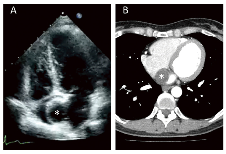
A 42-year-old woman with an unremarkable medical history was admitted with recent onset of dyspnea on exertion. Physical examination, electrocardiography, chest radiography, and blood tests revealed no abnormalities. Transthoracic echocardiography revealed a cyst-like structure attached to the interatrial septum, protruding into the right atrium. This mass measured 2.9 × 2.2 cm and showed well-defined margins (Fig. 1A). Computed tomography (CT) revealed a well-defined, homogeneous hypodense mass in the low interatrial septum (Fig. 1B). CT angiography revealed no feeding vessels from the coronary arteries.
Fig. 1.

(A) Preoperative transthoracic echocardiography scan and (B) contrast-enhanced computed tomography scan showing a cystic mass in the interatrial septum (asterisk).
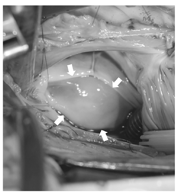
Surgical excision of the cystic tumor was planned, and the patient underwent standard median sternotomy and cannulation of the ascending aorta and the superior and inferior vena cava. Cardiac arrest was induced with cold blood cardioplegia. The right atrium was opened and a 2.5 cm round cyst with a smooth surface was identified in the fossa ovalis (Fig. 2). The mass was completely excised from the interatrial septum and the resultant atrial septal defect after cyst resection was repaired using an autologous pericardial patch. Weaning from extracorporeal circulation was uneventful. The cyst contained whitish-yellow colored mucous fluid. The patient’s postoperative course was uneventful, and she was discharged on the 10th postoperative day. The patient is asymptomatic without any evidence of recurrence over 2-year’s follow-up.
Fig. 2.

Intraoperative images showing a cystic mass (white arrows).
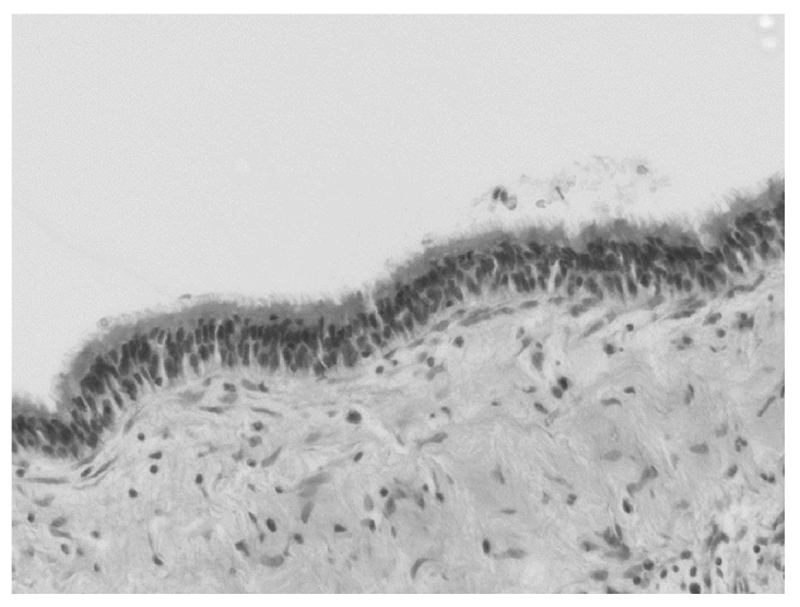
Histopathological examination of the resected cyst showed that it was lined with pseudostratified ciliated columnar epithelium, and the findings were consistent with a bronchogenic cyst without any evidence of malignancy (Fig. 3).
Fig. 3.

Histopathological examination showing a layer of ciliated columnar epithelium lining the inner side of the cyst, as well as acute inflammatory cells (hematoxylin & eosin stain, ×40).
Discussion
Bronchogenic cysts develop as congenital malformations following differentiation of the primitive foregut. Usually, these lesions occur in the mediastinum particularly within the lungs, and intracardiac bronchogenic cysts are extremely rare. Cardiac primordia are located in close proximity to the primitive bronchial tree. Abnormal budding of the primitive bronchial tree with bronchogenic cyst formation approximately during the 4 th week of embryogenic development can cause incorporation of the cyst within the primitive heart during formation of the heart wall, and this cyst subsequently manifests as an intracardiac bronchogenic cyst 1, 2). Bronchogenic cysts usually contain clear fluid and are lined with ciliated columnar or cuboidal epithelium and may also present with smooth muscle and cartilage.
Bronchogenic cysts are often discovered incidentally and are usually asymptomatic; however, they cause a variety of symptoms secondary to compression of adjacent structures. Compression of cardiac chambers can cause dyspnea, as observed in our case. Azeem et al. reported a rare case of a bronchogenic cyst presenting as acute coronary syndrome 3). Shiohira et al. reported a case of ventricular fibrillation secondary to this rare condition 4).
Echocardiography, CT, and magnetic resonance imaging are useful for diagnosis of bronchogenic cysts. The differential diagnosis includes thrombus, myxoma, papillary fibroelastoma, or metastasis. Bronchogenic cysts should be considered in the differential diagnosis of intracardiac cystic tumors.
Surgical excision of benign cardiac tumors is controversial. In a study performed by Kirmani et al., surgical resection of all mediastinal cysts was recommended, although approximately 33% of patients were asymptomatic 5). This recommendation could be based on the following factors:
Biopsy of such cysts is associated with the risk of embolization of the cystic fluid.
A few reports in the available literature have described potentially malignant bronchogenic cysts.
Surgical excision is the only modality available to confirm the diagnosis and exclude malignancy that may rarely develop in a bronchogenic cyst.
Furthermore, Yanagawa et al. suggested that most simple primary and some complex primary cardiac tumors should be surgically resected 6).
Patients undergoing surgical resection of intracardiac bronchogenic cysts show good prognosis.
Conclusions
Although bronchogenic cysts of the interatrial septum are extremely rare, they should be considered in the differential diagnosis of an intracardiac cystic tumor. Surgical resection is recommended for definitive diagnosis of such lesions.
Conflict of Interest
None declared.
References
- 1.Jiang H, Wang H, Wu H, Li X. Bronchogenic cyst of the interatrial septum. J Cardiothorac Surg, 8: 171-174, 2013. [DOI] [PMC free article] [PubMed] [Google Scholar]
- 2.Chen CC. Bronchogenic cyst in the interatrial septum with a single persistent left superior vena cava. J Chin Med Assoc, 69: 89-91, 2006. [DOI] [PubMed] [Google Scholar]
- 3.Azeem F, Finlay M, Rathwell C, Awad WI. A near fatal presentation of a bronchogenic cyst compressing the left main coronary artery. J Thorac Cardiovasc Surg, 135: 1395-1396, 2008. [DOI] [PubMed] [Google Scholar]
- 4.Shiohira S, Sasaki T, Maeda S, Kawabata M, Goya M, et al. Bronchogenic cyst of the atrioventricular septum presenting with ventricular fibrillation. HeartRhythm Case Rep, 3: 389-391, 2017. [DOI] [PMC free article] [PubMed] [Google Scholar]
- 5.Kirmani B, Kirmani B, Sogliani F. Should asymptomatic bronchogenic cysts in adults be treated conservatively or with surgery? Interact Cardiovasc Thorac Surg, 11: 649-659, 2010. [DOI] [PubMed] [Google Scholar]
- 6.Yanagawa B, Chan EY, Cusimano RJ, Reardon MJ. Approach to surgery for cardiac tumors: Primary simple, primary complex, and secondary. Cardiol Clin, 37: 525-531, 2019. [DOI] [PubMed] [Google Scholar]


