Abstract
Studies published in recent years have demonstrated that abnormal long noncoding RNA (lncRNA) antisense RNA to TP73 gene (TP73-AS1) expression is markedly associated with tumorigenesis, cancer progression and the prognosis of cancer patients. We aimed to explore the prognostic value of TP73-AS1 in multiple cancers. We comprehensively searched PubMed, Embase, Web of Science and the Cochrane Library (up to February 21, 2019). Hazard ratios (HRs), odds ratios (ORs) and the corresponding 95% confidence intervals (95% CIs) were calculated to estimate the association of TP73-AS1 with survival and clinicopathological features. The potential targets and pathways of TP73-AS1 in multiple cancers were summarized. Nineteen studies that involved thirteen types of cancers and 1329 cancer patients were identified as eligible for this meta-analysis. The results showed that high TP73-AS1 expression was significantly correlated with shorter overall survival (OS) (HR = 1.962, 95% CI 1.630-2.362) and disease-free survival (DFS) (HR = 2.050, 95% CI 1.293-3.249). The summary HRs of OS were 2.101 (95% CI 1.516-2.911) for gastric cancer (GC) and 1.920 (95% CI 1.253-2.942) for osteosarcoma. Subgroup analysis of OS demonstrated that the differential expression of TP73-AS1 in cancer tissues was a potential source of heterogeneity. Furthermore, increased TP73-AS1 expression was markedly associated with larger tumor size (OR = 2.759, 95% CI 1.759-4.330), advanced histological grade (OR = 2.394, 95% CI 1.231-4.656), lymph node metastasis (OR = 2.687, 95% CI 1.211-5.962), distant metastasis (OR = 4.145, 95% CI 2.252-7.629) and advanced TNM stage (OR = 2.633, 95% CI 1.507-4.601). The results of Egger’s test and sensitivity analysis verified the robustness of the original results. High TP73-AS1 expression can predict poor survival and poor clinicopathological features in cancer patients and TP73-AS1 might be a potential biomarker and therapeutic target.
Subject terms: Cancer, Prognostic markers
Introduction
Cancer is an essential public health problem and a major cause of death worldwide. In 2019, Approximately 1,762,450 cancer cases will be diagnosed and 606,880 cancer patients will die in America as estimated by the American Cancer Society1. Although great advances in diagnoses and treatments have improved survival, the prognosis of patients with advanced cancers remains poor2. The exploration of novel therapeutic targets and prognostic biomarkers is a top priority. LncRNAs, comprising more than 200 nucleotides, are engaged in many biological processes, such as cell proliferation, differentiation, the DNA damage response and chromosomal imprinting3,4. A large number of studies have suggested that lncRNAs are tightly linked with oncogenicity and cancer progression5–7.
An antisense RNA to the TP73 gene (TP73-AS1), also named KIAA0495/PDAM, is located in the 1p36 chromosome region that is frequently deleted in tumors and comprises tumor suppressor genes8. TP73-AS1 functions as a tumor suppressor by sponging miR-941 at nine high-affinity binding sites9. TP73-AS1 can suppress the progression of bladder cancer by epithelial-mesenchymal transition (EMT), and low TP73-AS1 expression predicts a shorter survival of bladder cancer patients10. However, TP73-AS1 also acts as a tumor promoter. For example, TP73-AS1 can promote hepatocellular carcinoma (HCC) cell proliferation via miR-200a-dependent HMGB1/RAGE regulation, and its high expression level is significantly associated with a poorer prognosis of HCC patients11. Knockdown of TP73-AS1 suppresses the proliferation and invasion of osteosarcoma cells by sponging miR-14212. TP73-AS1, which is upregulated in cholangiocarcinoma (CCA) tissues, predicts adverse phenotypes for CCA, and silencing TP73-AS1 attenuates CCA cell proliferation, migration and invasion13.
Therefore, we performed this meta-analysis based on studies focusing on the association of TP73-AS1 with survival and clinicopathological features. We aimed to explore the potential of TP73-AS1 as a novel biomarker and therapeutic target.
Materials and methods
Search strategy
Two authors (Yuan Zhong and Yang Yu) independently screened PubMed, Web of Science, the Cochrane Library and Embase up to February 21,2019. The search terms were used as follows: (“TP73-AS1” OR “P73 antisense RNA 1 T” OR “antisense RNA to TP73 gene” OR “KIAA0495” OR “PDAM”) AND (“cancer” OR “tumor” OR “carcinoma” OR “neoplasm”).
Selection criteria
Inclusion criteria were as follows: (1) TP73-AS1 expression was detected in cancer tissues; (2) Patients were divided into two groups based on TP73-AS1 expression levels; (3) The association between TP73-AS1 expression and the prognosis of patients with cancer was investigated; and (4) Sufficient data were extracted to compute the HR and 95% CI of survival or the OR and 95% CI of clinicopathological parameters. Exclusion criteria were as follows: (1) Nonhuman studies, reviews, editorials, expert opinions, letters, and case reports; (2) Studies lacking key data; and (3) Duplicate publications.
Data extraction and quality assessment
Two authors extracted the following requisite data in each qualified article: first author’s name, publication year, country, cancer type, sample type, sample size, detection method, cut-off value, the number of patients in the high and low TP73-AS1 expression groups, survival analysis method, the HR and 95% CI of TP73-AS1 for survival and data for age, gender, TNM stage, tumor size, lymph node metastasis, etc. If the HR was not accessible from the original text, we utilized Engauge Digitizer v.10.11 software to extract data from the Kaplan-Meier survival curve and calculated the HR with assistance from Tierney’s spreadsheet14. The HR with the 95% CI from multivariate analysis was preferred over that from the univariate analysis because the multivariate analysis considered the effects of other factors. Two authors independently assessed the quality of each eligible study under the guidance of the Newcastle-Ottawa Quality Assessment Scale (NOS)15.
Statistical analysis
Pooled HRs with 95% CIs were calculated to investigate the value of TP73-AS1 on the survival of patients with cancer. Pooled ORs with 95% CIs were used to evaluate the association between TP73-AS1 expression and clinicopathological features. The heterogeneity of the pooled results was explored by means of the Q test and I2 statistics. The fixed pooling model was selected when I2 < 50% and p > 0.10. Otherwise, the random pooling model was used. Subgroup analysis was further conducted to explore the potential sources of heterogeneity. Sensitivity analysis was performed to test whether each single study could influence the stability of the results. Egger’s test was utilized to assess a publication bias. A p value less than 0.05 was considered statistically significant.
Results
Characteristics of the included studies
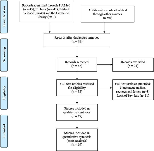
A total of 19 articles were eligible for this meta-analysis according to the selection criteria9–13,16–27. In total, 1329 patients with 13 types of cancer, including GC9,16–18, breast cancer13,19, ovarian cancer20,21, osteosarcoma12,22, brain glioma23, hepatocellular carcinoma (HCC)11, cholangiocarcinoma (CCA)13, colorectal cancer (CRC)27, pancreatic cancer24, lung adenocarcinoma (LAD)25, non-small cell lung cancer (NSCLC)19, bladder cancer10 and clear cell renal cell carcinoma (ccRCC)26, were analyzed. Twelve articles chose overall survival (OS) as the only survival outcome9,11–13,17–19,21–25, 2 articles used both OS and disease-free survival (DFS)16,26, and one article used both OS and progression-free survival (PFS)10. All studies were performed in China. TP73-AS1 expression levels were measured by quantitative real-time PCR (qRT-PCR). Patients were classified into high or low expression groups according to the cut-off value. More details are shown in Fig. 1 and Table 1.
Figure 1.
Flow diagram of this meta-analysis.
Table 1.
Characteristics of the included studies.
| Cancer | First author | Year | Country | Sample type | Sample size(n) | Detection method | Cut-off value | PT | Survival analysis | Multivariate analysis | Hazard ratios | NOS score |
|---|---|---|---|---|---|---|---|---|---|---|---|---|
| GC | Wei Zhang9 | 2018 | China | Tissue | 76 | qRT-PCR | NA | no | OS | No | K-M curve | 7 |
| GC | Yufeng Wang16 | 2018 | China | Tissue | 64 | qRT-PCR | NA | NA | OS, DFS | No | K-M curve | 6 |
| GC | Jianjun Peng17 | 2018 | China | Tissue | 58 | qRT-PCR | mean | no | OS | Yes | K-M curve | 6 |
| GC | Zhi Ding18 | 2018 | China | Tissue | 72 | qRT-PCR | median | no | OS | no | K-M curve | 6 |
| Breast cancer | Qiongyan Zou19 | 2017 | China | Tissue | 86 | qRT-PCR | median | NA | NA | No | NA | 8 |
| Breast cancer | Jia Yao13 | 2017 | China | Tissue | 36 | qRT-PCR | 2-fold | NA | OS | Yes | Text | 7 |
| Ovarian cancer | Xiaoqian Wang20 | 2018 | China | Tissue | 60 | qRT-PCR | median | NA | NA | No | NA | 8 |
| Ovarian cancer | Xiuyun Li34 | 2018 | China | Tissue | 62 | qRT-PCR | mean | no | OS | Yes | K-M curve | 6 |
| Osteosarcoma | Xi Chen35 | 2018 | China | Tissue | 132 | qRT-PCR | NA | no | OS | Yes | Text | 7 |
| Osteosarcoma | Guangling Yang12 | 2018 | China | Tissue | 46 | qRT-PCR | mean | no | OS | No | K-M curve | 8 |
| Brain glioma | Rong Zhang23 | 2017 | China | Tissue | 47 | qRT-PCR | median | NA | OS | Yes | Text | 7 |
| HCC | Shaling Li11 | 2017 | China | Tissue | 84 | qRT-PCR | median | NA | OS | no | K-M curve | 8 |
| Pancreatic cancer | Xianping Cui24 | 2019 | China | Tissue | 77 | qRT-PCR | NA | NA | OS | No | K-M curve | 7 |
| LAD | Chunfeng Liu25 | 2019 | China | Tissue | 80 | qRT-PCR | median | no | OS | Yes | K-M curve | 7 |
| NSCLC | Lin Zhang19 | 2018 | China | Tissue | 45 | qRT-PCR | median | NA | OS | No | K-M curve | 7 |
| Bladder cancer | Zhiyong Tuo10 | 2018 | China | Tissue | 128 | qRT-PCR | median | no | OS, PFS | No | K-M curve | 7 |
| ccRCC | Guanghua Liu26 | 2018 | China | Tissue | 40 | qRT-PCR | median | no | OS, DFS | No | K-M curve | 8 |
| CCA | Yue Yao13 | 2018 | China | Tissue | 75 | qRT-PCR | mean | no | NA | No | NA | 8 |
| CRC | Zeming Jia27 | 2019 | China | Tissue | 61 | qRT-PCR | NA | no | NA | No | NA | 7 |
Abbreviations: GC, gastric cancer; HCC, hepatocellular carcinoma; LAD, lung adenocarcinoma; NSCLC, non-small cell lung cancer; ccRCC, clear cell renal cell carcinoma; CCA, cholangiocarcinoma; CRC, colorectal cancer; PT, preoperative treatment; NA, Not available.
Association between TP73-AS1 and survival
TP73-AS1 was relatively upregulated in most cancer tissues compared to paired normal or non-cancerous tissues except for bladder cancer specimens. As shown in Fig. 2, high TP73-AS1 expression was significantly associated with poor OS (HR = 1.962, 95% CI 1.630-2.362) with mild heterogeneity (I2 = 34.9%) and short DFS (HR = 2.050, 95% CI 1.293-3.249). High TP73-AS1 expression only in bladder cancer indicated long OS (HR = 0.400, 95% CI 0.180-0.880).
Figure 2.
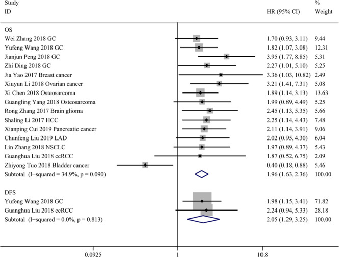
Forest plots for association of TP73-AS1 expression with overall survival (OS) and disease-free survival (DFS). Abbreviations: GC, gastric cancer; HCC, hepatocellular carcinoma; LAD, lung adenocarcinoma; NSCLC, non-small cell lung cancer; ccRCC, clear cell renal cell carcinoma.
Subgroup analysis of the association between TP73-AS1 and OS was performed by examining TP73-AS1 expression in cancer tissue (high or low), cancer type (digestive system or non-digestive system), analysis method (multivariate analysis or univariate analysis) and NOS score (≥7 or <7), as shown in Table 2. The result of each subgroup was not significantly changed. Subgroup analysis revealed that differential expression of TP73-AS1 in cancer tissues was the potential source of heterogeneity.
Table 2.
Subgroup analysis of the pooled HRs for OS.
| Subgroup analysis | Number of studies | Number of patients | HR (95% CI) | P value | Heterogeneity | |
|---|---|---|---|---|---|---|
| I2 | P value | |||||
| Overall | 15 | 1047 | 1.962 (1.630-2.362) | p <0.001 | 34.9% | 0.09 |
| Expression in cancer tissue | ||||||
| High | 14 | 919 | 2.151 (1.777-2.603) | p <0.001 | 0.0% | 0.971 |
| Low | 1 | 128 | 0.400 (0.181-0.884) | 0.024 | NA | NA |
| Cancer type | ||||||
| Digestive system | 6 | 431 | 2.124 (1.629-2.770) | p <0.001 | 0.0% | 0.671 |
| Non-digesstive system | 9 | 616 | 1.837 (1.230-2.744) | 0.003 | 54.6% | 0.024 |
| Analysis method | ||||||
| Multivariate analysis | 6 | 415 | 2.452 (1.816-3.310) | p <0.001 | 0.0% | 0.653 |
| Univariate analysis | 9 | 632 | 1.709 (1.350-2.164) | p <0.001 | 45.8% | 0.064 |
| NOS score | ||||||
| ≥7 | 11 | 791 | 1.803 (1.449-2.244) | p <0.001 | 39.2% | 0.088 |
| <7 | 4 | 256 | 2.437 (1.716-3.461) | p <0.001 | 0.5% | 0.389 |
Four studies focusing on GC showed that high TP73-AS1 expression markedly predicted poor OS (HR = 2.101, 95% CI 1.516-2.911). Two studies on osteosarcoma demonstrated that high TP73-AS1 expression was significantly correlated with short OS (HR = 1.920, 95% CI 1.253-2.942). More details are provided in Fig. 3.
Figure 3.
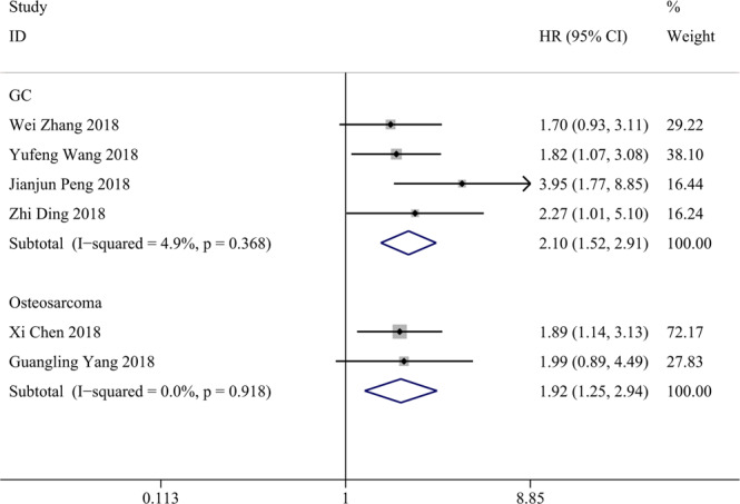
Forest plots for association of TP73-AS1 expression with overall survival (OS) in gastric cancer (GC) and osteosarcoma.
Association between TP73-AS1 and clinicopathological features
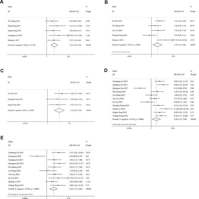
The results demonstrated that high TP73-AS1 expression was not significantly associated with the age or gender of cancer patients (age: OR = 1.082, 95% CI 0.817-1.433; gender: OR = 1.034, 95% CI 0.772-1.385). As shown in Fig. 4, high TP73-AS1 expression was significantly related to large tumor size (OR = 2.759, 95% CI 1.759-4.330), advanced histological grade (OR = 2.394, 95% CI 1.231-4.656), lymph node metastasis (OR = 2.687, 95% CI 1.211-5.962), distant metastasis (OR = 4.145, 95% CI 2.252-7.629) and advanced TNM stage (OR = 2.633, 95% CI 1.507-4.601).
Figure 4.
Forest plots for association of TP73-AS1 expression with clinicopathological features: (A) Tumor size; (B) Histological grade; (C) Distant metastasis; (D) Lymph node metastasis; (E) TNM stage.
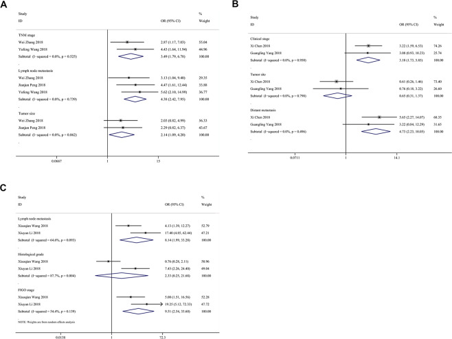
In Fig. 5, studies on GC showed that high TP73-AS1 expression was significantly associated with lymph node metastasis (OR = 3.792, 95% CI 1.793-8.018) and large tumor size (OR = 2.137, 95% CI 1.087-4.203) in GC patients. Two articles on breast cancer demonstrated that high TP73-AS1 expression was significantly linked with large tumor size (OR = 4.201, 95% CI 1.961-8.999), advanced TNM stage (OR = 5.764, 95% CI 2.637-12.598) and the absence of lymph node metastasis (OR = 0.234, 95% CI 0.057-0.966) but not with ER status (OR = 1.220, 95% CI 0.596-2.497) or PR status (OR = 1.700, 95% CI 0.828-3.491). Two studies on osteosarcoma supported that high TP73-AS1 expression was notably associated with advanced clinical stage (OR = 3.184, 95% CI 1.732-5.854) and distant metastasis (OR = 4.730, 95% CI 2.225-10.053) but not with tumor site (OR = 0.650, 95% CI 0.309-1.366). Two studies concerning ovarian cancer revealed an association of TP73-AS1 with lymph node metastasis (OR = 6.616, 95% CI 3.241-13.504) and histological grade (OR = 2.129, 95% CI 1.046-4.336). Moreover, two articles showed a significant association of TP73-AS1 with lymph node metastasis (OR = 8.139, 95% CI 1.990-33.282) and advanced FIGO stage (OR = 9.514, 95% CI 2.542-35.604) but not with histological grade (OR = 2.333, 95% CI 0.251-21.682).
Figure 5.
Forest plots for association of TP73-AS1 expression with clinicopathological features in 3 cancers: (A) Gastric cancer; (B) Osteosarcoma; (C) Ovarian cancer.
Publication bias and sensitivity analysis
Egger’s test was conducted to explore a possible publication bias. As shown in Fig. 6, the symmetrical Egger’s plot of all eligible studies for OS showed no significant publication bias(p = 0.633).
Figure 6.
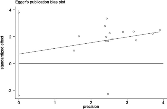
Egger’s test for publication bias of results of overall survival (OS).
Sensitivity analysis was performed to estimate the stability of the original OS results. The robustness of the meta-analysis conclusions was confirmed because the results were not altered significantly when any study was eliminated (Fig. 7).
Figure 7.
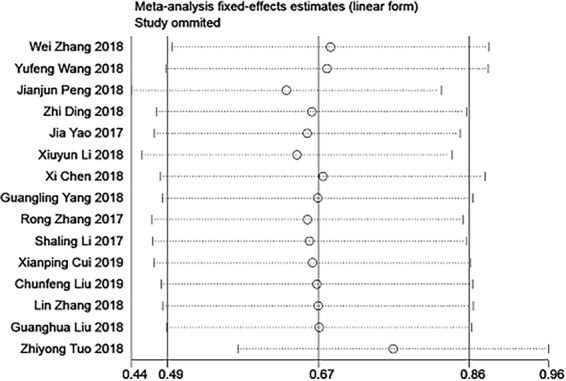
Sensitivity analysis for studies about OS by omitting each study sequentially.
Discussion
A large number of articles have identified that lncRNAs such as lncRNA PVT1, lncRNA HULC, and lncRNA CRNDE are closely related to the initiation and progression of various cancers and could serve as prognostic biomarkers28–30. TP73-AS1 was initially described in a study on oligodendroglial tumors, which revealed that TP73-AS1 was downregulated in tumor samples mainly via epigenetic modifications and chromosome 1p loss, and the knockdown of TP73-AS1 induced cisplatin resistance in glioma cells31. High TP73-AS1 expression was associated with larger tumor size, advanced WHO stage and a shorter OS in glioma patients23. Tuo et al. reported that low TP73-AS1 expression predicted unfavorable survival and diminished bladder cancer cell proliferation, migration and invasion but facilitated apoptosis10. In contrast, TP73-AS1 promoted oesophageal cancer cell proliferation and inhibited apoptosis32. Knockdown of TP73-AS1 inhibited the vasculogenic mimicry formation of triple-negative breast cancer cells by targeting the miR-490-3p/TWIST1 axis33. Liu et al. revealed that TP73-AS1, upregulated ccRCC tissues, predicted the prognosis of ccRCC patients and promoted ccRCC cell proliferation and invasion but inhibited apoptosis26. These discrepancies show the different roles of TP73-AS1 in multiple cancers.
In this meta-analysis, nineteen eligible studies with 1329 cancer patients were included. Pooled results indicated that TP73-AS1 overexpression was significantly associated with poor OS and DFS. The association of TP73-AS1 with OS was robust in either gastric cancer or osteosarcoma. Subgroup analysis of OS indicated no significant change in any subgroup. Furthermore, high TP73-AS1 expression was markedly related to larger tumor size, lymph node metastasis, distant metastasis, advanced TNM stage and histological grade but not with age or gender.
Furthermore, we payed attention to potential targets and pathways of TP73-AS1 in the included cancers (Table 3). TP73-AS1 functioned as an oncogenic lncRNA in most cancers except bladder cancer and CRC. In this meta-analysis, high TP73-AS1 expression was associated with long OS only in bladder cancer. Tuo et al. found that in bladder cancer overexpression of TP73-AS1 could inhibit cell growth, arrest cell cycle, reduce cell migration and invasion, promote cell apoptosis, and diminish EMT by inhibiting the expression of vimentin, snail, MMP-2, and MMP-9 and upregulating the expression of E-cadherin10. On the contrary, TP73-AS1 could promote tumor progression through regulating HMGB1/RAGE pathway in GC, brain glioma and HCC. TP73-AS1 could target miR-142 both in osteosarcoma and brain glioma. In addition, TP73-AS1 was linked to chemoresistance in both GC and glioblastoma. TP73-AS1 might not only act as a biomarker of chemosensitivity, but also as a novel therapeutic target.
Table 3.
Summary of potential targets and pathways of TP73-AS1.
| Cancer type | Expression | Potential target | Pathway | References |
|---|---|---|---|---|
| GC | Upregulated | NA | Cell proliferation, apoptosis, invasion and metastatic properties; tumorigenesis; cisplatin resistance; Bcl-2/caspase-3 pathway; EMT; WNT/β-catenin signaling pathway; HMGB1/RAGE signaling pathway; miR-194-5p/SDAD1 axis | 9,16–18 |
| Breast cancer | Upregulated | miR-200a; miR-490-3p | Cell invasion, migration and vasculogenic mimicry; TFAM; TP73-AS1/miR-200a/ZEB1 regulating loop; miR-490-3p/TWIST1 axis | 13,19,33 |
| Ovarian cancer | Upregulated | p21; EZH2 | Cell proliferation, apoptosis, invasion and migration; tumor growth; MMP2 and MMP9 | 20,21,36 |
| Osteosarcoma | Upregulated | miR-142 | Cell proliferation, migration and invasion; tumor growth; TP73-AS1/miR-142/Rac1 signaling pathway | 12,22 |
| Brain glioma | Upregulated | miR-142; miR-124 | Cell proliferation and invasion; temozolomide resistance; HMGB1/RAGE pathway; ALDH1A1; TP73-AS1/miR-124/iASPP axis | 23,37,38 |
| HCC | Upregulated | miR-200a | Cell proliferation; HMGB1/RAGE pathway and NF-кB expression | 11 |
| Pancreatic cancer | Upregulated | miR-141 | Cell migration, invasion and metastasis; BDH2 | 24 |
| LAD | Upregulated | NA | Cell proliferation, apoptosis, migration and invasion; tumor growth and metastasis; PI3K/AKT pathway | 25 |
| NSCLC | Upregulated | miR-449a | Cell growth and cycle progression; TP73-AS1/miR-449a/EZH2 axis | 19 |
| Bladder cancer | Downregulated | NA | Cell proliferation, apoptosis, migration and invasion; EMT | 10 |
| ccRCC | Upregulated | KISS1 | Cell proliferation, invasion and apoptosis; PI3K/Akt/mTOR pathway | 26 |
| CCA | Upregulated | NA | Cell proliferation, apoptosis and metastatic properties; tumor growth | 13 |
| CRC | Downregulated | miR-103; miR-194 | Cell proliferation, apoptosis migration and invasion; tumor growth; TP73-AS1/miR-103 axis; PTEN; TP73-AS1/miR-194/TGFα axis | 27,39 |
The limitations of this meta-analysis deserve to be mentioned. First, each study was conducted in China, which means that the results need to be verified by studies from other countries. Second, most studies had a small sample size (n < 100), which might result in inflated estimates of effect size. Third, the cut-off values were not available in 5 studies. Finally, we calculated the HRs from Kaplan-Meier curves in 12 studies, which may have affected the accuracy of the results.
Conclusion
High TP73-AS1 expression in multiple cancers predicted poor OS, poor DFS, larger tumor size, lymph node metastasis, distant metastasis, advanced TNM stage and histological grade. TP73-AS1 might be a novel prognostic biomarker and therapeutic target for cancers. More high-quality studies with a large sample size and a standardized methodologic design are required to further certify the prognostic value of TP73-AS1 in cancers.
Acknowledgements
This study was supported by the Project of Standard Diagnosis and Treatment of Key Disease of Jiangsu Province (BE2015722).
Author contributions
Yuan Zhong collected and analyzed the data, wrote the paper; Meng Zhao analyzed the data, revised the paper; Yang Yu collected the data; Quanpeng Li and Fei Wang conceived and designed this study; Yuan Zhong, Meng Zhao, Yang Yu, Quanpeng Li, Fei Wang, Peiyao Wu, Wen Zhang and Lin Miao reviewed the paper. All authors read and approved the final manuscript.
Data availability
All data are included in the article.
Competing interests
The authors declare no competing interests.
Footnotes
Publisher’s note Springer Nature remains neutral with regard to jurisdictional claims in published maps and institutional affiliations.
These authors contributed equally: Yuan Zhong, Meng Zhao and Yang Yu.
References
- 1.Siegel RL, Miller KD, Jemal A. Cancer statistics, 2019. CA Cancer J Clin. 2019;69:7–34. doi: 10.3322/caac.21551. [DOI] [PubMed] [Google Scholar]
- 2.Miller KD, et al. Cancer treatment and survivorship statistics, 2016. CA Cancer J Clin. 2016;66:271–289. doi: 10.3322/caac.21349. [DOI] [PubMed] [Google Scholar]
- 3.Schmitt AM, et al. An inducible long noncoding RNA amplifies DNA damage signaling. Nature genetics. 2016;48:1370–1376. doi: 10.1038/ng.3673. [DOI] [PMC free article] [PubMed] [Google Scholar]
- 4.Khorkova O, Hsiao J, Wahlestedt C. Basic biology and therapeutic implications of lncRNA. Advanced drug delivery reviews. 2015;87:15–24. doi: 10.1016/j.addr.2015.05.012. [DOI] [PMC free article] [PubMed] [Google Scholar]
- 5.Huang D, et al. NKILA lncRNA promotes tumor immune evasion by sensitizing T cells to activation-induced cell death. Nature immunology. 2018;19:1112–1125. doi: 10.1038/s41590-018-0207-y. [DOI] [PubMed] [Google Scholar]
- 6.Sun TT, et al. LncRNA GClnc1 Promotes Gastric Carcinogenesis and May Act as a Modular Scaffold of WDR5 and KAT2A Complexes to Specify the Histone Modification Pattern. Cancer discovery. 2016;6:784–801. doi: 10.1158/2159-8290.Cd-15-0921. [DOI] [PubMed] [Google Scholar]
- 7.Zhang Y, et al. Analysis of the androgen receptor-regulated lncRNA landscape identifies a role for ARLNC1 in prostate cancer progression. Nature genetics. 2018;50:814–824. doi: 10.1038/s41588-018-0120-1. [DOI] [PMC free article] [PubMed] [Google Scholar]
- 8.Kaghad M, et al. Monoallelically expressed gene related to p53 at 1p36, a region frequently deleted in neuroblastoma and other human cancers. Cell. 1997;90:809–819. doi: 10.1016/S0092-8674(00)80540-1. [DOI] [PubMed] [Google Scholar]
- 9.Hu H, et al. Recently Evolved Tumor Suppressor Transcript TP73-AS1 Functions as Sponge of Human-Specific miR-941. Mol Biol Evol. 2018;35:1063–1077. doi: 10.1093/molbev/msy022. [DOI] [PMC free article] [PubMed] [Google Scholar]
- 10.Tuo Z, Zhang J, Xue W. LncRNA TP73-AS1 predicts the prognosis of bladder cancer patients and functions as a suppressor for bladder cancer by EMT pathway. Biochem Biophys Res Commun. 2018;499:875–881. doi: 10.1016/j.bbrc.2018.04.010. [DOI] [PubMed] [Google Scholar]
- 11.Li S, et al. The long non-coding RNA TP73-AS1 modulates HCC cell proliferation through miR-200a-dependent HMGB1/RAGE regulation. J Exp Clin Cancer Res. 2017;36:51. doi: 10.1186/s13046-017-0519-z. [DOI] [PMC free article] [PubMed] [Google Scholar]
- 12.Yang G, Song R, Wang L, Wu X. Knockdown of long non-coding RNA TP73-AS1 inhibits osteosarcoma cell proliferation and invasion through sponging miR-142. Biomed Pharmacother. 2018;103:1238–1245. doi: 10.1016/j.biopha.2018.04.146. [DOI] [PubMed] [Google Scholar]
- 13.Yao Y, Sun Y, Jiang Y, Qu L, Xu Y. Enhanced expression of lncRNA TP73-AS1 predicts adverse phenotypes for cholangiocarcinoma and exerts oncogenic properties in vitro and in vivo. Biomed Pharmacother. 2018;106:260–266. doi: 10.1016/j.biopha.2018.06.045. [DOI] [PubMed] [Google Scholar]
- 14.Tierney JF, Stewart LA, Ghersi D, Burdett S, Sydes MR. Practical methods for incorporating summary time-to-event data into meta-analysis. Trials. 2007;8:16. doi: 10.1186/1745-6215-8-16. [DOI] [PMC free article] [PubMed] [Google Scholar]
- 15.Lo CK, Mertz D, Loeb M. Newcastle-Ottawa Scale: comparing reviewers’ to authors’ assessments. BMC medical research methodology. 2014;14:45. doi: 10.1186/1471-2288-14-45. [DOI] [PMC free article] [PubMed] [Google Scholar]
- 16.Wang Y, Xiao S, Wang B, Li Y, Chen Q. Knockdown of lncRNA TP73-AS1 inhibits gastric cancer cell proliferation and invasion via the WNT/beta-catenin signaling pathway. Oncol Lett. 2018;16:3248–3254. doi: 10.3892/ol.2018.9040. [DOI] [PMC free article] [PubMed] [Google Scholar]
- 17.Peng J. si-TP73-AS1 suppressed proliferation and increased the chemotherapeutic response of GC cells to cisplatin. Oncol Lett. 2018;16:3706–3714. doi: 10.3892/ol.2018.9107. [DOI] [PMC free article] [PubMed] [Google Scholar]
- 18.Ding Z, Lan H, Xu R, Zhou X, Pan Y. LncRNA TP73-AS1 accelerates tumor progression in gastric cancer through regulating miR-194-5p/SDAD1 axis. Pathol Res Pract. 2018;214:1993–1999. doi: 10.1016/j.prp.2018.09.006. [DOI] [PubMed] [Google Scholar]
- 19.Zou Q, et al. A TP73-AS1/miR-200a/ZEB1 regulating loop promotes breast cancer cell invasion and migration. J Cell Biochem. 2018;119:2189–2199. doi: 10.1002/jcb.26380. [DOI] [PubMed] [Google Scholar]
- 20.Wang X, Yang B, She Y, Ye Y. The lncRNA TP73-AS1 promotes ovarian cancer cell proliferation and metastasis via modulation of MMP2 and MMP9. J Cell Biochem. 2018;119:7790–7799. doi: 10.1002/jcb.27158. [DOI] [PubMed] [Google Scholar]
- 21.Li X, Wang X, Mao L, Zhao S, Wei H. LncRNA TP73AS1 predicts poor prognosis and promotes cell proliferation in ovarian cancer via cell cycle and apoptosis regulation. Mol Med Rep. 2018;18:516–522. doi: 10.3892/mmr.2018.8951. [DOI] [PubMed] [Google Scholar]
- 22.Chen Xi, Zhou Yu, Liu Shuping, Zhang Desheng, Yang Xi, Zhou Qing, Song Yueming, Liu Yuehong. LncRNA TP73‐AS1 predicts poor prognosis and functions as oncogenic lncRNA in osteosarcoma. Journal of Cellular Biochemistry. 2018;120(2):2569–2575. doi: 10.1002/jcb.27556. [DOI] [PubMed] [Google Scholar]
- 23.Zhang R, Jin H, Lou F. The Long Non-Coding RNA TP73-AS1 Interacted With miR-142 to Modulate Brain Glioma Growth Through HMGB1/RAGE Pathway. J Cell Biochem. 2018;119:3007–3016. doi: 10.1002/jcb.26021. [DOI] [PubMed] [Google Scholar]
- 24.Cui, X. P. et al. LncRNA TP73-AS1 sponges miR-141-3p to promote the migration and invasion of pancreatic cancer cells through the upregulation of BDH2. Biosci Rep, 10.1042/BSR20181937 (2019). [DOI] [PMC free article] [PubMed]
- 25.Liu, C., Ren, L., Deng, J. & Wang, S. LncRNA TP73-AS1 promoted the progression of lung adenocarcinoma via PI3K/AKT pathway. Biosci Rep39, 10.1042/BSR20180999 (2019). [DOI] [PMC free article] [PubMed]
- 26.Liu G, et al. LncRNA TP73-AS1 Promotes Cell Proliferation and Inhibits Cell Apoptosis in Clear Cell Renal Cell Carcinoma Through Repressing KISS1 Expression and Inactivation of PI3K/Akt/mTOR Signaling Pathway. Cell Physiol Biochem. 2018;48:371–384. doi: 10.1159/000491767. [DOI] [PubMed] [Google Scholar]
- 27.Jia Z, et al. Long non-coding RNA TP73AS1 promotes colorectal cancer proliferation by acting as a ceRNA for miR103 to regulate PTEN expression. Gene. 2019;685:222–229. doi: 10.1016/j.gene.2018.11.072. [DOI] [PubMed] [Google Scholar]
- 28.Chen X, et al. lncRNA PVT1 identified as an independent biomarker for prognosis surveillance of solid tumors based on transcriptome data and meta-analysis. Cancer Manag Res. 2018;10:2711–2727. doi: 10.2147/CMAR.S166260. [DOI] [PMC free article] [PubMed] [Google Scholar]
- 29.Chen X, et al. The Value of lncRNA HULC as a Prognostic Factor for Survival of Cancer Outcome: A Meta-Analysis. Cell Physiol Biochem. 2017;41:1424–1434. doi: 10.1159/000468005. [DOI] [PubMed] [Google Scholar]
- 30.Liang C, et al. Long non-coding RNA CRNDE as a potential prognostic biomarker in solid tumors: A meta-analysis. Clin Chim Acta. 2018;481:99–107. doi: 10.1016/j.cca.2018.02.039. [DOI] [PubMed] [Google Scholar]
- 31.Pang JC, et al. KIAA0495/PDAM is frequently downregulated in oligodendroglial tumors and its knockdown by siRNA induces cisplatin resistance in glioma cells. Brain Pathol. 2010;20:1021–1032. doi: 10.1111/j.1750-3639.2010.00405.x. [DOI] [PMC free article] [PubMed] [Google Scholar]
- 32.Zang W, et al. Knockdown of long non-coding RNA TP73-AS1 inhibits cell proliferation and induces apoptosis in esophageal squamous cell carcinoma. Oncotarget. 2016;7:19960–19974. doi: 10.18632/oncotarget.6963. [DOI] [PMC free article] [PubMed] [Google Scholar]
- 33.Tao W, Sun W, Zhu H, Zhang J. Knockdown of long non-coding RNA TP73-AS1 suppresses triple negative breast cancer cell vasculogenic mimicry by targeting miR-490-3p/TWIST1 axis. Biochem Biophys Res Commun. 2018;504:629–634. doi: 10.1016/j.bbrc.2018.08.122. [DOI] [PubMed] [Google Scholar]
- 34.Li X, Wang X, Mao L, Zhao S, Wei H. LncRNA TP73-AS1 predicts poor prognosis and promotes cell proliferation in ovarian cancer via cell cycle and apoptosis regulation. Molecular medicine reports. 2018;18:516–522. doi: 10.3892/mmr.2018.8951. [DOI] [PubMed] [Google Scholar]
- 35.Chen X, et al. LncRNA TP73-AS1 predicts poor prognosis and functions as oncogenic lncRNA in osteosarcoma. Journal of cellular biochemistry. 2019;120:2569–2575. doi: 10.1002/jcb.27556. [DOI] [PubMed] [Google Scholar]
- 36.Li Y, Jiao Y, Hao J, Xing H, Li C. Long noncoding RNA TP73-AS1 accelerates the epithelial ovarian cancer via epigenetically repressing p 21. Am J Transl Res. 2019;11:2447–2454. [PMC free article] [PubMed] [Google Scholar]
- 37.Xiao S, Wang R, Wu X, Liu W, Ma S. The Long Noncoding RNA TP73-AS1 Interacted with miR-124 to Modulate Glioma Growth by Targeting Inhibitor of Apoptosis-Stimulating Protein of p53. DNA Cell Biol. 2018;37:117–125. doi: 10.1089/dna.2017.3941. [DOI] [PubMed] [Google Scholar]
- 38.Mazor G, et al. The lncRNA TP73-AS1 is linked to aggressiveness in glioblastoma and promotes temozolomide resistance in glioblastoma cancer stem cells. Cell Death Dis. 2019;10:246. doi: 10.1038/s41419-019-1477-5. [DOI] [PMC free article] [PubMed] [Google Scholar]
- 39.Cai Y, et al. Long non-coding RNA TP73-AS1 sponges miR-194 to promote colorectal cancer cell proliferation, migration and invasion via up-regulating TGFalpha. Cancer Biomark. 2018;23:145–156. doi: 10.3233/CBM-181503. [DOI] [PubMed] [Google Scholar]
Associated Data
This section collects any data citations, data availability statements, or supplementary materials included in this article.
Data Availability Statement
All data are included in the article.


