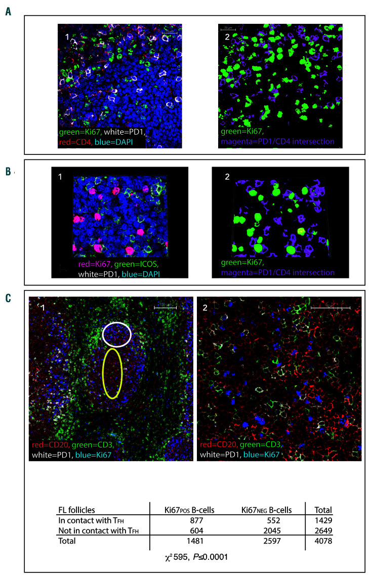
Figure 3.
Ki67pos cells are in close proximity to follicular helper T cells (TFH) in follicular lymphoma lymph nodes. (A1) Representative image of a neoplastic follicle showing Ki67pos cells (green) in close proximity to CD4pos (red), PD-1Hi (white) T cells. The scale bar represents 25 μm. (A2) Binary image of (A1), the binary layers of Ki67 (green) and the CD4-PD-1Hi intersection (magenta) are shown highlighting the close association of Ki67pos cells to PD-1Hi T cells. (B1) Representative image demonstrating that the majority of the PD-1Hi cells in contact with Ki67pos cells (red) are also positive for ICOS (green). (B2) This is highlighted in the binary layer 3D reconstruction of the same image, PD-1/ICOSpos (magenta) and Ki67 (green). Images representative of n=100 images from n=23 follicular lymphoma (FL) samples (4A), n=43 images from n=13 samples (4B). (C1) Ki67=blue, CD20=red, PD-1=white, CD3=green. Low power image (x10) showing Ki67pos and Ki67neg CD20pos B-cell co-localisation with PD1Hi CD3pos T cells in FL. Within the follicles there are areas of low proliferation (low Ki67=blue) where there are few PD1Hi (white) CD3pos T cells (green) - area highlighted by yellow oval, whereas in areas where there is high Ki67, there are more PD1Hi, CD3pos T cells (area highlighted by white circle) and they are frequently in contact with Ki67pos CD20pos FL B cells. Scale bar represents 100 μm. (C2) High power image (x60) in which the close correlation of Ki67pos (blue) B cells with PD1Hi (white) CD3pos (green) cells can be seen, whilst the CD20pos (red), Ki67neg cells are less frequently in contact with follicular helper T cells (TFH). Scale bar represents 50 μm. (C3) contingency tables showing that Ki67pos B cells are significantly more likely to be in contact with TFH than Ki67neg B-cells in FL (for all samples analyzed together [n=25 images from n=7 follicular lymphoma specimens] χ2 595, P<0.0001).

