



Abstract
While elective cardiac surgeries have been postponed to prevent coronavirus disease 2019 (COVID‐19) transmission and to reduce resource utilization, patients with urgent indications necessitating surgery may still be at risk of contracting the disease throughout their postoperative recovery. We present a case of an 81‐year‐old female who underwent urgent coronary artery bypass grafting and was readmitted following discharge to a nursing facility with a cluster of COVID‐19 cases. Despite symptomatology and imaging concerning for COVID‐19, two initial reverse transcription polymerase chain reaction (RT‐PCR) tests were negative but a third test was positive. This case emphasizes the risks of discharge location in the COVID‐19 era as well as the importance of clinical suspicion, early isolation practices for those presumed positive, and repeat testing, given the marginal sensitivity of available COVID‐19 RT‐PCR.
Keywords: CABG, COVID‐19
1. CASE REPORT
An 81‐year‐old female with insulin‐dependent diabetes, hypertension, hyperlipidemia, and chronic kidney disease initially presented with a non‐ST‐elevation myocardial infarction after a syncopal episode. The patient underwent three‐vessel coronary artery bypass grafting with left internal mammary artery to left anterior descending and reverse saphenous vein grafts to the obtuse marginal and right coronary arteries. The patient recovered without incident and she was discharged to a skilled nursing facility.
During her stay at the skilled nursing facility, several of the residents tested positive for coronavirus disease of 2019 (COVID‐19) prompting the patient to return home to avoid further exposure. She re‐presented to the emergency room on postoperative day 22 with a 4‐day history of fatigue, dyspnea on exertion, and poor appetite. Given her exposure history and symptomology, the patient was tested with real‐time reverse transcription polymerase chain reaction (RT‐PCR) for COVID‐19 with a negative result. On the second day of her admission, she developed a persistent fever to 38.0°C with cough and dyspnea with increased oxygen requirements. Given the concerns for pneumonia, patient was started on broad‐spectrum antibiotics with vancomycin and cefepime. Blood cultures and a respiratory viral panel were negative. Infectious disease was consulted and recommended a repeat test for COVID‐19 and a computed tomography (CT) scan of the chest. The second COVID‐19 RT‐PCR result was also negative. The CT scan showed peripheral ground‐glass opacities with interlobular septal thickening consistent with a “crazy paving pattern” highly suspicious for COVID‐19 infection (Figure 1). Given these findings and despite the negative testing, the patient was empirically isolated in a negative‐pressure room. The patient was tested for COVID‐19 a third time on hospital day 7 with a positive result. A retest 2 days later was also positive. Over the remainder of her hospital course, the patient was treated with supportive measures and monitoring for worsening respiratory function. Fortunately, she was weaned from her oxygen to room air with improvement in her fever, cough, and shortness of breath. She was discharged on day 10 of her hospitalization with instructions to self‐isolate for 14 days. On telemedicine follow‐up she continues to improve and her home oxygen saturations have remained stable on room air.
Figure 1.
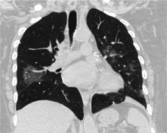
Computed tomography of the chest demonstrating bilateral patchy ground‐glass opacities with interlobular septal thickening consistent with a crazy paving pattern found in COVID‐19 infection
2. DISCUSSION
As the COVID‐19 pandemic unfolds, the impact on cardiac surgical patients raises complex questions concerning testing and screening. In this report, we describe the first documented case of a postoperative cardiac surgical patient infected with COVID‐19. While the exposure occurred at an outside facility, the importance of repeat testing and screening of patients based on exposure history and symptoms cannot be overstated. RT‐PCR has emerged as the test of choice for detection of viral nucleic acids in infected individuals. Notably, through studies originating in China at the onset of this pandemic, the sensitivity of RT‐PCR tests has been shown to be lacking. One study found that nearly 25% of COVID‐19 positive individuals were negative on their initial testing with some testing positive after their third or fourth repeat test. 1 Xiao et al 2 similarly reported that over 20% of COVID‐19 infected individuals tested positive on their third consecutive test, after two initial negative results. The sensitivity of RT‐PCR in several studies has been quoted at 71% and 83%, respectively, corresponding to a false negative rate of up to 30%. 3 , 4 Given the reported sensitivity of RT‐PCR for COVID‐19, repeat testing in patients with high clinical suspicion of COVID‐19 infection, and therefore high pre‐test probability, is of paramount importance.
Given these findings, early isolation and treatment of high‐risk individuals should be based on symptomology, exposure history, and CT findings. In this case, the patient initially presented with fatigue while progressing to fever, cough, shortness of breath, and diarrhea. In retrospective analyses, the most common symptoms attributable to COVID‐19 infection included fever, cough, shortness of breath, and fatigue in the vast majority of cases. 5 , 6 In postoperative cardiac surgical patients, it may be difficult to differentiate these common symptoms of a postoperative bacterial pneumonia from a new onset COVID‐19 infection. For this reason, exposure history and CT findings become vital in the COVID‐19 era. In this case, the patient was exposed to COVID‐19 positive individuals at her skilled nursing facility. If an exposure history had not been elicited, it is possible that repeat testing would not have been pursued.
Obtaining CT imaging should also be an integral component of screening for COVID‐19 in cardiac surgical patients. Typical CT findings include consolidation, vascular enhancement, air bronchus sign, and bilateral peripheral ground‐glass opacities with interlobular septal thickening consistent with a “crazy paving pattern.” 7 In our case, these features prompted continued isolation of our patient as presumed‐positive. While these findings can reinforce clinical suspicion, they may also be found in other viral syndromes making them fairly nonspecific. However, recent discoveries have revealed the efficacy of CT scanning in this patient population and several studies have shown CT imaging to enhance the sensitivity of RT‐PCR in individuals undergoing screening for COVID‐19. 3 , 8
Indeed, a negative RT‐PCR test does not exclude the possibility of COVID‐19 infection. Given the poor sensitivity of RT‐PCR, repeat testing is essential to identify COVID‐19 positive individuals. Thus, isolation and treatment practices should be guided by a combination of testing, exposure history, symptomology, and radiologic evidence. High levels of clinical suspicion based on these parameters should prompt early isolation in these “presumed‐positive” patients regardless of RT‐PCR results to prevent exposure of healthcare workers and patients.
Fisher B, Seese L, Sultan I, Kilic A. The importance of repeat testing in detecting coronavirus disease 2019 (COVID‐19) in a coronary artery bypass grafting patient. J Card Surg. 2020;35:1342–1344. 10.1111/jocs.14604
REFERENCES
- 1. Li Y, Yao L, Li J, et al. Stability issues of RT‐PCR testing of SARS‐CoV‐2 for hospitalized patients clinically diagnosed with COVID‐19 [published online ahead of print March 26, 2020]. J Med Virol. 2020. 10.1002/jmv.25786 [DOI] [PMC free article] [PubMed] [Google Scholar]
- 2. Xiao AT, Tong YX, Zhang S. False‐negative of RT‐PCR and prolonged nucleic acid conversion in COVID‐19: rather than recurrence [published online ahead of print April 9, 2020]. J Med Virol. 2020. 10.1002/jmv.25855 [DOI] [PMC free article] [PubMed] [Google Scholar]
- 3. Fang Y, Zhang H, Xie J, et al. Sensitivity of Chest CT for COVID‐19: comparison to RT‐PCR. Radiology. 2020:200432. 10.1148/radiol.2020200432 [DOI] [PMC free article] [PubMed] [Google Scholar]
- 4. Long C, Xu H, Shen Q, et al. Diagnosis of the coronavirus disease (COVID‐19): rRT‐PCR or CT? Eur J Radiol. 2020;126:108961. [DOI] [PMC free article] [PubMed] [Google Scholar]
- 5. Chen T, Dai Z, Mo P, et al. Clinical characteristics and outcomes of older patients with coronavirus disease 2019 (COVID‐19) in Wuhan, China (2019): a single‐centered, retrospective study [published online ahead of print April 11, 2020]. J Gerontol A Biol Sci Med Sci. 2020. 10.1093/gerona/glaa089 [DOI] [PMC free article] [PubMed] [Google Scholar]
- 6. Fu L, Wang B, Yuan T, et al. Clinical characteristics of coronavirus disease 2019 (COVID‐19) in China: a systematic review and meta‐analysis [published online ahead of print April 10, 2020]. J Infect. 2020. 10.1016/j.jinf.2020.03.041 [DOI] [PMC free article] [PubMed] [Google Scholar]
- 7. Dai H, Zhang X, Xia J, et al. High‐resolution Chest CT features and clinical characteristics of patients infected with COVID‐19 in Jiangsu, China. Int J Infect Dis. 2020;95:106‐112. [DOI] [PMC free article] [PubMed] [Google Scholar]
- 8. Feng H, Liu Y, Lv M, Zhong J. A case report of COVID‐19 with false negative RT‐PCR test: necessity of chest CT. Jpn. J Radiol. 2020;38. 10.1007/s11604-020-00967-9 [DOI] [PMC free article] [PubMed] [Google Scholar]