To the Editor: We read with great attention the Clinical Letter by Soldati et al, entitled “Is There a Role for Lung Ultrasound During the COVID‐19 Pandemic?”1 We must congratulate the authors: Dr Soldati is one of the most experienced and influential authorities in thoracic ultrasound. We agree with the authors that thoracic ultrasound plays a role in this pandemic and that there could be an opportunity to spread its use even more.2
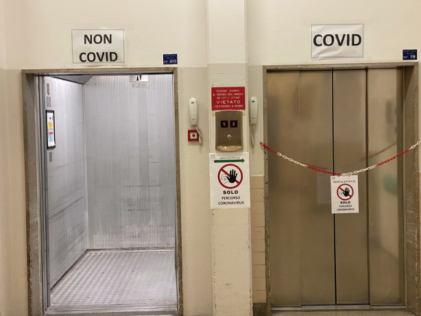
In our opinion, the most importance advantages of thoracic ultrasound are closely associated with the following facts. Since Italy was recognized as the second most affected country after China by the 2019 novel coronavirus disease (COVID‐19), in many of our hospitals, it was necessary to create 2 separate areas: COVID and non‐COVID areas (Figure 1). In this context, thoracic ultrasound is an alternative tool, easy to use, and quick to clean, and it can be used at the patient's bedside: moving patients from the COVID area is not necessary, thus avoiding exposing health workers who work outside this area (non‐COVID). Furthermore, thoracic ultrasound is more sensitive than chest radiography in diagnosing pneumonia of various etiologies.3, 4 However, we are cautious about a specific lung ultrasound (LUS) pattern for COVID‐19. Although the sensitivity of thoracic ultrasound is high, its specificity may not be the same.5 COVID‐19 infection is the most frequent interstitial lung disease at this time, so in the affected countries, the pretest probability is very high, but tomorrow, it could be not so high.
Figure 1.

Examples of COVID and non‐COVID areas.
In the first 2 months of 2020, several social media outlets showed tutorials, anonymous images, and clips showing the “typical” COVID‐19 LUS pattern. Although we think that Soldati et al are substantially right (thanks to their experience), we would like to be more cautious in “labeling” this pattern as highly specific.
The distinction between pulmonary edema and interstitial patterns from other etiologies is clear, but the difference between viral pneumonia and interstitial patterns from different etiologies may not be as obvious.6, 7 Furthermore, although the most frequent cause of interstitial pneumonia at this time is the severe acute respiratory syndrome coronavirus 2 (SARS‐CoV‐2) virus, other causes remain possible (atypical bacteria, flu viruses, etc).
The clinical pictures of the patients we are evaluating in this period are often overlapping. For example, we visited patients with thoracic ultrasound findings indicative of cardiogenic pulmonary edema, who, after the first negative pharyngeal test result, tested positive for the RNA polymerase chain reaction test on bronchoalveolar lavage. So, the potential misinformation about these tools is likely to do more harm than good to the reputation and appreciation of this exceptional instrument. It, therefore, seems premature to be able to affirm definitive data regarding the performance of thoracic ultrasound examinations until results are available on a large population of COVID‐19–positive patients.8
References
- 1. Soldati G, Smargiassi A, Inchingolo R, et al. Is there a role for lung ultrasound during the COVID‐19 pandemic [published online ahead of print March 20, 2020] J Ultrasound Med. 10.1002/jum.15284. [DOI] [PMC free article] [PubMed] [Google Scholar]
- 2. Brogi E, Bignami E, Sidoti A, et al. Could the use of bedside ultrasound reduce the number of chest x‐ray in the intensive care unit? Cardiovasc Ultrasound 2017; 15:23. [DOI] [PMC free article] [PubMed] [Google Scholar]
- 3. Maw AM, Hassanin A, Ho PM, et al. Diagnostic accuracy of point‐of‐care lung ultrasonography and chest radiography in adults with symptoms suggestive of acute decompensated heart failure: a systematic review and meta‐analysis. JAMA Netw Open 2019; 2:e190703. [DOI] [PMC free article] [PubMed] [Google Scholar]
- 4. Soldati G, Demi M. The use of lung ultrasound images for the differential diagnosis of pulmonary and cardiac interstitial pathology. J Ultrasound 2017; 20:91–96. [DOI] [PMC free article] [PubMed] [Google Scholar]
- 5. Long L, Zhao HT, Zhang ZY, Wang GY, Zhao HL. Lung ultrasound for the diagnosis of pneumonia in adults: a meta‐analysis. Medicine (Baltimore) 2017; 96:e5713. [DOI] [PMC free article] [PubMed] [Google Scholar]
- 6. Testa A, Soldati G, Copetti R, Giannuzzi R, Portale G, Gentiloni‐Silveri N. Early recognition of the 2009 pandemic influenza A (H1N1) pneumonia by chest ultrasound. Crit Care 2012; 16:R30. [DOI] [PMC free article] [PubMed] [Google Scholar]
- 7. Wang J, Xu H, Yang X, et al. Cardiac complications associated with the influenza viruses A subtype H7N9 or pandemic H1N1 in critically ill patients under intensive care. Braz J Infect Dis 2017; 21:12–18. [DOI] [PMC free article] [PubMed] [Google Scholar]
- 8. Vetrugno L, Bove T, Orso D, et al. Our Italian experience using lung ultrasound for identification, grading and serial follow‐up of severity of lung involvement for management of patients with COVID‐19. Echocardiogr‐J card 2020; 37:625–627. [DOI] [PMC free article] [PubMed] [Google Scholar]


