Dear Editor,
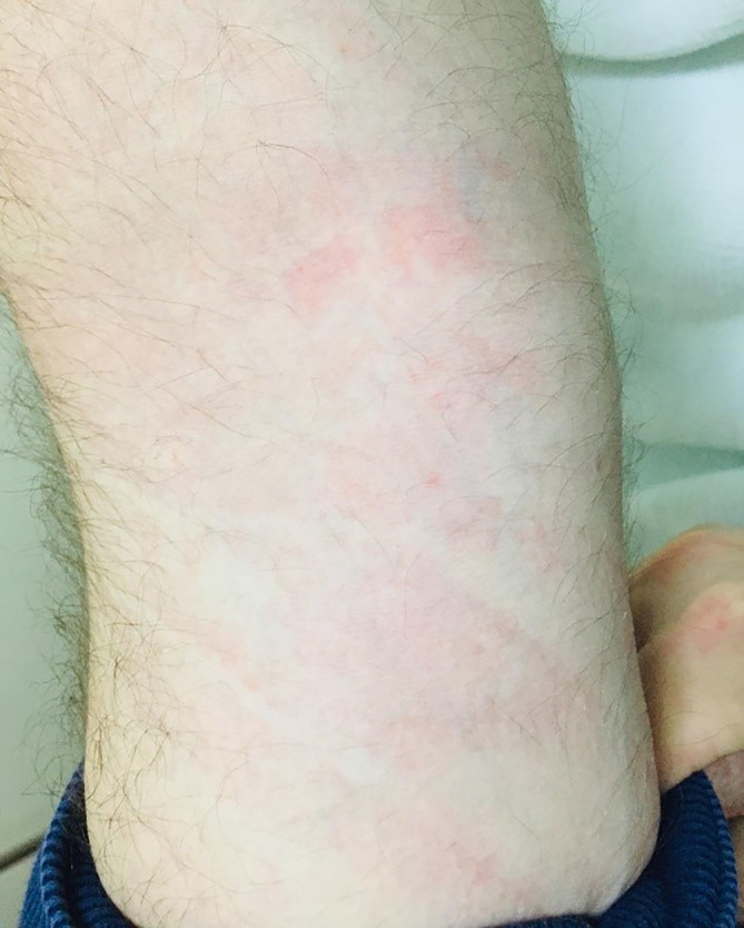
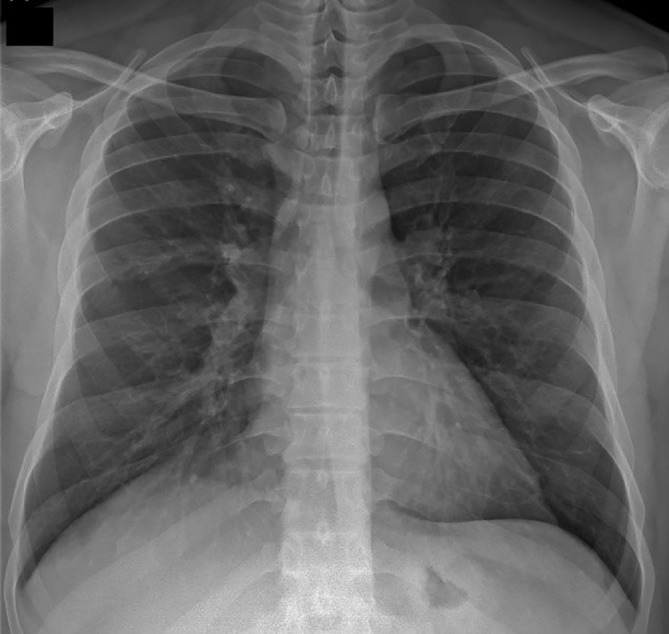
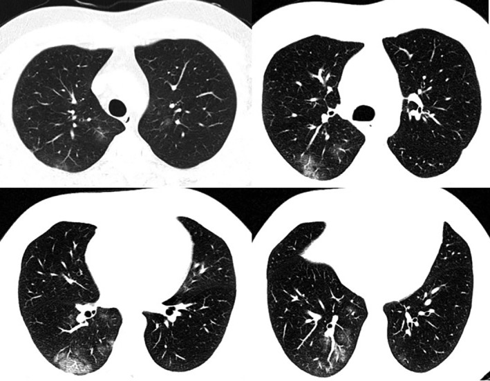
A 53‐year‐old male applied to the emergency room with complaints of generalized pruritus and rush which continued for about 48 hours. He had no previous history of atopic conditions including drug or food allergy, chronic urticaria, and so forth. He had quitted smoking 10 years ago after smoking 20 pack‐years of cigarettes. He had a history of being abroad 10 days ago. Body temperature was 36.6°C, heart rate was 80 bpm, blood pressure was 110/70 mm Hg, oxygen saturation breathing room air was 98%. Physical examination revealed edematous and itchy plaques throughout the body (Figure 1). Any other abnormal finding was not recorded in physical examination and auscultation of chest was also normal. In his posterior‐anterior chest graphy, right diaphragma was slightly elevated accompanied by infiltration of the lower part of the right lung (Figure 2). Computed tomography revealed patchy infiltrations bilaterally in both lungs (Figure 3). Complete blood count was within normal levels; white blood cell count: 9.200/μL, platelet count: 276.000/μL, hemoglobin:15.2 g/dL, hematocrit: 44.7%, lymphocyte count: 2500/μL (34.7%), eosinophil count: 0/μL (0.1%). C‐reactive protein was elevated to 9.12 mg/L. Serum biochemistry was within normal limits; blood urea nitrogen: 36 mg/dL, creatinin: 0.9 mg/dL, alanine aminotransferase:22 U/L, asparate aminotransferase:12 U/L, lactate dehydrogenase:195 U/L, sodium:138 mEq/L, potassium:3.88 mEq/L.
FIGURE 1.

Edema, itchy, and red multiple plaques in the right forearm
FIGURE 2.

Rise and retraction of the right diaphragmatic medial part in posterior‐anterior chest X‐ray
FIGURE 3.

Thorax tomography revealing infiltration areas with focal ground glass density in both lung parenchyma
The patient was hospitalized with a preliminary diagnosis of COVID‐19 pneumonia. Severe acute respiratory syndrome coronavirus‐2 (SARSCoV‐2) PCR result in the combined nasal‐throat swab sample taken at the patient's hospitalization was negative. However, with the strong possibility of radiological COVID‐19 pneumonia, a second swab sample was sent and the result was found positive. Treatment was started with the diagnosis of COVID‐19 pneumonia. On the fourth day of his admission, his skin lesions regressed and he was discharged on the fifth day of his admission. Written informed consent was obtained from the patient.
Urticaria is a dermatologic condition that presents with pruritic, well‐circumscribed, raised wheals. Its lifetime prevalence of approximately 20%. Urticaria that recurs within a period of less than 6 weeks is acute. 1 Most common causes of urticaria are allergens, food pseudoallergens, insect envenomation, drugs, and infections. Viruses (eg, rhinovirus, rotavirus, Epstein‐Barr, hepatitis A, hepatitis B, hepatitis C, herpes simplex, human immunodeficiency virus), bacteria (eg, urinary tract infections, streptococcus, mycoplasma, Helicobacter pylori), and some parasites are infectious agents associated with urticaria. 1
Coronaviruses are a large family of viruses. The most recently discovered coronavirus causes COVID‐19 which was unknown before the outbreak began in Wuhan, China, in December 2019. 2 As the COVID‐19 outbreak continues to evolve, we are learning more about this new virus every day.
Fever, tiredness, and dry cough are the most commonly encountered symptoms of COVID‐19, followed by aches and pains, nasal congestion, runny nose, sore throat, or diarrhea. On the contrary, some people become infected but stay asymptomatic as they do not feel unwell. 2
World Health Organization denotes that COVID‐19 should be suspected in (a) a case with acute respiratory illness (fever and at least one sign/symptom of respiratory disease, for example, cough, shortness of breath), and a history of travel to or residence in a location reporting community transmission of COVID‐19 disease during the 14 days prior to symptom onset; (b) a case with any acute respiratory illness and having been in contact with a confirmed or probable COVID‐19 case in the last 14 days prior to symptom onset should also be diagnosed as COVID‐19; and (c) a case with severe acute respiratory illness (fever and at least one sign/symptom of respiratory disease, for example, cough, shortness of breath; AND requiring hospitalization) together with the absence of an alternative diagnosis that fully explains the clinical presentation. Probable case for COVID‐19 is defined as (a) a suspect case for whom testing for the COVID‐19 virus is inconclusive; or (b) a suspect case for whom testing could not be performed for any reason. Confirmed case is a case with laboratory confirmation of COVID‐19 infection, irrespective of clinical signs and symptoms. 3
Full clinical spectrum of the disease COVID‐19 is yet unknown. Zhang et al reported the clinical characteristics of all hospitalized patients admitted to No. 7 Hospital of Wuhan from January 16 to February 3, 2020 with clinical diagnosis of viral pneumonia and confirmed test results for SARSCoV‐2. They have outlined past medical history and presenting symptoms of 140 patients infected with SARSCoV‐2. Sixteen patients had self‐reported medical history of drug hypersensitivity and two patients reported chronic urticaria. None of the subjects declared other allergic diseases including asthma, allergic rhinitis, food allergy, atopic dermatitis. Regarding symptomatology of the study population according to the report of Zhang et al none of the subjects presented with allergic reactions or skin lesions. 4 However, as COVID‐19 has spread quickly across the world, awkward signs and symptoms associated with the disease have been reported. Among them cutaneous manifestations including acute urticaria and inflammatory and vascular skin lesions have been reported (Table 1). 5 , 6 , 7 On the other hand, the development of skin findings during follow‐up in patients with COVID‐19 diagnosis may be related to the drugs used. 8
TABLE 1.
Skin findings detected in the course of COVID‐19 infection
| Author | Article type | Subject(s) | Presenting symptoms | Defined lesion |
|---|---|---|---|---|
| Henry et al. 5 | Case report | 27‐year‐old female | Odynophagia, diffuse arthralgia, and pruritic disseminated erythematous plaques eruption with particular face and acral involvement | Acute urticaria |
| van Damme et al. 6 | Case report | 71‐year‐old man and 39‐year‐old female |
Case 1: General weakness, pyrexia, and a cutaneous rash Case 2: Generalized, pruritic urticarial rash on her forearms, pyrexia with chills, myalgia and headache, rhinorrhea, dry cough, and dyspnea |
Acute urticaria |
| Bouaziz et al. 7 | Observational study | N/A | N/A | Inflammatory lesions (exanthema, chicken pox like vesicles, cold urticaria) and vascular lesions (violaceous macules with “porcelain‐like” appearance, livedo, non‐necrotic purpura, necrotic purpura, chilblain appearance with Raynaud's phenomenon, chilblain, eruptive cherry angioma) |
The present case has also presented with skin lesions. He, unlike other cases, had no respiratory or systemic complaints. The reason for investigating COVID‐19 in the case was that no reason to explain acute urticaria was found and he has been to a country where COVID‐19 was prevalent.
To conclude, it should be kept in mind that the presence of newly developing and unexplained skin findings may be a COVID‐19 finding. In addition, the development of skin findings during follow‐up in patients with COVID‐19 diagnosis may be related to the drugs used.
REFERENCES
- 1. Schaefer P. Acute and chronic urticaria: evaluation and treatment. Am Fam Physician. 2017;95(11):717‐724. [PubMed] [Google Scholar]
- 2. https://www.who.int/news-room/q-a-detail/q-a-coronaviruses. Accessed April 3, 2020.
- 3.World Health Organization (WHO). Coronavirus Disease 2019 (COVID‐19) Situation Report—72. https://www.who.int/emergencies/diseases/novel-coronavirus-2019/situation-reports. Accessed April 3, 2020.
- 4. Zhang JJ, Dong X, Cao YY, et al. Clinical characteristics of 140 patients infected with SARS‐CoV‐2 in Wuhan, China. Allergy. 2020. 10.1111/all.14238. [DOI] [PubMed] [Google Scholar]
- 5. Henry D, Ackerman M, Sancelme E, Finon A, Esteve E. Urticarial eruption in COVID‐19 infection. J Eur Acad Dermatol Venereol. 2020;34:e244‐e245. [DOI] [PMC free article] [PubMed] [Google Scholar]
- 6. van Damme C, Berlingin E, Saussez S, Accaputo O. Acute urticaria with pyrexia as the first manifestations of a COVID‐19 infection. J Eur Acad Dermatol Venereol. 2020. https://doi.org10.1111/jdv.16523. [DOI] [PMC free article] [PubMed] [Google Scholar]
- 7. Bouaziz JD, Duong T, Jachiet M, et al. Vascular skin symptoms in COVID‐19: a french observational study. J Eur Acad Dermatol Venereol. 2020. 10.1111/jdv.16544. [DOI] [PMC free article] [PubMed] [Google Scholar]
- 8. Schwartz RA, Janniger CK. Generalized pustular figurate erythema: a newly delineated severe cutaneous drug reaction linked with hydroxychloroquine. Dermatol Ther. 2020;33:e13380. [DOI] [PMC free article] [PubMed] [Google Scholar]


