

Abstract
“COVID toes” are a new phenomenon of pernio-like lesions that has been associated with coronavirus disease 2019 (COVID-19) infection. As dermatology practices reopen and increase patient volumes amid the waning of the coronavirus pandemic, it is important to consider the finding of pernio and pernio-like lesions in the context of both COVID-19 and its other possible etiologies. This contribution will discuss possible causes of pernio and pernio-like lesions and offer suggestions for appropriate diagnostic workup and management when indicated.
Introduction
Pernio, also known as perniosis or chilblains, characteristically presents as painful or pruritic erythematous macules or papules on the digits lasting more than 24 hours. It is likely the result of vasospasm of the superficial vasculature and secondary inflammatory reaction.1 It is most commonly seen on the feet but may be found on other sites like the hands, ears, and nose.
Primary pernio, also known as idiopathic pernio, is the most common cause of pernio and is usually associated with cool temperatures and vasospasm.2 Secondary pernio is associated with systemic inflammatory conditions like chronic cutaneous lupus erythematosus and sarcoidosis. Secondary pernio likely accounts for less than 7% of all pernio conditions.2 Other conditions, including myeloid leukemias and the coronavirus disease 2019 (COVID-19), have been associated with pernio-like skin lesions.3 , 4 Table 1 summarizes some distinguishing features of these etiologies.
Table 1.
Distinguishing features of pernio and pernio-like eruptions
| Feature | Primary (idiopathic) pernio | Chilblain lupus erythematosus | Chilblain-like leukemia cutis | Chilblain-like lesions of COVID-19 (COVID toes) |
|---|---|---|---|---|
| Exposure to cool temperature | Yes | Yes | No | No |
| Seasonal correlation | Yes | No | No | No |
| Common distribution | Feet, hands | Feet, hands | Feet, hands, face | Feet |
| Histology | Superficial and deep perivascular and perieccrine lymphocytic infiltrate with dermal edema | Lymphocytic infiltration of dermis | Leukemic cells | Combination of idiopathic pernio and CHLE, focal thrombi |
| Immunofluorescence | (−) | (+) Immunoglobulins and complement deposition at the dermoepidermal junction | (−) | Unknown |
| Irregular laboratory values | Various | Various | CBC Peripheral blood smear |
Unknown |
CHLE, chilblain lupus erythematosus; CBC, complete blood count.
(-) = negative, (+) = positive
Primary (idiopathic) pernio
Idiopathic pernio is typically associated with cold, but nonfreezing, temperatures. It often appears during the cool, wet seasons of the year, like late fall and early spring. Although the exact etiology is unknown, it is suspected that vasospasm and a secondary inflammatory reaction contribute to its progression.1 , 2 Along with the fingers and toes, primary pernio lesions may be found on the ears and face. It is more common in women of reproductive age, those with a low body mass index, those who smoke, and those with Raynaud phenomenon.1 , 2
Physical examination and history are typically sufficient for a diagnosis of primary pernio. Patients report appearance of lesions within about 24 hours of exposure to cool temperatures. On histopathology, perniosis classically presents with a superficial and deep lymphoid vasculitis, dermal edema, and perieccrine lymphocytic infiltrates.5 No laboratory tests are required for diagnosis.2
Management of primary pernio generally includes conservative measures like keeping the affected areas warm and dry, as well as smoking cessation. Idiopathic pernio typically will resolve in 1 to 3 weeks. For unresolving or more severe cases, topical corticosteroids or oral calcium channel blockers like nifedipine may be attempted, but success is variable.1 , 2 Patients with unresolving or severe pernio are at risk of developing systemic lupus erythematosus (SLE) in the following months and, therefore, should be monitored for systemic clinical manifestations.1 Additionally, in patients with unresolving, severe, or otherwise atypical pernio, it is important to consider secondary causes of pernio or a pernio-like syndrome as described in the following section.
Secondary pernio
Secondary pernio results from an underlying systemic cause. This accounts for a small proportion of all patients with pernio. Additionally, these are generally rare cutaneous manifestations of the systemic cause.
Chilblain lupus erythematosus (CHLE) is a form of cutaneous lupus erythematosus that presents with lesions of similar morphology and distribution to primary pernio.6 It may be sporadic or familial. The familial form of CHLE is associated with a mutation in TREX1.6 Like primary pernio, the lesions of CHLE may appear with cold temperatures; however, the lesions are not seasonal and typically persist longer than a few weeks. Acral lesions and characteristic histopathologic or immunofluorescence findings are necessary for a diagnosis of CHLE. If CHLE is suspected, biopsy and immunofluorescence should be performed. Biopsies of CHLE will show findings similar to idiopathic pernio with superficial and deep perivascular lymphocytic infiltrates; however, CHLE is more likely to show a widespread vacuolar interface with scattered necrotic keratinocytes in the lower part of the epidermis and is less likely to show perieccrine lymphocytic infiltration and significant dermal edema.5 , 7 Immunofluorescence can show deposition of immunoglobulins and complement proteins at the dermoepidermal junction in CHLE which is characteristically absent in idiopathic pernio.6 CHLE is often associated with discoid lupus erythematosus.6 Up to 18% of patients with CHLE will also develop SLE, although this may take years.6 Many autoantibodies, including antiphospholipid antibodies, are associated with CHLE; however, there are no significant correlations between presence of autoantibodies and the risk of development of SLE.6 Initial management of CHLE is the same as that of primary pernio. If the lesions are unresponsive, topical immunosuppressants, like tacrolimus, or systemic immunosuppressants, like mycophenolate mofetil, may be used.6 Patients should continue to be monitored for clinical manifestations of SLE.6
Chilblain-like lesions
Leukemia cutis is any specific cutaneous manifestation of leukemia that may be identified by the presence of leukemic cells within the skin lesion upon histological examination.8 Leukemia cutis may have many different morphologies, including a chilblain-like leukemia cutis. This is a rare manifestation which is most often associated with myeloid leukemias.3 , 9 In addition to a biopsy, chilblain-like leukemia cutis may be diagnosed by peripheral blood smear and complete blood counts which provide evidence of leukemia.8
COVID-19 has been associated with many different cutaneous manifestations, including a chilblain-like eruption on the feet, thus the nickname of “COVID toes.” This cutaneous manifestation seems to appear in younger patients.4 , 10 , 11 These patients typically do not have other clinical manifestations of the virus upon presentation and will not go on to develop further clinical manifestations.4 One study showed that about one-quarter of those with chilblain-like lesions will have other clinical manifestations of COVID-19.10 If patients do report clinical manifestations of COVID-19, they usually occur before the chilblain lesions.11 It has been suggested that patients with chilblain-like lesions may be contagious, and they should be counseled to use all social distancing precautions.4 , 11 Interestingly, patients with severe cases of COVID-19 have been found to develop ischemic lesions of the extremities, including acral regions.12 These patients demonstrate elevated antiphospholipid antibodies, similar to CHLE.12 This may suggest vascular dysfunction similar to that of pernio. The histology of COVID toes has shown similarities, as well. Superficial and deep perivascular lymphocytic infiltrates with perieccrine involvement, vacuolar interface, and scattered necrotic keratinocytes have been described.10 , 13 Focal thrombosis can be seen, which is not characteristic of CHLE or idiopathic pernio. Most lesions associated with COVID-19 do not require treatment and will resolve within a few days.12 At this time, the Centers for Disease Control and Prevention (CDC) do not specifically list the chilblain-like eruption or any cutaneous manifestation of COVID-19 as stand-alone symptomatology warranting nasopharyngeal swab testing14; however, the CDC does state that clinicians should use their judgment to determine whether a patient has symptomatology consistent with COVID-19 in deciding whether the patient should be tested.14
Figure 1 depicts suspected COVID toes in a 20-year-old man hospitalized for clinical manifestations of COVID-19. Interestingly, the patient’s reverse transcriptase–polymerase chain reaction (RT-PCR) test results were negative. This case illustrates a challenge related to the diagnosis of COVID-19: a significant risk of false-negative RT-PCR tests.15 For this reason, negative RT-PCR test results should be interpreted cautiously and in the context of the patient’s clinical manifestations and possible exposures to the virus.15 Alternative testing using serum antibodies may be a useful tool for confirming previous infection with COVID-19 and further establishing the link between pernio-like lesions known as COVID toes and infection with COVID-19. Antibody testing is best conducted at least 1 week after onset of clinical manifestations and has also been associated with a significant false-positive test result.16
Fig. 1.
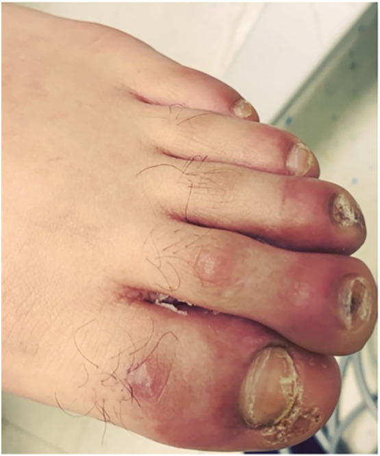
Pernio-like eruption in 20-year-old man admitted for COVID-19 clinical manifestations.
Conclusions
Pernio and pernio-like lesions represent a constellation of clinical conditions ranging from idiopathic to malignant and life-threatening. The first step in evaluation of pernio and pernio-like lesions should include obtaining a patient history for exposure to cold temperatures. If this is the case, the lesions are likely primary (idiopathic) pernio. If the pernio lesions are found in atypical locations, do not resolve after a few weeks of conservative management, or are not associated with cool temperatures, secondary pernio or causes of pernio-like lesions should be considered. Biopsy of a lesion and histologic examination can be helpful for diagnosis. Table 1 includes an overview of common histological findings, as well as other laboratory findings and associations that may be helpful in determining the etiology of pernio.
The clinical significance of chilblain-like lesions in COVID-19 is still to be determined; however, as dermatology clinics reopen, dermatologists are likely to encounter more pernio-like lesions than before. It is important to interpret these findings in the context of all etiologies of pernio. Negative RT-PCR or antibody tests should be interpreted cautiously, as the risk of a false negative test is well established. Physicians should continue to look for updates regarding the significance and management protocols for cutaneous manifestations of COVID-19.
Declaration of competing interest
The authors declare that they have no known competing financial interests or personal relationships that could have appeared to influence the work reported in this paper.
References
- 1.Nyssen A., Benhadou F., Magnée M. Chilblains. Vasa. 2020;49:133–140. doi: 10.1024/0301-1526/a000838. [DOI] [PubMed] [Google Scholar]
- 2.Cappel J.A., Wetter D.A. Clinical characteristics, etiologic associations, laboratory findings, treatment, and proposal of diagnostic criteria of pernio (chilblains) in a series of 104 patients at Mayo Clinic, 2000 to 2011. Mayo Clin Proc. 2014;89:207–215. doi: 10.1016/j.mayocp.2013.09.020. [DOI] [PubMed] [Google Scholar]
- 3.Affleck A.G., Ravenscroft J.C., Leach I.H. Chilblain-like leukemia cutis. Pediatr Dermatol. 2007;24:38–41. doi: 10.1111/j.1525-1470.2007.00330.x. [DOI] [PubMed] [Google Scholar]
- 4.Fernandez-Nieto D., Jimenez-Cauhe J., Suarez-Valle A. Characterization of acute acro-ischemic lesions in non-hospitalized patients: a case series of 132 patients during the COVID-19 outbreak. J Am Acad Dermatol. 2020;83:e61–e63. doi: 10.1016/j.jaad.2020.05.120. [DOI] [PMC free article] [PubMed] [Google Scholar]
- 5.Weedon D., Patterson J.W. 4th ed. Elsevier; New York: 2015. Weedon’s Skin Pathology. [Google Scholar]
- 6.Hedrich C.M., Fiebig B., Hauck F.H. Chilblain lupus erythematosus—a review of literature. Clin Rheumatol. 2008;27:949–954. doi: 10.1007/s10067-008-0942-9. [DOI] [PubMed] [Google Scholar]
- 7.Cribier B., Djeridi N., Peltre B. A histologic and immunohistochemical study of chilblains. J Am Acad Dermatol. 2001;45:924–929. doi: 10.1067/mjd.2001.117861. [DOI] [PubMed] [Google Scholar]
- 8.Wagner G., Fenchel K., Back W. Leukemia cutis – epidemiology, clinical presentation, and differential diagnoses. J Dtsch Dermatol Ges. 2012;10:27–36. doi: 10.1111/j.1610-0387.2011.07842.x. [DOI] [PubMed] [Google Scholar]
- 9.Tran C, McEwen G, Fraga GR. Chilblain-like leukaemia cutis. BMJ Case Rep. 2016;2016:10.1136/bcr-214838. [DOI] [PMC free article] [PubMed]
- 10.Kolivras A., Dehavay F., Delplace D. Coronavirus (COVID-19) infection-induced chilblains: a case report with histopathologic findings. JAAD Case Rep. 2020;6:489–492. doi: 10.1016/j.jdcr.2020.04.011. [DOI] [PMC free article] [PubMed] [Google Scholar]
- 11.Piccolo V., Neri I., Filippeschi C. Chilblain-like lesions during COVID-19 epidemic: a preliminary study on 63 patients. J Eur Acad Dermatol Venereol. 2020 doi: 10.1111/jdv.16526. in press. [DOI] [PMC free article] [PubMed] [Google Scholar]
- 12.Wei C., Friedman A.J. COVID-19 pandemic: are there unique cutaneous manifestations in patients infected with SARS-CoV-2? J Drugs Dermatol. 2020;19:554–555. [PubMed] [Google Scholar]
- 13.Andina D., Noguera-Morel L., Bascuas-Arribas M. Chilblains in children in the setting of COVID-19 pandemic. Pediatr Dermatol. 2020;9 doi: 10.1111/pde.14215. in press. [DOI] [PMC free article] [PubMed] [Google Scholar]
- 14.Centers for Disease Control and Prevention. Evaluating and testing persons for coronavirus disease 2019 (COVID-19). Available at: https://www.cdc.gov/coronavirus/2019-ncov/hcp/clinical-criteria.html. Accessed May 21, 2020.
- 15.West C.P., Montori V.M., Sampathkuar P. COVID-19 testing: the threat of false-negative results. Mayo Clin Proc. 2020;95:1127–1129. doi: 10.1016/j.mayocp.2020.04.004. [DOI] [PMC free article] [PubMed] [Google Scholar]
- 16.Kontou P.I., Braliou G.G., Dimou N.L. Antibody tests in detecting SARS-CoV-2 infection: a meta-analysis. Diagnostics. 2020;10 doi: 10.3390/diagnostics10050319. [DOI] [PMC free article] [PubMed] [Google Scholar]