Abstract
Introduction
Skeletal dysplasia is a condition associated with various abnormalities of the skeleton and comprises multiple groups of disorders. Antenatal ultrasonographic assessment of the skeletal dysplasia requires a robust and systematic assessment of the long bones, fetal thorax, skull, spine, pelvis, hands and the feet. Large number of diseases, their overlapping phenotypic features and the lack of systematic approach lead to diagnostic inefficiency. A precise molecular diagnosis also requires an elaborate antenatal sonographic assessment to reach a final diagnosis.
Case report
A fetus with micromelia, thoracic dysplasia and polydactyly was detected on prenatal sonography. An algorithmic approach of this rare combination on prenatal sonography is highlighted.
Discussion
Fetal micromelia is a relatively common entity which can be subclassified into mild and severe types. The lethal nature of the condition requires assessment of the thoracic biometry which may further narrow down the diagnostic possibilities. The red flags or highlighting features of various conditions like polydactyly, hitch-hiker thumb deformity, ovoid tibia and absent fibula may lead to a specific diagnosis.
Conclusion
A background knowledge of various types of micromelia, their lethal nature, associations and specific features of various differential skeletal dysplasia will always be useful, if employed in a systematic manner.
Keywords: Fetus, micromelia, thoracic dysplasia, polydactyly, Jeune syndrome
Introduction
Antenatal sonographic diagnosis of skeletal dysplasia is a diagnostic challenge because of the lack of a systematic approach as well as due to more than 300 different conditions underlying this heterogenous group of skeletal abnormality.1,2 Identification of constellation of the phenotypic features and reaching a differential diagnosis will be helpful in deciding the fetal prognosis, proper patient counselling and in explaining the recurrence risk. Here we report a case of lethal skeletal dysplasia detected on ultrasound (US) comprising of mild micromelia, hypoplastic thorax and polydactyly. A provisional diagnosis of asphyxiating thoracic dystrophy (ATD, Jeune syndrome) was achieved using a step-wise diagnostic algorithm.
Case report
A 26-year-old primigravida at 39 weeks of gestational age was referred to our hospital. Patient was un-booked and a recent outside US report suggested fetal anomaly in the form of shortening of fetal long bones. Per-abdomen examination showed uterus corresponding to term gestation, with detectable fetal heart sound.
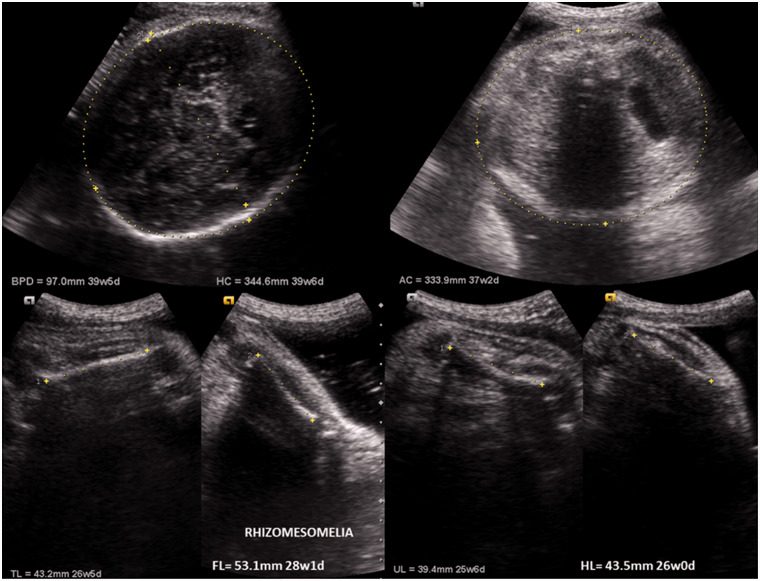
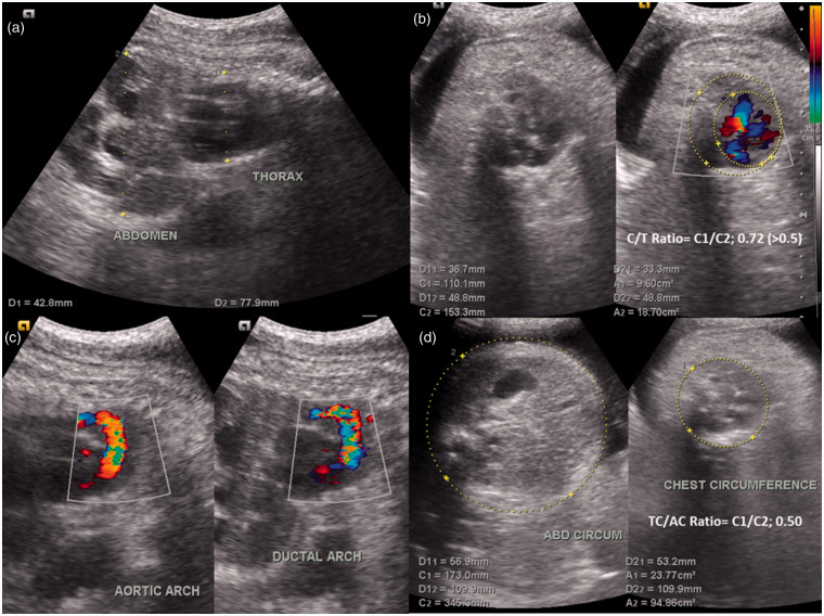
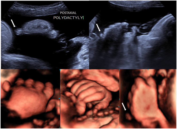
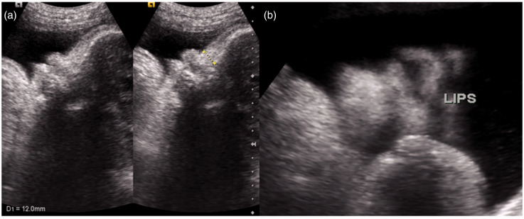
The patient was sent for US to confirm the previously reported sonographic findings and to assess the present fetal status. US showed a single live intrauterine fetus of 39 weeks + 6 days. Features of mild micromelia were evident in the form of shortening of all segments of all the long bones with approximately 10 weeks of discordance (Figure 1). All the segments of long bones were present with normal curvature and mineralization without fracture. Fetal chest was long and narrow with severely reduced thoracic circumference measuring 17.3 cm which was much below 5th centile for gestational age indicative of pulmonary hypoplasia.2–4 Cardio-thoracic (C/T) ratio was increased measuring 0.72 (>0.5) (Figure 2). Since the thoracic to abdominal circumference (TC/AC) ratio was 0.5 (<0.6) and femur length to abdominal circumference (FL/AC) ratio 0.15 (<0.16), strong possibility of perinatal lethal dysplasia was kept.2,4,5 The ribs were normal in morphology and echotexture. Cardiac assessment was within the normal limits with a regular heart rate. Fetal hands showed post-axial polydactyly (Figure 3) without syndactyly (fusion of digits) and clinodactyly (deviation of a finger). Feet were normal in morphology. Fetal head, spine and pelvis showed no remarkable abnormality. Clavicle and scapula were normally visible. Mild coarsening of the face was evident in the form of wide and depressed nasal bridge and mild frontal bossing (Figure 4). No facial cleft or other cranio-facial abnormality like micrognathia was noted (Figure 4). Features of polyhydramnios were present with amniotic fluid index of 26 cm. There was history of second-degree consanguinity. No skeletal abnormality was evident in mother and father, and they were of average normal height.
Figure 1.
Antenatal ultrasound images of fetus showing shortening of all long bones (micromelia).
Figure 2.
Antenatal ultrasound images (a and c) sagittal view, (b and d) transverse view of fetus, showing hypoplastic thorax with increased cardiothoracic ratio (CT ratio) and decreased thoracic to abdominal circumference (TC/AC) ratio.
Figure 3.
Antenatal 2D and 3D ultrasound images of fetus showing post-axial polydactyly of both hands.
Figure 4.
Antenatal ultrasound images of fetus showing broad and flat nasal bridge (a) and absence of cleft lip (b).
On the basis of US features, Jeune syndrome (ATD) and Ellis-van Crevald syndrome (chondroectodermal dysplasia, EVC) were kept as the differential diagnosis.
The parents were counselled about the poor fetal prognosis and advised for post-delivery fetal examination, radiographs, autopsy (if still-born) and chromosome analysis. The fetus was still-born; however, the parents did not give consent for the post-mortem fetal work-up.
Discussion
Micromelia, narrow thorax, short ribs with or without polydactyly are classified into a group of heterogenous skeletal disorders which includes ATD (Jeune syndrome), chondroectodermal dysplasia (EVC syndrome) and four types of the short-rib polydactyly syndromes (SRPS).6 These conditions are rare and follows autosomal recessive pattern of inheritance, showing 25% recurrence in the subsequent pregnancies. The incidence of EVC syndrome is 1 per 60,000 in general population while that of ATD is 1 in 1,00,000 to 1,30,000 live births.7,8 SRPS are uncommon with type 2 SRPS being the rarest of all.6 These disorders show overlapping of major features as described; however, the ATD and EVC show milder phenotype in the form of mild micromelia and variable lethal nature.
ATD or Jeune syndrome is characterized by proportional shortening of the limbs with long and small thorax, trident pelvis with normal spine morphology. Polydactyly is an unpredictable feature seen in 16% cases, while renal and liver disease may occur occasionally (in 30% cases).9 The disease is often lethal, owing to its narrow thorax leading to respiratory failure. Prominent forehead, absent nasal bone and polyhydramnios are commonly associated with ATD. Mutations in chromosomes 3, 11 and 15 have been identified in ATD with IFT80 gene defects (intra-flagellar transport protein).8,9
Chondro-ectodermal dysplasia (EVC syndrome) is characterized by mild micromelia (may show acro-mesomelia), small thorax with polydactyly of the hand in all cases and feet in few of them. Fifty per cent of the cases show congenital heart defects (atrial or ventricular septal defect) and Dandy-walker malformation.7,10 Normal spine morphology, trident pelvis, proportional limb-shortening and small thorax make this disorder difficult to distinguish from ATD. Mutations in EVC genes on chromosome 4 are attributed to this syndrome.7
SRPS are a heterogenous group of lethal skeletal dysplasia with severe micromelia and comprising of four major types: Saldino-Noonan (type 1), Majewski (type 2), Verma-Naumoff (type 3) and Beemar-Langer (type 4). The exact incidence and molecular basis of SRPS are not exactly elucidated, however are found to be linked with aberrations in chromosome 4. The classical features of SRPS are severely short and narrow fetal thorax, very short ribs, polydactyly, normal vertebrae. Associated genito-urinary, gastrointestinal abnormalities and facial clefts are noted in all types of SRPS. Short-ovoid tibia is a classical feature of type 2 SRPS, while abnormal femur is seen in types 1 and 3.6
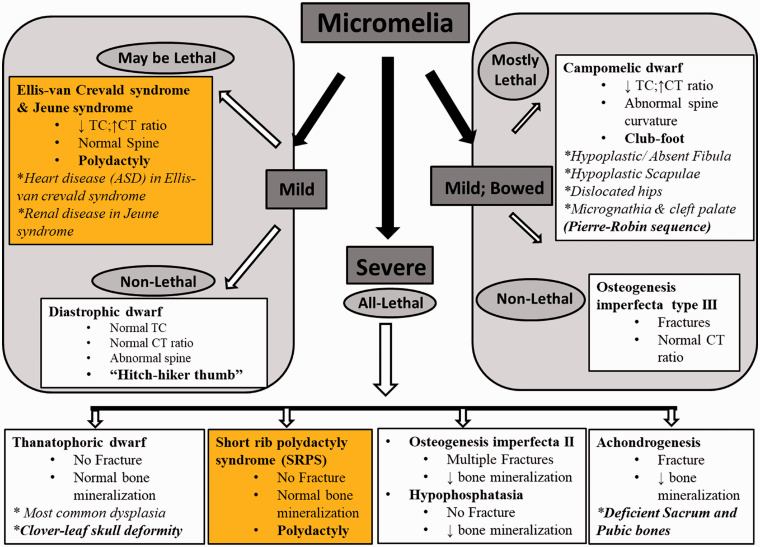
Figure 5 tabulates the various diseases associated with fetal micromelia and classifies them under three major divisions: mild, mild with bowing and severe micromelia. Mild micromelia without abnormal bone curvature is seen in ATD, EVC and diastrophic dwarfism where the latter is non-lethal with presence of ‘hitch-hiker thumb’ deformity as a diagnostic feature. Bowing of the long bones shifts the classification to another side with two major differential diagnoses, i.e. campomelic dwarf and osteogenesis imperfecta type 3 (OI type 3). The identifying features of campomelic dwarfism are club-foot deformity with absent fibula and hypoplastic scapula. The disease is also called as bent-bone dysplasia due to abnormal spinal curvature. OI type 3 is a non-lethal dysplasia without thoracic hypoplasia. Fractures in long bones are visualized. All diseases under severe micromelia category have perinatal lethal outcome due to severe thoracic hypoplasia. They are classified according to the degree of bone mineralization and fractures. Polydactyly is only seen in SRPS.
Figure 5.
Algorithmic approach to fetal micromelia. TC: thoracic circumference; CT ratio: cardiothoracic ratio.
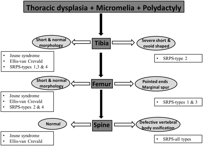
Prenatal sonographic visualization of fetal micromelia, thoracic dysplasia and polydactyly is approached systematically as described in Figure 6. Tibia and femur are evaluated for morphology where severely short and ovoid tibia is noted in SRPS type 2 and abnormal pointed femur in SRPS types 1 and 3. Final assessment of spine is crucial, as defective vertebral body ossification is noted in all types of the SRPS while absent in ATD and EVC as seen in this case.
Figure 6.
Algorithmic approach to combined fetal micromelia with thoracic dysplasia and polydactyly.
Conclusion
Prenatal sonographic approach to fetal micromelia requires a background knowledge about this entity, disorders related to it, criteria of lethality as well as enhanced precision of the imaging specialist. A systematic and multidisciplinary approach is required where a combination of sonographic, genetic, histopathological and autopsy studies are employed in a systematic manner.
Acknowledgments
None.
Contributors
SA and AA researched literature and conceived the manuscript. AA acquired the images. AA wrote the first draft of the manuscript. SA and AA wrote the final version of the manuscript. All authors reviewed and approved the final version of the manuscript.
Declaration of Conflicting Interests
The author(s) declared no potential conflicts of interest with respect to the research, authorship, and/or publication of this article.
Ethics Approval
The authors certify that they have obtained all appropriate patient consent forms. In the form, the patient(s) has/have given his/her/their consent for his/her/their images and other clinical information to be reported in the journal. The patients understand that their names and initials will not be published and due efforts will be made to conceal their identity, but anonymity cannot be guaranteed.
Funding
The author(s) received no financial support for the research, authorship, and/or publication of this article.
Guarantor
AA.
References
- 1.Superti-Furga A, Bonafé L, Rimoin DL. Molecular-pathogenetic classification of genetic disorders of the skeleton. Am J Med Genet 2001; 106: 282–293. [PubMed] [Google Scholar]
- 2.Krakow D, Lachman RS, Rimoin DL. Guidelines for the prenatal diagnosis of fetal skeletal dysplasias. Genet Med 2009; 11: 127–133. [DOI] [PMC free article] [PubMed] [Google Scholar]
- 3.Shaheen R, Levine D. The fetal chest. In: Rumack CM, Wilson SR, Charboneau JW, et al. (eds). Diagnostic ultrasound, 4th ed Philadelphia, PA: Elsevier/Mosby, 2011, pp. 1273–1293. [Google Scholar]
- 4.Dighe M, Fligner C, Cheng E, et al. Fetal skeletal dysplasia: an approach to diagnosis with illustrative cases. Radiographics 2008; 28: 1061–1077. [DOI] [PubMed] [Google Scholar]
- 5.Yoshimura S, Masuzaki H, Gotoh H, et al. Ultrasonographic prediction of lethal pulmonary hypoplasia: comparison of eight different ultrasonographic parameters. Am J Obstet Gynecol 1996; 175: 477–483. [DOI] [PubMed] [Google Scholar]
- 6.Jutur PS, Kumar CP, Goroshi S. Case report: short rib polydactyly syndrome-type 2 (Majewski syndrome). Indian J Radiol Imaging 2010; 20: 138–142. [DOI] [PMC free article] [PubMed] [Google Scholar]
- 7.Shilpy S, Nikhil M, Samir D. Ellis Van Creveld syndrome. J Indian Soc Pedod Prev Dent 2007; 25(Suppl S1): 5–7. [PubMed] [Google Scholar]
- 8.Mistry KA, Suthar PP, Bhesania SR, et al. Antenatal diagnosis of Jeune syndrome (asphyxiating thoracic dysplasia) with micromelia and facial dysmorphism on second-trimester ultrasound. Pol J Radiol 2015; 80: 296–299. [DOI] [PMC free article] [PubMed] [Google Scholar]
- 9.Tonni G, Panteghini M, Bonasoni M, et al. Prenatal ultrasound and MRI diagnosis of Jeune syndrome type I (asphyxiating thoracic dystrophy) with histology and post-mortem three-dimensional CT confirmation. Fetal Pediatr Pathol 2013; 13: 123–132. [DOI] [PubMed] [Google Scholar]
- 10.Schramm T, Gloning KP, Minderer S, et al. Prenatal sonographic diagnosis of skeletal dysplasias. Ultrasound Obstet Gynecol 2009; 1: 160–170. [DOI] [PubMed] [Google Scholar]


