

Abstract
Patient: Female, 52-year-old
Final Diagnosis: Foreign body in the abdominal cavity
Symptoms: Abdominal pain • hip pain
Medication:—
Clinical Procedure: Laparotomy
Specialty: Surgery
Objective:
Unusual clinical course
Background:
Intra-abdominal impalement injuries caused by a penetrating foreign body are rare and often fatal. The mechanism of injury is usually associated with vascular and organ damage, and the course is dynamic, with high morbidity and mortality. Post-traumatic presence of glass pieces in the peritoneal cavity after an old impalement injury is rare.
Case Report:
A 52-year-old woman sustained a 4-cm laceration in her lumbar region after falling on a glass table that shattered. After a physical examination and wound exploration in the emergency room, no foreign body was found. The laceration was sutured without X-ray imaging. She was admitted to the Surgical Department 9 months later for diagnosis of lower abdominal pain. In a CT scan of the abdominal cavity, a 19-cm fragment of glass was found intraperitoneally, inter-looped in the pelvic cavity. A laparotomy was performed, during which the foreign body was found and removed. No abdominal organs were injured. Further outpatient treatment was normal.
Conclusions:
Potentially minor abdominal impalement injuries can cause serious organ damage. Every patient, even if asymptomatic, and even after trivial injury with a small skin wound, must be suspected of having a hidden foreign body. Accurate visual, manual, and instrumental wound exploration is always necessary. Imaging exams are an important diagnostic method when the presence of a post-traumatic foreign body is suspected.
MeSH Keywords: Foreign Bodies, Glass, Wounds and Injuries
Background
Intra-abdominal impalement injuries are rare and often fatal [1]. The extent of damage caused depends on the energy of the injury, the track of penetration, and the structure and size of the foreign body [2]. This mechanism of injury has a high rate of serious complications, such as damage to major vessels, gastrointestinal tract, and genitourinary system, as well as infections. Deep wounds can cause life-threatening bleeding [3]. Therefore, in most cases, acute symptoms occur shortly after the event. Extremely rare cases of long-term presence of a post-traumatic foreign body in the abdomen after an impalement injury have been reported. We report a case of pelvic and abdominal trauma caused by a sharp glass shard, in which the patient had no specific symptoms. Nine months after the initial injury, the patient only had periodic hypogastrium and hip pain.
Case Report
In September 2018, a 52-year-woman fell on a glass table, which broke into pieces, causing her to fall on the broken glass. She sustained a 4-cm laceration on the left lower lumbar region as a result of the fall. Soon after the incident, she was seen in the Emergency Department. The medical documentation from that hospital visit is scant. The patient was discharged from the Emergency Department less than 1 hour after arrival.
No surgical consultation was documented. According to the patient, a physician performed an inspection of the laceration and no foreign body was found. No additional imaging exams were performed. The wound was washed with antiseptic and sutured. An anti-tetanus injection was administered. During the next 9 months, due to persisting pain of the right hip, she was seen by several doctors, including an orthopedist, who performed an X-ray of the right hip (Figure 1). The radiologist and orthopedist reported no abnormalities in the examination. The patient was next referred to a doctor of physiotherapy and was qualified for physiotherapy treatment. Before starting the treatment, the patient’s GP decided to refer her to a surgeon. In May 2019, she was admitted to our Surgery Department for an abdominal CT scan. The patient was a reliable source of information without a prior history of any mental health diseases. Her past medical history included hyper-tension, well-controlled asthma, and overweight, with a body mass index of 26. She had no melena, vomiting, or gross hematuria. An abdominal examination revealed palpation pain in the right hypogastrium, a protrusion in the right groin, and a scar on the left side of the lower back (Figure 2). The CT scan (Figure 3A–3C) showed the presence of a foreign body (190×38 mm and 11 mm thick) located in the pelvic cavity between the intestinal loops, running from the left hip bone obliquely downwards, anteriorly to the bladder, superiorly from the pubic symphysis, to the right groin. Blood tests did not reveal elevated inflammatory parameters.
Figure 1.

X-ray of right hip.
Figure 2.

Scar on the left side of the low back.
Figure 3.

(A–C) CT scans and a 3D computed tomography reconstruction showing a sharp fragment of glass inside the pelvic cavity.
The patient was qualified for elective laparotomy. Before the surgery, she had an indwelling catheter inserted into the bladder, and tetanus prophylaxis was administered. The abdominal cavity was opened through a lower median incision inspected. A large fragment of glass was found in the pelvic cavity, surrounded by granulomatous tissues. Upon further investigation, no localized peritonitis or organ damage were noted. The foreign body was removed without complications (Figure 4). The operative field was irrigated with saline solution. There were no complications in the postoperative period.
Figure 4.
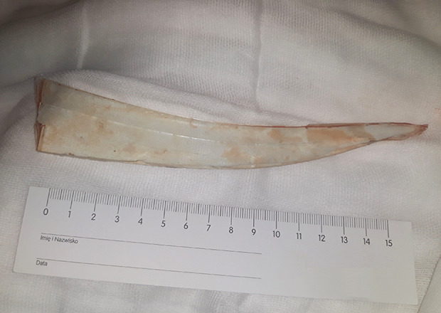
Shard of glass removed from the peritoneal cavity.
She was discharged home on the 4th postoperative day. Further outpatient treatment was normal.
Discussion
Foreign bodies may be found in the peritoneal cavity after impalement injuries, abdominal surgery, or translocations from the visceral lumen due to long-term migration or perforation of swallowed objects or those inserted via the rectum. The overwhelming majority of such cases involve ingested foreign bodies such as toothpicks or chicken bones [4]. Chronic perforation is most often caused by blunt foreign bodies and usually occurs in the sigmoid colon [5]. Intrauterine contraceptive devices have been reported to migrate through the uterus intraperitoneally [6,7].
Impalement injuries are one of the most difficult challenges for trauma surgeons due a high incidence of visceral or vascular injuries with significant morbidity and mortality [2]. Visible post-traumatic foreign bodies in the wound, hemodynamic instability, deterioration of the patient, and bleeding all raise concern of an intra-abdominal process and surgical exploration is needed. After the event, our patient was stable. She had slight pain, and a small wound without significant bleeding and the foreign body was not physically detectable. Although a 19-cm sharp glass fragment was in the peritoneal cavity for 9 months, there was no organ damage or migration. Such a case is highly unusual because most impalement injuries present acutely severe pain and injury to intra-abdominal structures.
In the literature, we only found 4 other cases of long-term presence of a piece of glass in the peritoneal cavity with delayed presentation after an impalement injury [8–11] (Table 1). All described cases had a common mechanism of injury – impact on a stationary object (type I by clinical classification of impalement injury) [12]. In our case and 2 other cases, the patient fell on a glass table and suffered a small wound in the back. Probably, the structure of the broken glass in the form of a sharp triangular fragment facilitated deep penetration into the body. After the event, patients were in good condition and did not raise suspicion of serious abdominal injury. Unfortunately, physical examination and wound exploration were insufficient to identify a foreign body, and only diagnostic imaging prompted by chronic abdominal pain showed the presence of a foreign body.
Table 1.
Clinical features, treatment, and visual outcomes for previously reported long-term retention of a glass foreign body (FB) in the abdominal cavity after impalement injury.
| Case number | Literature | Sex | Age at injury | Site (penetrating wound location) | Dimensions of FB (length× width× thickness) in cm | Symptoms | Time interval between injury and surgery (months) | Operation method | Organs damage |
|---|---|---|---|---|---|---|---|---|---|
| 1 | Davidov et al. 1999 [8] | Male | 25 | Left side of the lower back | 6.2×0.8×0.3 | Abdominal pain, macrohematuria, lumbar pains and decreased function of the left kidney | 2 | Lumbotomy and pyelotomy | Left kidney trauma |
| 2 | Crawford et al. 2008 [9] | Male | 60 | Left upper abdominal wall | 9.0×2.0×0.2 | Abdominal pain | 16 | Hand-assisted laparoscopic colectomy | Delayed colonic perforation |
| 3 | Rosat et al. 2015 [10] | Male | 66 | Left upper abdominal wall | 16×1.0×1.0 | Abdominal pain | 14 | Laparotomy, closed colonic perforation with a lineal stapler | Delayed colonic perforation |
| 4 | Johnston et al. 2007 [11] | Female | 60 | Right lower back, just above the natal cleft | 8,5 (length) | Abdominal pain, nausea, bloating | 20 | Laparotomy, Hartmann’s procedure | Bowel perforation |
In 2 of the 3 patients, delayed presentation of bowel damage occurred and resection was necessary [9,11]. In 1 patient, delayed closure of a colonic perforation with a linear stapler without resection was performed [10] and the late results were good.
Our case is unique because of 3 features. First, the mechanism of the injury is extremely rare. Second, the symptoms that were seen for 9 months after the injury where very unspecific and slight, and the patient only had chronic lower abdominal pain. Third, the piece of glass left the organs and blood vessels undamaged, although it is the largest piece of glass described in the literature that remained long-term in the peritoneal cavity without any organ damage.
Conclusions
Even a small torso wound in an asymptomatic patient must be suspected of having a foreign body and serious visceral injury. In any such case, visual, manual, and instrumental wound examination is always necessary. If exploration of the wound is troublesome and painful for the patient and full accuracy cannot be guaranteed, the procedure should be performed under anesthesia. Imaging tests are an important diagnostic method when a post-traumatic foreign body is suspected, especially when the fascia is damaged. Radiological diagnostics is also needed as a check for residual foreign bodies after cleaning the wound.
Acknowledgments
The authors thank all the general surgery staff for their cooperation.
Footnotes
Conflict of interests
None.
References:
- 1.Horowitz MD, Dove DB, Eismont FJ, Green BA. Impalement injuries. J Trauma. 1985;25:914–16. doi: 10.1097/00005373-198509000-00017. [DOI] [PubMed] [Google Scholar]
- 2.Mohan R, Ram DU, Baba YS, et al. Transabdominal impalement: Absence of visceral or vascular injury a rare possibility. J Emerg Med. 2011;41:495–98. doi: 10.1016/j.jemermed.2008.03.033. [DOI] [PubMed] [Google Scholar]
- 3.Oya S, Miyata K, Yuasa N, et al. Impalement injury to the left buttock with massive bleeding: A case report. Nagoya J Med Sci. 2013;75:147–52. [PMC free article] [PubMed] [Google Scholar]
- 4.Yalcin S, Karnak I, Ciftci AO, et al. Foreign body ingestion in children: An analysis of pediatric surgical practice. Pediatr Surg Int. 2007;23:755–61. doi: 10.1007/s00383-007-1958-y. [DOI] [PubMed] [Google Scholar]
- 5.Henderson FF, Gaston EA. Ingested foreign body in the gastrointestinal tract. Arch Surg. 1938;36:66–95. [Google Scholar]
- 6.Browning JJ, Bigrigg MA. Recovery of the intrauterine contraceptive device from the sigmoid colon. Three case reports. Br J Obstet Gynaecol. 1988;95:530–32. doi: 10.1111/j.1471-0528.1988.tb12813.x. [DOI] [PubMed] [Google Scholar]
- 7.Brunner SM, Comman A, Gaetzschmann P, et al. Laparoscopic removal of a perforating intrauterine device mimicking chronic appendicitis. J Laparoendosc Adv Surg Tech A. 2008;18:609–10. doi: 10.1089/lap.2007.0155. [DOI] [PubMed] [Google Scholar]
- 8.Davidov MI, Liadov AA. Glass foreign body in the kidney. Urologiia. 1999;(6):36–38. [PubMed] [Google Scholar]
- 9.Crawford DL, McVay WB. Delayed presentation of colonic impalement injury by picture frame glass fragment treated using hand-assisted laparoscopic colectomy. Surg Laparosc Endosc Percutan Tech. 2008;18:619–21. doi: 10.1097/SLE.0b013e3181889d53. [DOI] [PubMed] [Google Scholar]
- 10.Rosat A, Sánchez JM, Chocarro C, Barrera M. Impalement injury by glass shard with delayed colonic perforation. Pan Afr Med J. 2015;21:330. doi: 10.11604/pamj.2015.21.330.7676. [DOI] [PMC free article] [PubMed] [Google Scholar]
- 11.Johnston SA, Lisle DA, Borrowdale RC. A “painful” perforation. Med J Aust. 2007;186:46. doi: 10.5694/j.1326-5377.2007.tb00791.x. [DOI] [PubMed] [Google Scholar]
- 12.Missliwetz J. Fatal impalement injuries after falls at construction sites. Am J Forensic Med Pathol. 1995;16:81–83. doi: 10.1097/00000433-199503000-00018. [DOI] [PubMed] [Google Scholar]