

Abstract
Vertigo is a medical condition where a person feels as if they or the objects around them are moving when they are not. Any disturbance in the functioning of vestibular organ may cause vertigo. This study aims to find the role of cervical Vestibular evoked myogenic potential in diagnosing BPPV. The study performed was a prospective study of 50 patients with the age distribution from 20 to 60 years of age, attending the OPD in ENT department of MY Hospital, Indore with complaints of Vertigo. c-VEMP test was performed. The results were evaluated to diagnose BPPV. Of all the patients with the complaints of vertigo maximum belonged to the 5th decade of life. Of all the patients, females were found to be affected more than males making the male female ratio to be 1:1.28. In our study Dix–Hallpike test was found to be positive in 38 (74%) patients. On performing cervical VEMP, 15 (30%) patients of 50 patients showed abnormal latencies and amplitudes suggesting abnormal saccular functioning on affected side. Of these 15 abnormal VEMPs, 13 patients were positive on performing Dix–Hallpike test i.e. were diagnosed to have BPPV. VEMP showed a positive correlation with Dix–Hallpike test in diagnosis of vertigo of postural origin and thus can be used as a diagnostic tool for BPPV as VEMP is found to have high specificity.
Keywords: Vertigo, BPPV, VEMP, Dix–Hallpike test
Introduction
Vertigo is a symptom, not a disease. Effective diagnosis and management of vertigo begins with understanding what the symptom may represent. The vestibular organ in the inner ear has the most important role in maintaining balance of the body. Any disturbance in the functioning of vestibular organ may cause vertigo.
Positional vertigo is a sensation of spinning that occurs after the head of the patient is moved to a new position with respect to gravity and is the most common cause of Vertigo [1].
A detailed history and proper examination of the patient is required to diagnose Benign Paroxysmal Positional vertigo. Examination mainly includes the Dix–Hallpikes Positional test with a newly invented neurophysiological objective technique—Vestibular Evoked Myogenic Potential (VEMP) to diagnose the cases of Benign Paroxysmal Positional Vertigo (BPPV).
VEMP test is a small but important part of vestibulometry and it evaluates a very small part of vestibular system [2]. VEMP is used to determine the function of the otolithic organs (utricle and saccule) of the inner ear. It complements the information provided by caloric testing and other forms of vestibular apparatus testing. There are two different types of VEMPs [2]:
Cervical VEMP (c-VEMP)—measures the integrity of the saccule and inferior vestibular nerve.
Ocular VEMP (o-VEMP)—measures the integrity of the utricle and superior vestibular nerve.
Thus from this study, it is hoped that we become more familiar with the the role of Vestibular Evoked Myogenic Potential (VEMP) in the diagnosis of BPPV.
Aims and Objective
This study aims to assess the diagnostic potential of VEMP in patients with positional vertigo and to compare the diagnostic potential of VEMP and Dix–Hallpike test in patients of positional vertigo.
Materials and Methods
Present study was a prospective study which consisted of an assessment of 50 cases of vertigo whose examination was conducted in the ENT Department of MY Hospital, Indore in the period from April 2018 to July 2019.
Inclusion Criteria
The patients with the history of vertigo as a predominant symptom or associated symptoms of other complaints like dizziness, vomiting, headache, hearing loss etc. will be taken up for the study.
The age group taken for study is between 20 and 60 years.
The patients giving consent for being the part of the study.
Exclusion Criteria
Age group below 20 and above 60 years.
The patients with neurological deficits—Hemiplagia, Quadriplagia, Stroke (CVA).
The patients not willing to be the part of the study.
A proper detailed medical history with primary focus on type of vertigo was obtained to include onset of symptoms, severity, duration and associated factors. Dix–Hallpike tests was performed to diagnose positional vertigo. c-VEMP were performed in all patients and its co-relation with results of Dix–Hallpike tests was calculated.
Cervical vestibular evoked myogenic potentials is a test to check for the saccadic pathway which is been recorded from the SCM. The prolongation of the positive peak i.e. increase in latency or decreased amplitude means that c-VEMP is abnormal, that is there is saccular dysfunction.
It was performed after the patient was made to lie down. VEMP machine of the Neurosoft company was used (Fig. 1); and electrodes were placed—active electrodes were placed over the midpoint of the SCM muscle, reference electrode was placed on the sternal end of Sternocleidomastoid muscle and the ground electrode was placed over forehead (Fig. 2). The Sternocleidomastoid (SCM) muscle of the ipsilateral side (i.e. the SCM on the side being tested) is tonically contracted, which is achieved by asking the patient to voluntarily raise the head by about 30° above the bed without support (but chin should not touch the chest).
Fig. 1.
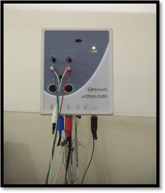
VEMP machine
Fig. 2.

Placement of leads to record c-VEMP
Click sounds of 0.1 ms at a rate of 5.1/s at an intensity of 95–105 dB nHL were presented to the ipsilateral ear by air conduction insert ear phones [2]. Electrodes measure the response from vestibular system, and then we look whether the response is symmetrical or not. The normalized peak-to-peak (p1 − n1) amplitudes and the latencies of the first positive (p1) and negative (n1) peaks were measured. Because background muscle activities could interfere with the VEMP amplitudes, the inter-peak amplitudes should be normalized [3, 4] (Figs. 3 and 4).
Fig. 3.

Normal c-VEMP
Fig. 4.

Abnormal c-VEMP
The asymmetry ratio was calculated for comparison between the right and left ears, using the formula described by Murofushi et al. [5].
Au: p1 − n1 (the peak-to-peak amplitude of the unaffected ear), Aa: p1 − n1 (the peak-to-peak amplitude of the affected ear).
An asymmetry of more than 35% and side ratio more than 2.1 was considered abnormal and is indicative of abnormally weaker saccular function on the side showing lower amplitude.
Observation and Results
In our study it was found that maximum patients, 18 (36%) of 50 patients with complain of vertigo lie in the 5th decade of life. Females were found to be affected more than males making the male to female ratio to be 1:1.08. BPPV was diagnosed on basis of history and performing Dix–Hallpike test.
From all the patients taking part in the study, on performing Dix–Hallpike test, 38 (74%) patients showed positive results with presence of nystagmus and dizziness. Of all, 26 (52%) patients had positive results for unilateral right side, 6 (12%) patients were positive for unilateral left side while only 6 (12%) patients were positive for bilateral side (Fig. 5).
Fig. 5.

Distribution on basis of side involved in BPPV
On performing cervical VEMP, 15 (30%) patients of 50 patients showed abnormal latencies and amplitudes suggesting abnormal saccular functioning on affected side (Fig. 6). Of these 15 abnormal VEMPs, 13 patients were positive on performing Dix–Hallpike test i.e. the test was found to have high specificity but low sensitivity for diagnosing BPPV (Table 1).
Fig. 6.

Results of VEMP
Table 1.
Relation of VEMP to Dix–Hallpike test
| VEMP | Dix–Hallpike test | Total | ||
|---|---|---|---|---|
| Negative | Positive | |||
| Abnormal | No. of patients | 2 | 13 | 15 |
| Percentage | 16.7% | 34.2% | 30.0% | |
| Normal | No. of patients | 10 | 25 | 35 |
| Percentage | 83.3% | 65.8% | 70.0% | |
| No. of patients | 12 | 38 | 50 | |
| Percentage | 100.0% | 100.0% | 100.0% | |
Sensitivity of the VEMP test was calculated to be 34.21%, based on the formula (Table 1).
While VEMP test was was to be more specific. Specificity of the test was calculated and found to be 83.33% using the formula (Table 1).
Discussion
In the present study it was found that maximum patients, 18 (36%) of 50 patients with complain of vertigo lie in the 5th decade of life. Of all the patients, females were found to be affected more than males making the male to female ratio to be 1:1.08.
Mauricio et al. [6], in 2017 also found that, females (69.8%) were more commonly affected with BPPV than males (30.2%), and the calculated gender ratio was found to be 1:2.311. In contrast to the above findings by Patangay and Ansari [7] in 2016, found that males (62.7%) were commonly affected than females (37.2%) making the ration to be 1.68:1. As compared to previous studies, the present study exhibits BPPV with male female ratio of 1:1.08, exhibiting female predominance, which could be explained as more awareness among females to seek treatment.
The diagnosis of BPPV affecting the posterior semicircular canal can be established by a history of episodic vertigo with changes in head position and the presence of characteristic nystagmus provoked by the Dix–Hallpike test according to a guideline from the American Academy of Otolaryngology-Head and Neck Surgery [8]. In our study Dix–Hallpike test was found to be positive in 38 (74%) patients which can be co-related to the study done by Caldas et al. [9], in which, BPPV with dizziness and positioning nystagmus occurred in 1033 cases (81.3%), making BPPV the most common cause of vertigo attending ENT OPD. In our study it was found that, 26 (52%) patients had positive results for unilateral right side, 6 (12%) patients were positive for unilateral left side i.e. total of 32 (64%) patients of 50 had unilateral ear involvement, while only 6 (12%) of all patients were positive for bilateral side; while in a study by Caldas et al. [9], the disease was unilateral in 91.8% of cases, and bilateral in 8.2% of cases of BPPV with dizziness and positioning nystagmus. Lopez-Escámez et al. [10], also found a right sided predominance (75.9%) and agreed to the widely accepted hypothesis that this predominance is due to the preferential right-side lying position during sleep in susceptible subjects.
Yang et al. [11] in his study in 2008, found that VEMP latencies are increased in BPPV patients. In our study, it was found that on performing cervical VEMP, 13 (34.2%) patients with BPPV, showed abnormal latencies and amplitudes suggesting abnormal saccular functioning on affected side. The results of our study show high specificity and low sensitivity of VEMP for diagnosing BPPV. The results of our study is similar to the study done by Hong [12], who found that of the 53 patients with BPPV, 13(24.5%) showed abnormal VEMP responses on the affected side when compared with their age-related control subgroup. Yetiser et al. [3], in his study to analyze the effect of VEMP in patients with benign paroxysmal positional vertigo (BPPV), also found that of 100 patients in his study, twenty-four patients (23.5%) had a gross VEMP abnormality. Kim et al. [13], in his study reported abnormalities in about 20% of cVEMP in patients of BPPV, Pascual et al. [14] in his study in 2017, found that BPPV group had abnormal cervical VEMPS in 49.25% of patients compared to 16.67% in the control group which concluded that utricular and saccular dysfunction in BPPV patients proved by VEMPS is higher than in healthy individuals. In the most recent study by Semmanaselvan et al. [15], to find VEMP abnormalities in individuals with Posterior canal Benign Paroxysmal Positional Vertigo (BPPV) found that cervical VEMP abnormalities in 8/36 (22.22%) affected ears with BPPV respectively and concluded that individuals with Posterior canal BPPV may have otoconia dislodgement or macular degeneration of utricle, saccule, both utricle and saccule unilaterally, or bilaterally.
Conclusion
BPPV is the most common cause of vertigo in people attending ENT OPD. It was found that vertigo is more commonly present in females and is more prevalent in 4th and 5th decade of life. Dix–Hallpike test is the subjective diagnostic positional test for BPPV and found that BPPV is more likely to invole right ear, the finding may be related to the habit of sleeping on the right side in the general population. VEMP showed a positive correlation with Dix–Hallpike test in diagnosis of vertigo of postural origin and thus can be used as a diagnostic tool for BPPV as VEMP is found to have high specificity.
Funding
This study was not funded by any source.
Compliance with Ethical Standards
Conflict of interest
The authors declare that they have no conflict of interest.
Ethical Approval
All procedures performed in studies involving human participants were in accordance with the ethical standards of the institutional and/or national research committee and with the 1964 Helsinki declaration and its later amendments or comparable ethical standards.
Human and Animal Rights
This article does not contain any studies with animals performed by any of the authors.
Informed Consent
Informed consent was obtained from all individual participants included in the study.
Footnotes
Publisher's Note
Springer Nature remains neutral with regard to jurisdictional claims in published maps and institutional affiliations.
References
- 1.Walker HK, Hall WD, Hurst JW, editors. Clinical methods: the history, physical, and laboratory examinations. 3. Boston: Butterworths; 1990. [PubMed] [Google Scholar]
- 2.Biswas A. Clinical audiovestibulo-metry for otologists and neurologists. Chapter 11. 5. New Delhi: Bhalani Publishing House; 1995. pp. 447–493. [Google Scholar]
- 3.Yetiser S, Ince D, Gul M. An analysis of vestibular evoked myogenic potentials in patients with benign paroxysmal positional vertigo. Ann Otol Rhinol Laryngol. 2014;123:686–695. doi: 10.1177/0003489414532778. [DOI] [PubMed] [Google Scholar]
- 4.Salviz M, Yuce T, Karatas A, Balikci HH, Ozkul MH. Diagnostic value of frequency—associated vestibular-evoked myogenic potential responses in Meniere’s disease. Audiol Neurootol. 2015;20:229–236. doi: 10.1159/000375395. [DOI] [PubMed] [Google Scholar]
- 5.Murofushi T, Iwasaki S, Ushio M. Recovery of vestibular evoked myogenic potentials after a vertigo attack due to vestibular neuritis. Acta Otolaryngol. 2006;126:364–367. doi: 10.1080/00016480500417189. [DOI] [PubMed] [Google Scholar]
- 6.Mauricio CE, Alanis-Nunez AJ, Andres S, Teresa GG. Dizziness and vertigo in emergency: a population-based analysis. J Otol Rhinol. 2018;7:1. doi: 10.4172/2324-8785.1000. [DOI] [Google Scholar]
- 7.Patangay KK, Ansari R. Benign paroxysmal positional vertigo: our experience. Indian J Otolaryngol Head Neck Surg. 2014;68(1):39–41. doi: 10.1007/s12070-014-0818-z. [DOI] [PMC free article] [PubMed] [Google Scholar]
- 8.Nguyen-Huynh AT. Evidence-based practice: management of vertigo. Otolaryngol Clin North Am. 2012;45(5):925–940. doi: 10.1016/j.otc.2012.06.001. [DOI] [PMC free article] [PubMed] [Google Scholar]
- 9.Caldas MA, Ganança CF, Ganança FF, Ganança MM, Caovilla HH. Clinical features of benign paroxysmal positional vertigo. Braz J Otorhinolaryngol. 2009;75(4):502–506. doi: 10.1590/S1808-86942009000400006. [DOI] [PMC free article] [PubMed] [Google Scholar]
- 10.Lopez-Escámez JA, Gámiz MJ, Fi-ana MG, Perez AF, Canet IS. Position in bed is associated with left or right location in benign paroxysmal positional vertigo of the posterior semicircular canal. Am J Otolaryngol. 2002;23:263–266. doi: 10.1053/ajot.2002.124199. [DOI] [PubMed] [Google Scholar]
- 11.Yang WS, Kim SH, Lee JD, Lee WS. Clinical significance of vestibular evoked myogenic potentials in benign paroxysmal positional vertigo. Otol Neurotol. 2008;29(8):1162–1166. doi: 10.1097/MAO.0b013e31818a0881. [DOI] [PubMed] [Google Scholar]
- 12.Hong SM. Vestibular evoked myogenic potentials in patients with benign paroxysmal positional vertigo involving each semicircular canal. Am J Otolaryngol. 2008;29(3):184–187. doi: 10.1016/j.amjoto.2007.07.004. [DOI] [PubMed] [Google Scholar]
- 13.Kim EJ, et al. Persistent otolith dysfunction even after successful repositioning in benign paroxysmal positional vertigo. J Neurol Sci. 2015;358(1–2):287–293. doi: 10.1016/j.jns.2015.09.012. [DOI] [PubMed] [Google Scholar]
- 14.Pascual PM, Merino PA. Otolithic damage study in patients with benign paroxysmal positional vertigo with vestibular evoked myogenic potentials. Acta Otorrinolaringol Esp. 2019;70(3):131–135. doi: 10.1016/j.otorri.2018.04.003. [DOI] [PubMed] [Google Scholar]
- 15.Semmanaselvan K, Vignesh SS, Muthukumar R, Jaya V. Vestibular evoked myogenic potentials after epleys manoeuvre among individuals with benign paroxysmal positional vertigo. Indian J Otolaryngol Head Neck Surg. 2019;71(2):195–200. doi: 10.1007/s12070-019-01581-6. [DOI] [PMC free article] [PubMed] [Google Scholar]