Since the outbreak of the novel coronavirus disease 2019 (COVID‐19), reports concerning suspicious COVID‐19 skin manifestations have been progressively increasing. Morbilliform, varicelliform or urticarial rashes were described first.1 Later, acral erythematous or purpuric lesions were reported.2–5
At the Araba University Hospital in Spain, which covers a population of 340 000 people, we designed a descriptive study of 74 patients. We recruited all patients presenting with suspicious acral manifestations for COVID‐19 from 7 to 22 April 2020. The average temperature during this period was 14 °C. Patients with changes in their pharmacological drugs during the previous month were excluded. Owing to the pandemic, data on most patients reached us via teledermatology. Age, sex, medical history, occupation and clinical characteristics were recorded for each patient. The results are shown in Table 1.
Table 1.
| Result, % | |
| Lesion morphology | |
| EP | 76.4 |
| PM | 40.54 |
| Both EP and PM | 16.21 |
| Erosion | 10.8 |
| Swelling | 16.21 |
| Distribution | |
| Hands | 8.1 |
| Feet | 95.94 |
| Both | 4.05 |
| Laterality | |
| Unilateral | 31.08 |
| Bilateral | 68.91 |
| Symmetry | |
| Symmetrical | 51.35 |
| Asymmetrical | 44.59 |
| Unknown | 4.05 |
| Symptoms | |
| Pruritus | 32.4 |
| Pain | 27 |
| Asymptomatic | 48.6 |
| Extracutaneous manifestations | |
| Frequency | 29.6 |
| Type | |
| Respiratory | 50 |
| General | 50 |
| Latency period, days | 16.15 |
| COVID‐19 symptoms | |
| Symptoms present | |
| Cough | 52.38 |
| Fever | 33.33 |
| Asthenia/myalgia | 28.57 |
| Diarrhoea/nausea/vomiting | 19 |
| Dyspnoea | 9.52 |
| Anosmia/ageusia | 4.76 |
EP, erythematous papules; PM, purpuric macules.
Of 74 patients, 42 (56.8%) were male. Mean age was 19.66 years (median 14.5 years, range 3–100 years). A small percentage (5.4%) were healthcare workers or had close contact with such workers, while 24.32% reported close contact with a person with confirmed or clinically diagnosed COVID‐19.
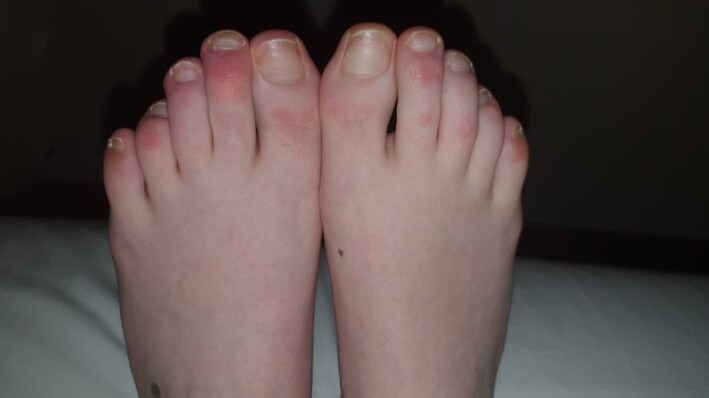
Most patients had erythematous papules (76.4%), similar to chilblains (Fig. 1), while 40.54% had purpuric macules. Nearly all patients showed foot involvement (95.94%) and the hands were affected in 8.1%. Bilateral (68.91%) and symmetrical (51.35%) were the most usual distribution patterns. The dorsa of the toes/fingers was the main affected location (74.3% on toes and 100% on fingers).
Figure 1.

Typical acral cutaneous findings suspicious for COVID‐19: erythematous chilblain‐like plaques with an asymmetrical distribution in the dorsum of toes.
Extracutaneous symptoms were found in 21 patients (29.6%), of which 50% also had clinical respiratory symptoms (cough and dyspnoea). In 66.7% of the cases, cutaneous manifestations developed after extracutaneous symptoms with a mean latency of 16.15 days. Two patients developed pneumonia (2.70%), both preceding the cutaneous symptoms.
In our area, COVID‐19 PCR, which has a sensitivity of about 70%, was performed on 17 516 people and 4649 were positive. Owing to the limited availability of resources only 11 patients in our study underwent PCR, and 1 had a positive result. Six patients underwent blood investigations (including autoimmunity), which did not show relevant alterations; this is in line with a previous report.2.
A skin biopsy was taken from a lesion on the toe of one patient who had a negative serology test for COVID‐19, and histological examination revealed a lymphocytic perivascular and perieccrine infiltrate. Neither vascular occlusion nor intravascular thrombi were seen. Direct immunofluorescence study was negative. These findings are compatible with those previously described.2, 3
The aetiology of these lesions remains unclear. A microangiopathic and inflammatory process is thought to occur.2, 4–6 Alternatively, activation of complement, leading to inflammation and thrombi formation has been proposed.6 However, neither our case nor the others previously published have described thrombi.2 More recent articles3, 4 have proposed a delayed antigen–antibody immunological reaction, which could explain their development in asymptomatic and paucisymptomatic patients.
Interestingly, we noticed an increase in the number of acral lesions 25 days after the start of lockdown. Conversely, last April we did not have similar lesions registered. Thus, we wonder whether some factors related to quarantine might have been involved, such as lack of sun exposure and consequent low levels of vitamin D.
We hope this paper will encourage sturdier studies. If the results validate our findings, acral cutaneous manifestations will represent a useful clue to identify COVID‐19 in asymptomatic and paucisymptomatic patients.
Acknowledgement
We wish to thank the collaboration of all our colleagues in the Dermatology Department of the Araba University Hospital, as well as the pathologists (Dr Malo‐Díez and Dr Martínez‐Aracil). We also thank the primary care physicians, physicians from the emergency department, pediatricians and other specialists from our hospital and from the primary care centres in our area who collaborated and helped us to perform this study.
Contributor Information
A. Saenz Aguirre, Department of Dermatology Hospital Universitario Araba Vitoria Spain
F. J. De la Torre Gomar, Department of Dermatology Hospital Universitario Araba Vitoria Spain
P. Rosés‐Gibert, Department of Dermatology Hospital Universitario Araba Vitoria Spain
J. Gimeno Castillo, Department of Dermatology Hospital Universitario Araba Vitoria Spain
Z. Martinez de Lagrán Alvarez de Arcaya, Department of Dermatology Hospital Universitario Araba Vitoria Spain
R. Gonzalez‐Perez, Department of Dermatology Hospital Universitario Araba Vitoria Spain
References
- Recalcati S. Cutaneous manifestations in COVID‐19: a first perspective. J Eur Acad Dermatol Venereol 2020; 34: e212–13. [DOI] [PubMed] [Google Scholar]
- Kolivras A, Dehavay F, Delplace D et al. Coronavirus (COVID‐19) infection‐induced chilblains: a case report with histopathological findings. JAAD Case Rep 2020; 6: 489–92. [DOI] [PMC free article] [PubMed] [Google Scholar]
- Recalcati S, Barbagallo T, Frasin LA et al. Acral cutaneous lesions in the time of COVID‐19. J Eur Acad Dermatol Venereol 2020; 34: e346–7. [DOI] [PMC free article] [PubMed] [Google Scholar]
- Landa N, Mendieta‐Eckert M, Fonda‐Pascual P et al. Chilblain‐like lesions on feet and hands during the COVID‐19 pandemic. Int J Dermatol 2020; 59: 739–43. [DOI] [PMC free article] [PubMed] [Google Scholar]
- López‐Robles J, de la Hera I, Pardo J et al. Chilblain‐like lesions: a case series of 41 patients during the COVID‐19 pandemic. Clin Exp Dermatol 2020. [DOI] [PMC free article] [PubMed] [Google Scholar]
- Magro C, Mulvey JJ, Berlin D et al. Complement associated microvascular injury and thrombosis in the pathogenesis of severe COVID‐19 infection: a report of five cases. Transl Res 2020. [DOI] [PMC free article] [PubMed] [Google Scholar]


