Abstract
The left renal vein (LRV) passing behind the abdominal aorta is termed as a retroaortic LRV (RLRV) and it is a relatively uncommon condition. Since the left kidney is preferred in the setting of live donor kidney transplantation, urologists must be familiar with the anomalies of the LRV. There are four variants of RLRV mentioned in the literature. However, we came across two newer variants of RLRV in two donors for renal transplantation. Both donors underwent successful left donor nephrectomy.
INTRODUCTION
It is mandatory to evaluate the renal vasculature during the pretransplant workup of a patient awaiting donor nephrectomy. Although much importance is given for identifying anatomical variations of the renal arteries, which are more common, venous anomalies are also seen in some of these donors. A retroaortic left renal vein (RLRV) is one such entity. The RLRV is a rare variant, according to the previous studies, the incidence of a retroaortic renal vein was reported to be 0.6%–3.7%.[1,2] There are four subtypes of the RLRV which include as follows: (1) Type 1, where the RLRV joins the inferior vena cava (IVC) in the orthotopic position; (2) Type 2, where the RLRV joins the gonadal and ascending lumbar veins before joining the IVC at the level of the fourth to fifth lumber vertebral bodies; (3) Type 3 is a RLRV which consists of both anterior and RLRVs (circumaortic) before joining the IVC and (4) Type 4, where the ventral preaortic limb of the left renal vein (LRV) is obliterated and the dorsal limb persists as a RLRV, coursing obliquely and caudal behind the aorta to join the left common iliac vein.[3]
Although RLRV has more diagnostic significance during pretransplant workup, it has a deep association with renovascular aneurysm, varicocele, inguinal or flank pain, and hematuria. We present the radiological images of two newer variants of RLRV not mentioned in literature.
CASE REPORT
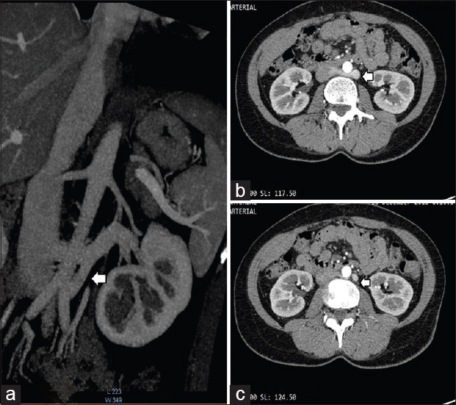
Forty-year-old female during donor workup showed the LRV passing caudally and bifurcating into two [Figure 1a], one segment draining into the IVC at L4–L5 [Figure 1b] and other segment draining into the left common iliac vein [Figure 1c]
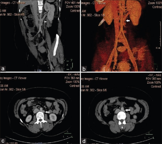
Thirty-three-year-old female during spousal donor workup showed the left RLRV passing caudally and bifurcating into two [Figure 2a and b], cranial segment draining into IVC at the level of L3–L4 [Figure 2c] and the caudal segment draining separately into IVC about 2 cm distally [Figure 2d].
Figure 1.

Left renal vein passing caudally and bifurcating into two (a), one segment draining into the inferior vena cava at L4–L5 (b) and other segment draining into the left common iliac vein (c)
Figure 2.

Left retroaortic left renal vein passing caudally and bifurcating into two (a and b), cranial segment draining into the inferior vena cava at the level of L3–L4 (c) and the caudal segment draining separately into the inferior vena cava about 2 cm distally (d)
DISCUSSION
During development of the IVC there are anastomotic communications between subcardinal and supracardinal channels, which form a collar of veins encircling the aorta.[4] The ventral portion of the circumaortic collar persists as the normal LRV. If the dorsal portion of this collar persists, the LRV is posterior to the aorta, forming an RLRV. The most common urological symptom of RLRV is hematuria due to compression of the LRV between the abdominal aorta and vertebrae (posterior nutcracker phenomenon). Although four various type of RLRV have been described, the two renal vein anomalies in the two cases presented do not belong to any of these categories. Therefore, we propose Type V and VI as the two newer variants of RLRV presented in our series.
CONCLUSION
Two rare cases of double RLRV not mentioned in literature has been presented.
Declaration of patient consent
The authors certify that they have obtained all appropriate patient consent forms. In the form the patient(s) has/have given his/her/their consent for his/her/their images and other clinical information to be reported in the journal. The patients understand that their names and initials will not be published and due efforts will be made to conceal their identity, but anonymity cannot be guaranteed.
Footnotes
Financial support and sponsorship: Nil.
Conflicts of Interest: There are no conflicts of interest.
REFERENCES
- 1.Kawai K, Tanaka T, Watanabe T. A rare anomaly of left renal vein drainage into the left common iliac drainage into the left common iliac vein: A case report. Int J Surg Case Rep. 2016;20:4–6. doi: 10.1016/j.ijscr.2015.12.050. [DOI] [PMC free article] [PubMed] [Google Scholar]
- 2.Arslan H, Etlik O, Ceylan K, Temizoz O, Harman M, Kavan M. Incidence of retro-aortic left renal vein and its relationship with varicocele. Eur Radiol. 2005;15:1717–20. doi: 10.1007/s00330-004-2563-2. [DOI] [PubMed] [Google Scholar]
- 3.Prakash C, Ali S, Khan SM. Anomalous left renal vein coursing behind aorta and draining into the left common iliac vein: A rare variant. Int J Case Rep Images. 2014;5:553–7. [Google Scholar]
- 4.Karaman B, Koplay M, Ozturk E, Basekim CC, Ogul H, Mutlu H, et al. Retroaortic left renal vein: Multidetector computed tomography angiography findings and its clinical importance. Acta Radiol. 2007;48:355–60. doi: 10.1080/02841850701244755. [DOI] [PubMed] [Google Scholar]


