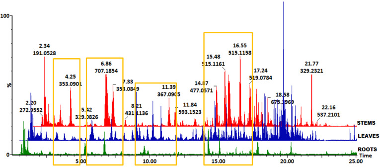
Figure 1.
Representative UHPLC-QTOF-MS base peak intensity (BPI) chromatograms showing the separation of secondary metabolites in extracts of B. pilosa roots (green), leaves (blue), and stems (red). The yellow rectangles indicate the chromatographic regions where hydroxycinnamic acid derivatives are present across the three tissue types with some visible differences in intensities.

