Abstract
Zalypsis® (PM00104) is a novel tetrahydroisoquinoline alkaloid related to trabectedin (Ecteinascidin 743; Et743). Et743 and PM00104 have similar A- and B-rings but differ in their C-rings. The present study demonstrates that Et743 and PM00104 differ in at least two ways: in their DNA binding properties and NER dependency for cellular targeting. DNase I footprinting shows that the two drugs bind DNA differentially. We also found that, in contrast to Et743, the antiproliferative activity of PM00104 does not depend on transcription-coupled nucleotide excision repair (NER). Accordingly, PM00104 induces γ-H2AX foci with the same efficiency in NER-deficient or proficient cells. Moreover, the formation of γ-H2AX foci is replication-dependent for PM00104 whereas it is both transcription- and replication-dependent in the case of Et743. These findings demonstrate the importance of the C-ring structure of tetrahydroisoquinoline ecteinascidin derivatives for NER targeting. Finally, PM00104 exerts antiproliferative activity at nanomolar concentrations and induces γ-H2AX response in two Ewing sarcoma cell lines, suggesting that γ-H2AX could serve as a pharmacodynamic biomarker for the clinical development of PM00104.
Keywords: Nucleotide excision repair, DNA double-strand breaks, histone γ-H2AX, Et743, PM00104, Ewing Sarcoma
Introduction
PM00104 (Zalypsis®) is a novel semi-synthetic tetrahydroisoquinoline alkaloid. This family of compounds includes Jorumycin, obtained from a pacific nudibranch, the family of Renieramycins, which are isolated from sponges, and the Ecteinascidins isolated from a carribean tunicate. These compounds are potent cytotoxic agents and display high antitumor and antimicrobial activities (1, 2). Consistent with other members of this family, PM00104 exhibits strong in vitro and in vivo antitumor activity in a wide variety of solid and hematological tumor cell lines and human transplantable breast, gastric, prostate and renal xenografted tumors. PM00104 has now entered Phase I clinical trials for the treatment of solid tumors and lymphoma1.
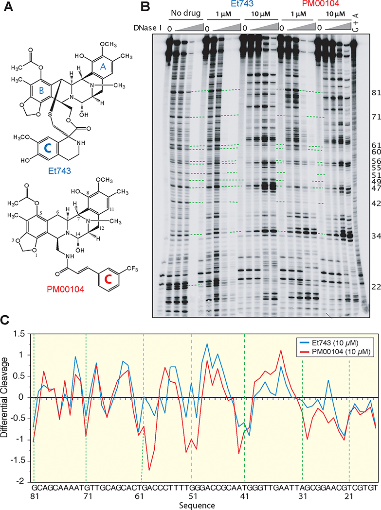
PM00104 is chemically related to Ecteinascidin 743 (Trabectedin) (Fig. 1A). However, PM00104 was chemically engineered whereas Et743 is a natural product initially purified from extracts of the tunicate Ecteinascidia turbinata in 1990 (3). Structurally, PM00104 and Et743 consist of similar A and B ring structures but differ in their C-ring (Fig. 1A). This chemical difference is important since the C-ring of Et743 has been proposed to directly interact with the NER endonuclease, XPG (4, 5).
Figure 1. Comparative DNA binding profiles of PM00104 and Et743.
A, Chemical structures of Et743 and PM00104. B, Representative DNase I footprinting experiment. The DNA substrate [5’-end-labeled PvuII/HindIII fragment of pBluescript SK (–) phagemid DNA (pSK)] was reacted in the absence (No drug) or presence of 1 μM or 10 μM Et743 or PM00104 for 30 min at 37°C. Samples were then subjected to either no treatment (0) or treatment with increasing concentrations of deoxyribonuclease I (DNase I) for 1 min at 25°C. Reactions were stopped with 15 mM EDTA (final concentration). DNA fragments were separated in denaturing polyacrylamide gels. Lane G + A, purine ladder after formic acid treatment. Numbers on the right correspond to the DNA sequence indicated in panel C. C, Differential DNase I cutting in the presence of 10 μM PM00104 (red line) and Et743 (blue line). Negative and positive values correspond to drug-protected sites and enhanced cleavage, respectively. Vertical scales are in units of ln(fa)-ln(fc), where fa is the fractional cleavage at any bond in the presence of the drug and fc is the fractional cleavage of the same bond in the control, given closely similar extents of overall digestion. The results are displayed on a logarithmic scale and the DNA sequence is indicated at the bottom. Numbers on the right of panel B correspond to that sequence.
Ecteinascidin 743 (Et743) is registered as Yondelis® and approved for the treatment of soft tissue sarcomas. Et743 is also being studied in Phase III trial for ovarian cancer and in Phase II trials for breast, prostate, and pediatric sarcomas1. Et743 alkylates the N2-position of guanines in the DNA minor groove (6, 7). NMR-based studies indicates that the A- and B-subunits of Et743 are responsible for DNA recognition and bonding (8), while the C-ring is projected out of the minor groove, making limited contacts with the DNA (6, 7, 9). A unique characteristic of Et743 is its selective poisoning of the transcription-coupled nucleotide excision repair (TC-NER) (5, 10, 11). This was revealed by the finding that a colon carcinoma cell line (HCT116-ER-5) selected for resistance to Et743 was deficient for the NER factor, XPG (5), and that TC-NER-deficient cells were resistant to Et743 (5, 10, 11). This mechanism of action sets Et743 apart from the “classical” DNA alkylating agents like platinum derivatives that tend to be inactive in TC-NER overexpressing cells (12, 13).
The aim of the present study was to compare PM00104 and Et743 with regard to DNA binding and NER dependency, and to uncover pharmacodynamic biomarkers for the clinical development of PM00104. To that effect, we report our investigation of the sequence-specific DNA binding of PM00104 in comparison with Et743, and the contribution of NER for the antiproliferative activity of the drugs. Finally, based on the clinical data available showing activity of PM00104 in sarcomas and the recent finding that Et743 induces γ-H2AX (14–16), we determined whether the induction of DNA damage by PM00104 could be measured using γ-H2AX as a pharmacodynamic marker (17) in human Ewing sarcoma cell lines.
MATERIALS AND METHODS
Drugs, chemicals, and enzymes
Ecteinascidin 743 (Et743) and PM00104 were provided by Pharmamar (Madrid, Spain). Drug stock solutions were made in dimethyl sulfoxide (DMSO) at 10 mM, and further dilutions were made in DMSO immediately before use. T4 polynucleotide kinase, and polyacrylamide-bis were purchased from Invitrogen (Carlsbad, CA). DNA Quick Spin columns were purchased from Roche Diagnostics (Indianapolis, IN). [γ−32P]dATP was purchased from New England Nuclear (Boston, MA). Aphidicolin was purchased from Sigma chemical company (St Louis, MO).
Cell culture
All cell lines were maintained in Dulbecco’s modified Eagle’s medium containing 10% fetal calf serum. Xeroderma pigmentosum group D (XPD) and their stably complemented counterparts XPD-c were provided by Dr. Kenneth Kraemer (Basic Research Laboratory, CCR, NCI, NIH, Bethesda, MD). XPF, XPG and wild type GM00637 were obtained from the Coriell cell repository (Camden, NJ). Human colon carcinoma HCT116 and their Et743-resistant derivative were generated as described (5). Ewing Sarcoma cells TC32 and TC71 were obtained from Dr. Javed Khan (Advanced Technology Center, NCI, NIH, Gaithersburg, MD).
DNase I footprinting
The 161-base pair DNA fragment from pBluescript SK(–) phagemid DNA (Stratagene, La Jolla, CA) was generated by PCR amplification as described (18). Reaction products were separated by electrophoresis in 1.5% agarose gels made in 1x Tris-borate EDTA buffer. The 161-bp fragment (pSK) was eluted from the gel slice using the Qiaex II kit (Qiagen, Inc., Valencia, CA). The pSK DNA was 5’-end-labeled by using T4 polynucleotide kinase with [γ−32P]dATP for 1 h at 37oC. Reactions were stopped by incubating the samples for 10 min at 70oC. To retain the 5’-radiolabel in only one of the strands, samples were cleaved with HindIII restriction endonuclease in supplied NE buffer 2 (New England Biolabs, Beverly, MA) for 1 h at 37°C. Labeled mixtures were subsequently centrifuged through mini Quick Spin DNA columns to remove the unincorporated radiolabel.
For the deoxyribonuclease I (DNase I) footprinting experiments, labeled pSK was reacted in the absence (Control) or presence of 1 μM or 10 μM Et743 or PM00104 in 10 mM Tris-Cl, pH 7.5, 50 mM KCl, 5 mM MgCl2, 0.1 mM EDTA, and 15 μg/ml BSA, (final concentrations). After incubation for 30 min at 37oC, samples were subjected to either no treatment (–) or treatment with several concentrations of deoxyribonuclease I (DNase I) (0.005, 0.01, 0.05, or 0.1 unit for control and 0.05, 0.1, 0.5, or 1 unit for drug treated) in a 10 μl reaction, for 1 min at 25oC (6). Reactions were stopped with 15 mM EDTA (final concentration) and DNA sequencing loading buffer (80% formamide, 10 mM NaOH, 1 mM EDTA, 0.1% xylene cyanol, and 0.1% bromophenol blue). Samples were heated to 90oC and immediately loaded into DNA sequencing gels (7% polyacrylamide; 19:1 acrylamide:bis) containing 7 M urea in TBE buffer. Electrophoreses were at 2500 V (60 W) for 4 h. Imaging and quantitation were performed with a PhosphorImager using ImageQuant software (Molecular Dynamics, Sunnyvale, CA).
3-(4,5-Dimethylthiazol-2-yl)-2,5-diphenyltetrazolium bromide (MTT) assays
Cells were seeded at a density of 1,000 per well in 96-well microtiter plates. Et743 or PM00104 were added the next day. After 72 hours, MTT was added to each well (0.5 mg/mL; Sigma Aldrich, St Louis, MO), and plates were maintained at 37°C for 4 hours. The medium was then discarded and DMSO was added to each well to lyse the cells. Absorbance was measured at 450 nm using a multiwell spectrophotometer (Emax, Molecular Devices, Sunnyvale, CA).
Confocal microscopy
Cells were grown one day before drug treatment in Nunc chamber slides (Nalgene, Rochester, NY). Following treatment, the medium was aspirated out and cells were washed in PBS. Cells were immediately fixed and permeabilized by a 20 min incubation at room temperature with 2% paraformaldehyde and a 5 min incubation in ice-cold 70% ethanol. To block nonspecific binding, cells were incubated with 8% bovine serum albumin for 1 h at room temperature. Cells were stained for 90 min with mouse anti-γ-H2AX antibody (Upstate Biotechnology, Lake Placid, NY), diluted 1/1,000. Fluorescent secondary antibody (Alexa-488, Molecular probes, Carlsbad, CA, 1/800) was then added for 45 min. All incubations were in 1% bovine serum albumin, at room temperature and followed by three PBS washes. Nuclei were stained with propidium iodide (0.05 mg/ml) and RNase A (0.5 mg/ml) for 10 min at 37°C. Slides were mounted using Vectashield mounting liquid (Vector Labs, Burlingame, CA) and visualized using a Nikon Eclipse TE-300 confocal laser scanning microscope system. Images were captured and stored as TIFF files. For each sample in each experiment, 50 to 200 cells were scored. The quantification of staining intensities was made with Adobe Photoshop 7.0 and normalized to the number of analyzed cells.
RESULTS
PM00104 exhibits differential DNase I footprinting in comparison with Et743
Because PM00104 and Et743 are derived from a similar chemical scaffold (A-B rings), which is known to form covalent bonding with DNA (6, 19) but differ in their appended C-rings (for structures, see Fig. 1A), we evaluated whether the differences in C ring affected DNase I footprinting (6). As illustrated in Figure 1B, PM00104 produced a clear footprint with suppression of cleavage at the drug binding sites and enhancement of cleavage in the flanks of the drug-bound DNA (6, 20). This indicates that PM00104 is a sequence-specific DNA binding agent. Comparison of the DNA cleavage patterns obtained in the presence of PM00104 and Et743 showed DNA binding at several similar sites with differences in the relative intensities (Fig. 1B and C). At 1 μM, the DNase I footprinting of PM00104 appeared generally stronger for PM00104 than for Et743 (Fig. 1B). At 10 μM, both PM00104 and Et743 gave clear footprints. Some binding sites, however, were stronger for PM00104 (Fig. 1C). These results demonstrate that PM00104 binds DNA at specific sites and exhibits both similarities and differences compared to Et743.
The antiproliferative activity of PM00104 is independent of NER
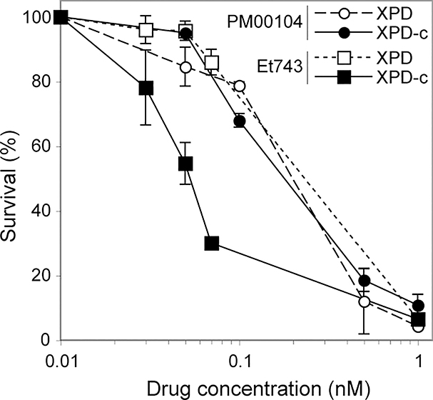
Because Et743 is selective for TC-NER-proficient cells (5, 10), we tested the anti-proliferative activity of PM00104 in NER-deficient XPD cells and their XPD-complemented counterparts, XPD-c (Fig. 2). As previously described (5), XPD-complemented cells are about 4-fold more sensitive to Et743 than XPD cells. Remarkably, NER functionality had no impact on the activity of PM00104 (Fig. 2). The XPD-deficient and -complemented cell lines were equally sensitive to PM00104 with IC50 in the subnanomolar range (0.2 nM). These results suggested that, in contrast to Et743, PM00104 acts independently of NER.
Figure 2. The antiproliferative activity of PM00104 is not influenced by the XPD-NER status.
Cells were exposed to the indicated concentrations of Et743 or PM00104 for 72 hours and analyzed by MTT assay. Open symbols correspond to the NER-deficient XPD cells and filled symbols to the XPD-complemented counterpart, XPD-c. Squares and circles represent Et743 and PM00104, respectively.
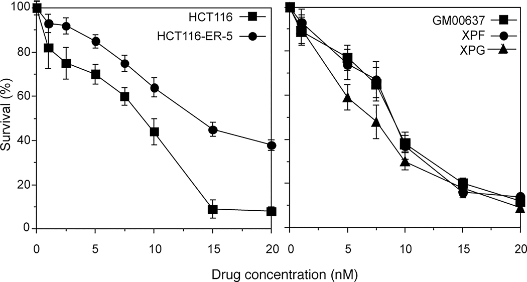
Next, we tested the antiproliferative activity of PM00104 in the Et743-resistant cell line (HCT116-ER-5) derived from the colon carcinoma cell line HCT116 (5). Resistance of the HCT116-ER-5 cells has been linked to inactivation of the NER nuclease XPG (5). When tested for PM00104’s antiproliferative activity, HCT116-ER5 exhibited a slight resistance compared to the parental HCT116 (Fig. 3, left panel). However, because those HCT116-ER-5 cells were selected in the presence of Et743, it is possible that other alterations, besides XPG inactivation could contribute to their partial resistance to PM00104. To test this further, we analyzed two other NER-deficient cell lines, XPG and XPF previously reported to be resistant to Et743 (5). Those XPG and XPF cells showed no resistance to PM00104 when compared to wild type fibroblasts GM00637 (Fig. 3, right panel). Thus the combination of these results indicates that, by contrast to Et743, PM00104’s antiproliferative activity is independent of NER.
Figure 3. Comparative antiproliferative activity of PM00104 in Et743-resistant cells.
Left: HCT116-ER-5 cells selected for resistance to Et743 (5) are cross-resistant to PM00104. Right: XPF and XPG fibroblasts (both NER deficient) are not cross-resistant to PM00104. Cells were exposed to the indicated concentrations of PM00104 for 72 hours and analyzed by MTT assay.
Induction of replication-dependent γ-H2AX foci by PM00104
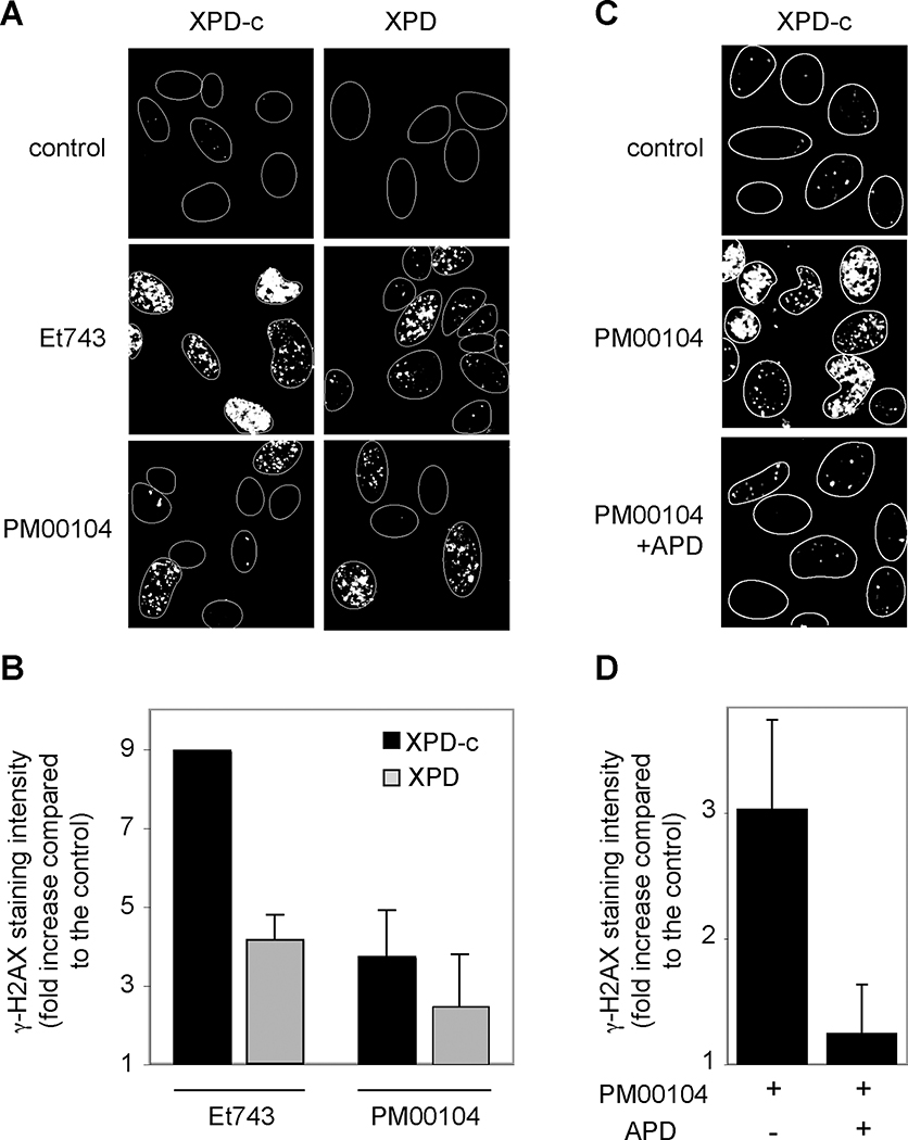
Phosphorylation of H2AX, one of the histone H2A variants, on serine 139 (referred to as γ-H2AX) is one of the most sensitive and reliable markers for DNA damage response (17, 21). Because Et743 was recently found to induce γ-H2AX response (14–16), and we found the Et743-induced γ-H2AX response more intense in NER-proficient cells (14, 15), we investigated whether PM00104 also induced γ-H2AX foci. We compared the induction of γ-H2AX in XPD and XPD-complemented cells. Figure 4A shows that PM00104 and Et743 induced γ-H2AX foci in both XPD and XPD-complemented cells. However, Et743 induced significantly more γ-H2AX foci in XPD-complemented than in XPD cells, whereas PM00104 had the same efficiency in both cell lines. Figure 3B shows the quantification of several experiments, and demonstrates that the intensity of γ-H2AX staining after Et743 was increased by 9.0 ± 0.1 fold and 4.1 ± 0.6 fold in XPD-c and XPD cells, respectively. In contrast, the effect of PM00104 on γ-H2AX induction was statistically similar in XPD-complemented and XPD cells (3.7 ± 0.7 and 2.3 ± 0.7 fold, respectively). Those results demonstrate that the induction of γ-H2AX foci by PM00104 in NER-proficient or -deficient cells is comparable to the induction observed after Et743 treatment of NER-deficient cells.
Figure 4. γ-H2AX foci induced by PM00104 are independent of NER and are replication-associated.
A and B, γ-H2AX foci in XPD and XPD-c cells treated with 10 nM Et743 or PM00104 for 6 hours. A, Representative pictures. B, Fold increase of the intensity of γ-H2AX staining normalized to non-treated cells (AU, arbitrary units). Values are averages of five independent experiments. Bars are SEM. C and D, Inhibition of PM00104-induced γ-H2AX by the DNA polymerase inhibitor, aphidicolin (APD). XPD-c cells were pre-incubated with 1μM APD for 15 minutes. PM00104 (10 nM) was then added for 6 additional hours. C, Representative pictures. D, Fold increase of the intensity of γ-H2AX staining normalized to untreated cells (AU, arbitrary units). Values are the average of three independent experiments. Bars are SEM.
Single cell analyses for γ-H2AX response to PM00104 consistently showed that some cells remained negative while neighboring cells were positive for γ-H2AX (see Fig. 4A). We had previously observed a similar phenomenon for γ-H2AX induction by camptothecins and the indenoisoquinolines (inhibitors of topoisomerase I), and related this selective response to the conversion of the topoisomerase I cleavage complexes into replication-induced DNA damage in S-phase cells (18, 22). To investigate whether PM00104-induced γ-H2AX foci were also replication-dependent, cells were treated with PM00104 in the presence or absence of the DNA polymerase inhibitor aphidicolin (22). The representative pictures shown in Figure 4C demonstrate that aphidicolin almost completely inhibited the appearance of γ-H2AX foci. Quantitation of repeated independent experiments shows marked inhibition of the γ-H2AX response (Fig. 4D) in cells treated simultaneously with PM00104 and aphidicolin. From these results we conclude that PM00104-induced γ-H2AX foci are replication-dependent, and probably correspond to replication-associated DNA damage.
Activity of PM00104 in Ewing Sarcoma cells
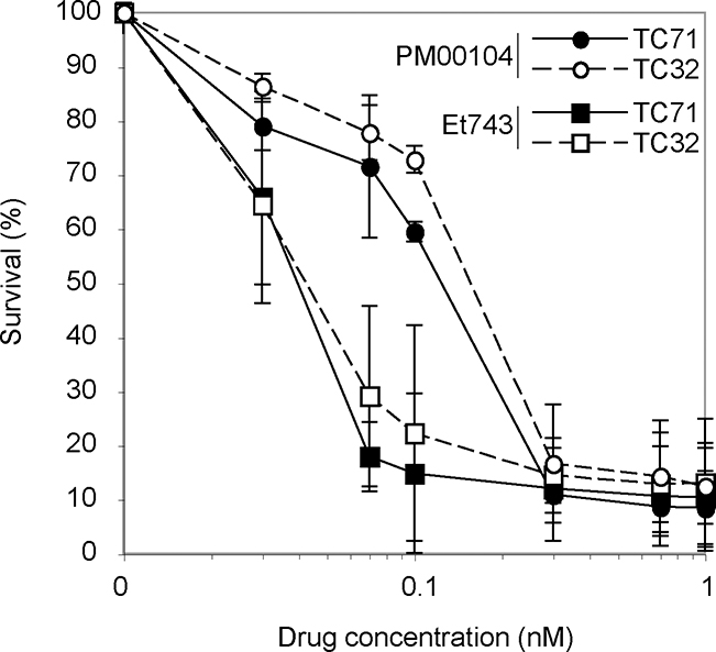
Because Et743 and PM00104 are both active in soft tissue sarcomas, we compared the potency of PM00104 and Et743 in Ewing sarcoma cell lines. First, we performed MTT assays in two Ewing sarcoma cell lines, TC32 and TC71. As shown on Figure 5, both Et743 and PM00104 are highly potent in both cell lines with IC50 at the subnanomolar range. The IC50 were 0.04 nM and 0.15 nM for Et743 and PM00104, respectively.
Figure 5. Antiproliferative activity of PM00104 in Ewing sarcoma cells.
Ewing sarcoma TC32 and TC71 cells were incubated with the indicated concentrations of PM00104 and Et743 for 72 hours and analyzed by MTT assay. Closed and open symbols correspond to TC71 and TC32 cells, respectively. Circles and squares represent PM00104 and Et743, respectively.
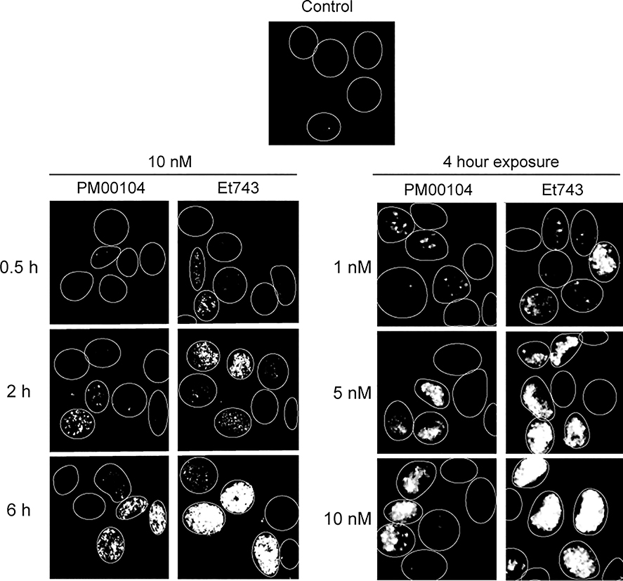
Because γ-H2AX is in development as a pharmacodynamic biomarker (17, 18), we looked for the induction of γ-H2AX foci in the Ewing sarcoma cells treated with PM01004 or Et743. Time-course experiments with 10 nM drug concentrations showed that both drugs induced γ-H2AX foci within two hours of exposures (Fig. 6, left panels). With PM00104, the γ-H2AX foci appeared later, and like with Et743, the γ-H2AX signal induced by PM00104 increased with drug exposure. Dose-response experiments after 4 hour drug treatments, showed that 1 nM PM00104 was sufficient to induce detectable γ-H2AX (representative pictures in Fig. 6, right panels). The γ-H2AX signal increased with drug concentrations for both drugs. At equal concentration, the γ-H2AX staining was consistently more intense with Et743 than with PM00104. Those experiments demonstrate that γ-H2AX foci are induced both by PM00104 and Et743 and that Et743 is generally more potent to induce γ-H2AX foci than PM00104 in Ewing sarcoma cells.
Figure 6. Induction of γ-H2AX at pharmacological concentrations of PM00104 and Et743 in Ewing sarcoma cells.
Representative pictures of γ-H2AX staining in TC71 cells incubated with Et743 or PM00104 for the indicated times and concentrations.
DISCUSSION
PM00104 is in Phase I clinical trials. Because, it is structurally related to Et743, we compared both compounds and investigated whether histone γ-H2AX could be used as a biomarker for clinical pharmacodynamic assessment. The present findings demonstrate that PM00104 differs from Et743 in, at least two ways: in their DNA binding properties and NER dependency for cellular targeting. We also show that PM00104 is effective in various cell lines including Ewing sarcoma cells at nanomolar concentrations and that histone γ-H2AX is induced by PM00104 at pharmacological concentrations.
Previous studies have implicated the A and B rings of Et743 as being primarily responsible for its minor groove DNA binding while the C-ring protrudes from the DNA minor groove (4–6, 11) (see Fig. 1A). Accordingly, we find that alteration of the C-ring, as in PM00104, does not prevent the drug’s ability to bind DNA (see Fig. 1B–C). Furthermore, PM00104 and Et743 exhibit overall similarity and sequence-specific differences in their DNA footprinting profiles (Fig. 1B). David-Cordonnier et al. (23) also showed that analogs of Et743 modified on the C-ring retain DNA binding ability. Our DNase I footprinting experiments (see Fig. 1) demonstrate sequence-specific differences between PM00104 and Et743, which is in agreement with the results of David-Cordonnier et al. who also observed slight differences in the binding properties of three other C-ring-modified analogs of Et743 (23). Thus, our present findings suggest that chemical differences in the C ring do influence PM00104’s interactions with DNA. It is plausible that such differences between PM00104 and Et743 may translate into differential effects on chromatin and on the interactions of PM00104 with other nuclear proteins and transcription factors.
Several studies have suggested the importance of the Et743’s C-ring for cellular activity. For instance, replacing the C-ring of Et743 by a tetrahydro-β-carboline reduces antitumor activity (24). Saframycin A, which is structurally related to ecteinascidins but lacks the C unit has lower antitumor activity than ecteinascidins (7, 24). However, modifications of the C-ring can be done without total loss of biological activity. For instance, Phthalascidin (Pt650) is as efficient as Et743 in a panel of 36 cancer cell lines (25). Other analogs of Et743 display very different biological activities (23). Here we show that PM00104 retains potent antiproliferative activity at nanomolar concentrations.
Our next question was whether the NER selectivity of Et743 (5, 10) was shared by PM00104. This was of particular interest because the C-ring of Et743, which protrudes outside of the DNA minor groove (7) has been proposed to make the key contact with the NER complex (4, 5). We compared the activities of PM00104 and Et743 in an isogenic pair of XPD-deficient and XPD-complemented cells and in two other cell lines deficient for NER, which we had previously used to establish the TC-NER-dependent selectivity of Et743 (5). Our results show that the activity of NER had no impact on the antiproliferative activity of PM00104 (see Figs. 2 and 3). Those findings provide further evidence for the importance of Et743’s C-ring for specific NER poisoning (4).
The similar activity of PM00104 in NER-proficient and -deficient cells suggest that the repair of the DNA lesions generated by PM00104 is not dependent on the NER pathway. In that respect, PM00104 differs from both Et743 (which tends to be more effective in NER-proficient cells; see above) and from platinum derivatives. Indeed, platinum adducts are repaired by the NER, and a common cause of resistance to platinum derivatives is due to increased NER activity in tumor cells (12, 13). It is therefore, plausible that PM00104 could be used in platinum-resistant tumors or in combination with platinum derivatives.
Histone γ-H2AX, the phosphorylated form of H2AX, has recently emerged as potential clinical biomarker because it can be detected with high sensitivity and specificity using antibodies and immunofluorescence (14–17, 22). Our study demonstrates γ-H2AX activation at pharmacological concentrations of PM00104. We recently reported that the increased activity of Et743 in NER-proficient cells was correlated to an increased formation of γ-H2AX in those cells (14) (see Fig. 4). Here we show that the induction of γ-H2AX foci by PM00104 is independent of NER (see Fig. 4). We also show that γ-H2AX activation is detectable at nanomolar pharmacological concentrations of PM00104 in Ewing sarcoma cells (see Fig. 6). Thus, PM00104 behaves as an “Et743-like” molecule without the NER-dependent component. Lacking the NER-dependent component of Et743 may provide some benefit for PM00104. Indeed, loss of heterozygosity and impaired expression of several NER factors have been reported in sporadic solid tumors (5, 26) and in sarcoma (27). In such tumors, PM00104 may remain potentially as active as Et743. Also, PM00104 might be less toxic toward the normal cells, which are NER-proficient. Finally, from a translational standpoint, our experiments indicate that γ-H2AX can be considered as a pharmacodynamic biomarker for the upcoming clinical development of PM00104.
ACKOWLEDGEMENTS
This work has been supported by the Intramural Research Program, Center for Cancer Research, National Cancer Institute, NIH. We wish to thank the PharmaMar team, and in particular Dr. Carmen Cuevas and Dr. José Fernandez Sousa-Faro for kindly providing PM00104 and Et743. We wish to thank the Laboratory of Molecular Pharmacology research team and in particular Dr. Kurt Kohn for suggestions during the course of the present studies.
Abbreviations
- Et743
Ecteinascidin 743
- DSB
DNA double-strand breaks
- γ-H2AX
phosphorylated histone H2AX
- NER
Nucleotide Excision Repair
Footnotes
REFERENCES
- 1.Lane JW, Estevez A, Mortara K, Callan O, Spencer JR, Williams RM. Antitumor activity of tetrahydroisoquinoline analogues 3-epi-jorumycin and 3-epi-renieramycin G. Bioorg Med Chem Lett 2006;16:3180–3. [DOI] [PMC free article] [PubMed] [Google Scholar]
- 2.Scott JD, Williams RM. Chemistry and biology of the tetrahydroisoquinoline antitumor antibiotics. Chem Rev 2002;102:1669–730. [DOI] [PubMed] [Google Scholar]
- 3.Rinehart KL, Holt TG, Fregeau NL, et al. Bioactive compounds from aquatic and terrestrial sources. J Nat Prod 1990;53:771–92. [DOI] [PubMed] [Google Scholar]
- 4.Herrero AB, Martin-Castellanos C, Marco E, Gago F, Moreno S. Cross-Talk between Nucleotide Excision and Homologous Recombination DNA Repair Pathways in the Mechanism of Action of Antitumor Trabectedin. Cancer Res 2006;66:8155–62. [DOI] [PubMed] [Google Scholar]
- 5.Takebayashi Y, Pourquier P, Zimonjic DB, et al. Antiproliferative activity of ecteinascidin 743 is dependent upon transcription-coupled nucleotide-excision repair. Nat Med 2001;7:961–6. [DOI] [PubMed] [Google Scholar]
- 6.Pommier Y, Kohlhagen G, Bailly C, Waring M, Mazumder A, Kohn KW. DNA sequence- and structure-selective alkylation of guanine N2 in the DNA minor groove by ecteinascidin 743, a potent antitumor compound from the Caribbean tunicate Ecteinascidia turbinata. Biochemistry 1996;35:13303–9. [DOI] [PubMed] [Google Scholar]
- 7.Zewail-Foote M, Hurley LH. Ecteinascidin 743: a minor groove alkylator that bends DNA toward the major groove. J Med Chem 1999;42:2493–7. [DOI] [PubMed] [Google Scholar]
- 8.Moore BM II, Seaman FC, Hurley LH. NMR-based model of an ecteinascidin 743-DNA adduct. J Am Chem Soc 1997;119:5475–6. [Google Scholar]
- 9.Sakai R, Rinehart KL, Guan Y, Wang AH. Additional antitumor ecteinascidins from a Caribbean tunicate: crystal structures and activities in vivo. Proc Natl Acad Sci U S A 1992;89:11456–60. [DOI] [PMC free article] [PubMed] [Google Scholar]
- 10.Erba E, Bergamaschi D, Bassano L, et al. Ecteinascidin-743 (ET-743), a natural marine compound, with a unique mechanism of action. Eur J Cancer 2001;37:97–105. [DOI] [PubMed] [Google Scholar]
- 11.Zewail-Foote M, Li VS, Kohn H, Bearss D, Guzman M, Hurley LH. The inefficiency of incisions of ecteinascidin 743-DNA adducts by the UvrABC nuclease and the unique structural feature of the DNA adducts can be used to explain the repair-dependent toxicities of this antitumor agent. Chem Biol 2001;8:1033–49. [DOI] [PubMed] [Google Scholar]
- 12.Furuta T, Ueda T, Aune G, Sarasin A, Kraemer KH, Pommier Y. Transcription-coupled nucleotide excision repair as a determinant of cisplatin sensitivity of human cells. Cancer Res 2002;62:4899–902. [PubMed] [Google Scholar]
- 13.Reed E ERCC1 and clinical resistance to platinum-based therapy. Clin Cancer Res 2005;11:6100–2. [DOI] [PubMed] [Google Scholar]
- 14.Guirouilh-Barbat J, Pommier Y. Et-743 induced DNA double strand breaks: a link between histone gamma-H2AX, Transcription Coupled Nucleotide Excision Repair and Mre11 endonuclease (Abstract number 4642). 97th Annual Meeting of the American Association for Cancer Research; 2006; Washington, DC. USA,; 2006. p. 1090. [Google Scholar]
- 15.Guirouilh-Barbat J, Redon C, Pommier Y. Transcription-coupled DNA double-strand breaks are mediated via the nucleotide excision repair and the Mre11-Rad50-Nbs1 complex. Mol Biol Cell 2008;19:3969–81. [DOI] [PMC free article] [PubMed] [Google Scholar]
- 16.Soares DG, Escargueil AE, Poindessous V, et al. Replication and homologous recombination repair regulate DNA double-strand break formation by the antitumor alkylator ecteinascidin 743. Proc Natl Acad Sci U S A 2007. [DOI] [PMC free article] [PubMed] [Google Scholar]
- 17.Bonner WM, Redon CE, Dickey JS, et al. gammaH2AX and cancer. Nat Rev Cancer 2008;8:957–67. [DOI] [PMC free article] [PubMed] [Google Scholar]
- 18.Antony S, Agama KK, Miao ZH, et al. Novel indenoisoquinolines NSC 725776 and NSC 724998 produce persistent topoisomerase I cleavage complexes and overcome multidrug resistance. Cancer Res 2007;67:10397–405. [DOI] [PubMed] [Google Scholar]
- 19.Guan Y, Sakai R, Rinehart KL, Wang AH. Molecular and crystal structures of ecteinascidins: potent antitumor compounds from the Caribbean tunicate Ecteinascidia turbinata. J Biomol Struct Dyn 1993;10:793–818. [PubMed] [Google Scholar]
- 20.Bailly C, Cuthbert AW, Gentle D, Knowles MR, Waring MJ. Sequence-selective binding of amiloride to DNA. Biochemistry 1993;32:2514–24. [DOI] [PubMed] [Google Scholar]
- 21.Redon C, Pilch D, Rogakou E, Sedelnikova O, Newrock K, Bonner W. Histone H2A variants H2AX and H2AZ. Curr Opin Genet Dev 2002;12:162–9. [DOI] [PubMed] [Google Scholar]
- 22.Furuta T, Takemura H, Liao ZY, et al. Phosphorylation of histone H2AX and activation of Mre11, Rad50, and Nbs1 in response to replication-dependent DNA double-strand breaks induced by mammalian DNA topoisomerase I cleavage complexes. J Biol Chem 2003;278:20303–12. [DOI] [PubMed] [Google Scholar]
- 23.David-Cordonnier MH, Gajate C, Olmea O, et al. DNA and non-DNA targets in the mechanism of action of the antitumor drug trabectedin. Chem Biol 2005;12:1201–10. [DOI] [PubMed] [Google Scholar]
- 24.Sakai R, Jares-Erijman EA, Manzanares I, Silva Elipe MV, Rinehart KL. Ecteinascidins: Putative Biosynthetic Precursors and Absolute Stereochemistry. J Am Chem Soc 1996;118:9017–23. [Google Scholar]
- 25.Martinez EJ, Corey EJ, Owa T. Antitumor activity- and gene expression-based profiling of ecteinascidin Et 743 and phthalascidin Pt 650. Chem Biol 2001;8:1151–60. [DOI] [PubMed] [Google Scholar]
- 26.Castro MA, Mombach JC, de Almeida RM, Moreira JC. Impaired expression of NER gene network in sporadic solid tumors. Nucleic Acids Res 2007;35:1859–67. [DOI] [PMC free article] [PubMed] [Google Scholar]
- 27.Le Morvan V, Longy M, Bonaiti-Pellie C, et al. Genetic polymorphisms of the XPG and XPD nucleotide excision repair genes in sarcoma patients. Int J Cancer 2006;119:1732–5. [DOI] [PubMed] [Google Scholar]


