

Editor
The presence of a worldwide outbreak of chilblain‐like lesions (CLL) contemporarily to COVID‐19 pandemic has been largely reported, not only on social media, but also in the literature. 1 , 2 , 3 , 4
SARS‐CoV‐2 has been hypothesized as the aetiologic agent of CLL, on the basis of the temporal correlation between the ‘burst’ of skin manifestations and the viral pandemic, even though we have scarce evidence of swab‐confirmed infections. Authors have therefore suggested some pathogenetic mechanisms such as a delayed immune‐mediated reaction to the virus in genetically predisposed patients 1 or an early IFN‐I response in young patients, muting early viral replication but also inducing microangiopathic changes. 2
The dermatologic community is currently focusing on the paediatric population, 1 having observed acute acral areas of erythema‐oedema in asymptomatic or mildly symptomatic young patients 4 and is speculating on clinical implications. On the contrary acral, true ischaemic lesions have been described in COVID‐19 as a consequence of clotting disorders, but only in severely affected patients, hospitalized in intensive care units. 4 , 5 , 6
We would like to report a case of SARS‐CoV‐2‐related acro‐ischaemia, peculiar for several reasons: i) the patient presented real acral ischaemia that progressed towards necrosis; ii) she was otherwise completely asymptomatic; iii) she was on regular medication with warfarin for atrial fibrillation.
A 74‐year‐old female was referred to our emergency service because of the presence of livedoid macules that had initially appeared on her hands 6 days before. She reported only pain, whilst she had no complaint referable to currently known COVID‐19 symptomatology.
Her medical history included chronic venous leg ulcers, atrial fibrillation on regular medication with warfarin and a recent hospitalization due to congestive heart failure one month before. During this hospitalization, in accordance with the directives of the Regional Health Service, she was tested for SARS‐CoV‐2 even though she was completely asymptomatic, and resulted positive. She was later discharged and was tested twice at home, resulting negative both times; the last test was 20 days before our evaluation.
On examination, we observed blanching of fingers, dusky red macules, digital infarcts and an ischaemic necrosis of the left third fingertip (Fig. 1).
Figure 1.
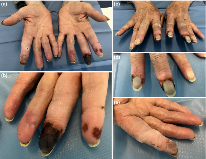
SARS‐CoV‐2‐related acro‐ischaemia. (a, b, c, d, e) Blanching of fingers, dusky red macules, digital infarcts and an ischaemic necrosis of the left third fingertip.
We requested power Doppler ultrasound examination and vascular surgery assessment, but the patient opted for discharge against medical advice. We contacted her by phone 6 days later, but she refused any further visits, claiming improvement.
The present case, to the best of our knowledge, is the first case of true acral necrosis in an asymptomatic SARS‐CoV‐2 infected. This observation raises several questions and considerations.
Firstly, it would be important to determine the pathogenesis of vascular damage to understand why the patient developed acral necrosis 20 days after her second test had resulted negative (i.e. the patient could be considered healed). Our case seems to support the theory of a delayed immune‐mediated reaction to the virus. 1
Moreover, until now we have been used to observing benign acral lesions that progress towards complete recovery; therefore, it is important to determine whether the necrotic outcome is related to any risk factor such as the advanced age of the patient, a genetic predisposition or whether it could be related to her well‐established venous impairment, the latter being less likely as we did not observe a worsening of the leg ulcers.
We would like to draw clinicians’ attention to the possibility that acral lesions may also be observed in the elderly and that these could have a necrotic outcome.
Finally, we underline that, whilst necrosis is considered a primary lesion of COVID‐19, 4 , 5 it can also present with a late onset, suggesting that a longer period of follow‐up is needed also in the healed population to detect late complications.
Conflicts of interest
The authors have no conflict of interest to disclose.
Funding sources
None.
References
- 1. Piccolo V, Neri I, Filippeschi C et al. Chilblain‐like lesions during COVID‐19 epidemic: a preliminary study on 63 patients. J Eur Acad Dermatol Venereol 2020. Apr 24. 10.1111/jdv.16526. [DOI] [PMC free article] [PubMed] [Google Scholar]
- 2. Kolivras A, Dehavay F, Delplace D et al. Coronavirus (COVID‐19) infection–induced chilblains: A case report with histopathologic findings. JAAD Case Rep 2020; 6:489–492. [DOI] [PMC free article] [PubMed] [Google Scholar]
- 3. Fernandez‐Nieto D, Jimenez‐Cauhe J, Suarez‐Valle A et al. Characterization of acute acro‐ischemic lesions in non‐hospitalized patients: a case series of 132 patients during the COVID‐19 outbreak. J Am Acad Dermatol 2020. Apr 24. pii: S0190–9622(20)30709‐X. 10.1016/j.jaad.2020.04.093. [DOI] [PMC free article] [PubMed] [Google Scholar]
- 4. Galván Casas C, Català A, Carretero Hernández G et al. Classification of the cutaneous manifestations of COVID‐19: a rapid prospective nationwide consensus study in Spain with 375 cases. Br J Dermatol. 2020. Apr 29. 10.1111/bjd.19163. [DOI] [PMC free article] [PubMed] [Google Scholar]
- 5. Zhang Y, Cao W, Xiao M et al. [Clinical and coagulation characteristics of 7 patients with critical COVID‐2019 pneumonia and acro‐ischemia]. Zhonghua Xue Ye Xue Za Zhi 2020. Mar 28;41: E006. [DOI] [PubMed] [Google Scholar]
- 6. Zhang Y, Xiao M, Zhang S et al. Coagulopathy and antiphospholipid antibodies in patients with Covid‐19. N Engl J Med 2020; 382(17):e38. [DOI] [PMC free article] [PubMed] [Google Scholar]
Acknowledgement
The patient in this manuscript has given written informed consent to publication of her case details.
All authors have agreed to the contents of the manuscript in its submitted form.