Abstract
A novel Gram-stain-positive, rod-shaped, endospore-forming bacterium, which we designated as strain 03113T, was isolated from greenhouse soil in Beijing, China. Phylogenetic analysis based on 16S rRNA gene sequences showed strain 03113T is in the genus Bacillus and had the highest similarity to Bacillus solani CCTCC AB 2014277T (98.14%). The strain grew at 4 °C–50 °C (optimum 37 °C), with 0–10% (w/v) NaCl (optimum 5%), and in the range of pH 3.0–12.0 (optimum pH 8.0). Menaquinone was identified as MK-7, and the major polar lipids were diphosphatidylglycerol, phosphatidylglycerol, and phosphatidylethanolamine. The main major cellular fatty acids detected were anteiso-C15:0 (51.35%) and iso-C15:0 (11.06%), which are the predominant cellular fatty acids found in all recognized members of the genus Bacillus. The 16S rRNA gene sequence and core-genome analysis, the average nucleotide identity (ANI), and in silico DNA—DNA hybridization (DDH) value between strain 03113T and the most closely related species were 70.5% and 22.6%, respectively, which supported our conclusion that 03113T represented a novel species in the genus Bacillus. We demonstrated that type strain 03113T (=ACCC 03113T=JCM 33017T) was a novel species in the genus Bacillus, and the name Bacillus telluris sp. nov. was proposed. Strain 03113T secreted auxin IAA and carried the nitrogenase iron protein (nifH) gene, which indicated that strain 03113T has the potential to fix nitrogen and promote plant growth. Bacillus telluris sp. nov. 03113T is a potential candidate for the biofertilizers of organic agriculture areas.
Keywords: Bacillus telluris sp. nov., genome analysis, plant-growth promoting rhizobacterium
1. Introduction
The genus Bacillus was first described by Cohn in 1872, and it is a genus of ubiquitous soil microorganisms [1]. It is comprised of endospore-forming, rod-shaped bacteria that are members of the phylum Firmicutes [2]. At the time of writing, there were 379 species in the genus Bacillus recorded on LPSN (www.bacterio.net/bacillus.html; Nov 2019). Based on their genetic similarity, Bacillus species can be classified into several groups, which include Bacillus cereus–Bacillus anthracis–Bacillus thuringiensis, Bacillus clausii–Bacillus halodurans, Bacillus coahuilensis–Bacillus sp. NRRLB-14911, and Bacillus subtilis–Bacillus licheniformis–Bacillus pumilus [3]. In addition, species in the genus Bacillus have a wide range of physiological and biochemical characteristics from psychrophilic to thermophilic, acidophilic to alkaliphilic, and some are halophilic [4], which allow them to live in a wide range of extreme habitats, such as desert sands, hot springs, and Arctic soils. In addition, the genus Bacillus is an extremely diverse group of bacteria that includes both the causative agent of anthrax (B. anthracis) [5,6] and several species that synthesize important antibiotics. In addition to medical uses, bacillus spores, due to their extreme tolerance of both heat and disinfectants, are used to test heat sterilization techniques and chemical disinfectants. Bacilli are also used in the detergent manufacturing industry for their ability to synthesize important enzymes.
In this study, we report a novel bacterial strain, 03113T, which was isolated from the greenhouse soil of Wangsiying, Chaoyang District, Beijing, China. Based on the phenotypic characteristics and phylogenetic analysis, strain 03113T represents a novel species in the genus Bacillus.
2. Materials and Methods
2.1. Bacterial Strains, Growth Conditions, and Cultivation
Strain 03113T was isolated from greenhouse soil from Wangsiying, Chaoyang District, Beijing, China (40°09′N, 116°42′E). We preserved the sample in freeze-dried milk ampoules at 4 °C and 20% (v/v) glycerol at −80 °C [7]. The type strains of species closely related to strain 03113T were used as reference strains under the same conditions for comparative taxonomic analysis, which included B. solani CCTCC AB 2014277T, B. praedii CCTCC AB 2015208T, and B. dabaoshanensis CCTCC AB 2013260T. All strains were maintained and cultivated in TSA or TSB (DifcoTM) medium plates at 30 °C, unless otherwise stated.
2.2. Phenotypic Characterization
Biochemical characteristics of strain 03113T were investigated. Growth at eight different temperatures (4, 15, 25, 30, 37, 40, 45, and 50 °C) was tested on TSA plates. The pH values (pH 3.0, 4.0, 5.0, 6.0, 7.0, 8.0, 9.0, 10.0, 11.0, and 12.0, with increments of 1.0 pH unit) were tested in LB medium. Growth at various NaCl concentrations was tested over the range 0%−12% (w/v) NaCl (at intervals of 1%) by incubating at 30 °C [8]. Gram staining was performed using the Gram-stain kit [9]. Cell morphology was observed by light microscopy (CX21; Olympus) and transmission electron microscopy. Endospores were examined according to the Schaeffer–Fulton staining method [10]. Motility was examined on motility agar [11]. Catalase activity was determined by investigating bubble production with 3% (v/v) H2O2, and oxidase activity was determined using 1% (v/v) tetramethyl-p-phenylenediamine. The basic biochemical characteristics were investigated on API-20NE, API 50CH (BioMérieux) [12], and BIOLOG GEN III MicroPlate (BIOLOG), according to the manufacturer’s instructions. The type strains of B. solani CCTCC AB 2014277T, B. praedii CCTCC AB 2015208T, and B. dabaoshanensis CCTCC AB 2013260T were used as reference strains.
2.3. Chemotaxonomic Analysis
For the measurement of chemotaxonomic characteristics, the menaquinone system was analyzed as described by Collins et al. [13] using reversed-phase HPLC [14]. The analysis of polar lipids by two-dimensional TLC was performed according to the method described by Minnikin et al. [15]. The cellular fatty acid is a useful and functional tool to identify species in the genus Bacillus and related genera. After 48 h of incubation at 30 °C on TSA, cellular fatty acids were extracted and analyzed using the method described by Sasser [16] and identified with the MIDI Sherlock Microbial Identification System (Library RTSA6 6.0, MIDI Sherlock Software Package, Version 6.0; Agilent 6890N).
2.4. Phylogenetic 16S rRNA Gene Analysis
Genomic DNA was extracted from a single colony of the novel strain grown on TSA plates at 30 °C for 2 d using Bacteria DNA Kit (Tiangen, Beijing, China), according to the manufacturer’s protocol. The 16S rRNA gene was amplified by PCR and sequenced using the universal primers 27F(5’-AGAGTTTGATCCTGGCTCAG-3’) and 1492R(5’-GGTTACCTTGTTACGACTT-3’) [17]. Pairwise 16S rRNA gene sequence similarities were calculated using the EzTaxon-e database (http://eztaxon-e.ezbiocloud.net/) [18]. The CLUSTAL_W algorithm was used for sequence alignments using the neighbour-joining [19,20] and maximum-likelihood [21] methods that were implemented with Mega 7.0 software for phylogenetic analysis. Evolutionary distances were computed by using the Kimura two-parameter model [22]. The robustness of the tree branches was estimated by bootstrap analysis with 1000 replications [23]. The GenBank/EMBL/DDBJ accession number of 16S rRNA sequence is MN907472.
2.5. Complete Genome Sequencing and Analysis
To confirm the results of the 16S rRNA gene sequence similarity analysis, the complete genome sequence of the novel species was performed. The genome was sequenced by Personal Biotechnology Co., Ltd (Shanghai, PR China). Genomes of the most closely related species chosen above were retrieved from the GenBank database in NCBI. Reads of each data set were filtered by using AdapterRemoval (ver. 2.1.7) [24], and high-quality paired-end reads were assembled using A5-MiSeq v20150522 [25]. The open reading frames (ORFs) were predicted by GeneMarkS (ver. 4.32 April 2015) [26]. The tRNA genes were predicted by tRNAscan-SE 94 (ver. 1.3.1) and the rRNA genes by Barrnap (0.9-dev) 95 (https://github.com/tseemann/barrnap) [27]. Calculations of average nucleotide identity (ANI) were performed using JSpecies software (http://www.imedea.uib.es/jspecies). In silico DNA—DNA hybridization (DDH) estimates were performed using Genome-to-Genome Distance Calculator (GGDC) with the BLAST+ (recommended) method [28]. The partial genome files were uploaded to the GGDC 2.0 web interface (http://ggdc.dsmz.de/ggdc.php#), and Formula 2 was used as recommended for the calculation of DDH values. As a further extension of genome-based phylogeny, the GGDC website was used to establish the phylogenomic tree of strain 03113T and other closely related Bacillus species.
2.6. Analysis of Core Orthologous Genes
To identify orthologous genes among the strains in Bacillus species, 13 Bacillus strains were selected for the core genome analysis based on their biological control properties. The 13 bacteria included B. solani CCTCC AB 2014277T, B. praedii CCTCC AB 2015208T, B. glycinifermentans GO-13, B. acidicola FJAT-2406, B. salacetis SKP7-4, B. shackletonii LMG 18435, B. circulans NBRC 13626, B. foraminis Bac44, B. persicus DSM 25386, B. oceanisediminis CGMCC 1.10115, B. firmus NCTC 10335, B. gottheilii FJAT-2394, and 03113T. The Bacterial Pan Genome Analysis (BPGA) pipeline [29] was used for the pan-genome analyses. The clustering tool USEARCH was used to cluster protein families. The OrthoFinder [30,31] was used to perform an all-versus-all BLAST search based on nucleotide gene sequences of strain 03113T and other related strains of the genus Bacillus to identify clusters of orthologous genes (OGs). Those OGs shared among all taxa and present in a single copy per genome were selected. They were aligned with Mafft [32] and subsequently concatenated. A phylogenetic tree based on orthologous proteins of the Bacillus genus was constructed by RA×ML version 8.2.12, based on the maximum-likelihood method.
2.7. Plant Growth-Promoting Characteristics
The performance of secreting plant growth hormone indoleacetic acid (IAA) of strain 03113T was measured by the PC Salkowski colorimetric method described by Glickmann and Dessaux [33]. The qualitative and quantitative analyses of siderophore production were conducted by the method described by Machuca and Milagres [34]. Phosphate solubilization was measured on inorganic and organic phosphate media [35]. All experimental analyses were performed in triplicate to ensure reproducibility. The results were expressed as the mean value of these determinations.
3. Results and Discussion
3.1. Phenotypic Characterization of 03113T
The colonies of strain 03113T were Gram-stain-positive and rod-shaped with a size range of 1−2 mm in diameter (Figure S1a). The size of the cells was observed by light microscopy. The cells produced ellipsoidal endospores that were positioned terminally (Figure S1b), and the cells were motile. Catalase and oxidase activity were positive. According to API 50CH tests, reactions of galactose, sorbose, rhamnose, dulcitol, α-metyl-D-glucoside, arbutin, esculin, melibiose, sucrose, trehalose, and D-turanose were positive but the other three reference strains were negative. With API 20NE, strain 03113T was positive for lysine, but the other three reference strains were negative. The phenotypic properties differentiating between strain 03113T and its closest phylogenetic neighbors are shown in Table 1.
Table 1.
Differential phenotypic characteristics of strain 03113T and closely related strains in the genus Bacillus.
| Characteristic | 1 | 2 | 3 | 4 |
|---|---|---|---|---|
| Optimal growth conditions | ||||
| Temperature for growth (°C) | 37 | 30–37 | 35 | 30 |
| pH for growth | 8.0 | 7.0 | 9.0 | 9.0 |
| NaCl concentration for growth (%, w/v) | 5 | 1 | 0 | 4 |
| The Acid produced from (API 50CH) | ||||
| L-arabinose | + | + | + | − |
| Esculin | + | + | − | − |
| API 20NE | ||||
| β-galactosidase | + | + | − | − |
| Lysine | + | − | − | − |
| Lohn gelatin | + | + | + | − |
| Utilization (Biolog GEN III) | ||||
| dextrin | + | + | − | + |
| d-maltos, d-trehalose, sucrose, d-turanose, d-raffinose | + | − | − | − |
| N-acetyl-d-glucosamine | − | + | + | − |
| N-acetyl-β-d-mannosamine, acetic acid | − | + | − | − |
| Stachyose, d-mannose | w | − | − | − |
| N-acetyl-d-galactosamine | − | w | − | − |
| α-d-glucose | + | − | w | − |
| d-fructose | + | + | w | − |
| Inosine, d-serine, glycerol, d-glucose-6-PO4, d-fructose-6-PO4, nalidixic acid, lithium chloride, aztreonam, lincomycin | − | + | + | + |
| Troleandomycin | − | − | + | − |
| L-aspartic acid, L-glutamic acid, L-histidine, L-pyroglutamic acid, L-serine, L-lactic acid, sodium butyrate | - | + | + | − |
Strains: 1, 03113T; 2, B. dabaoshanensis CCTCC AB 2013260T; 3, B. solani CCTCC AB 2014277T; 4, B. praedii CCTCC AB 2015208T. All strains were negative for sodium thiosulfate, tryptophan, d-cellobiose, gentiobiose, α-d-lactose, d-melibiose, β-methyl-d-glucoside, d-salicin, N-acetyl neuraminic acid, d-galactose, 3-methyl glucose, L-fucose, L-rhamnose, fusidic acid, d-sorbitol, d-mannitol, myo-inositol, d-aspartic acid, minocycline, pectin, d-galacturonic acid, glucuronamide, mucic acid, quinic acid, p-hydroxy-phenylacetic acid, citric acid, α-keto-glutaric acid, d-malic acid, γ-amino-butryric acid, α-hydroxy-butyric acid, β-hydroxy-D, L-butyric acid, α-keto-butyric acid, propionic acid, and formic acid. All strains were positive for sodium lactate and potassium tellurite. All data were from the present study. +, positive; w, weakly positive; −, negative.
3.2. Analysis of Isoprenoid Quinones, Polar Lipids, and Cellular Fatty Acids
The main isoprenoid quinone of strain 03113T was identified as MK-7. The polar lipids detected were diphosphatidylglycerol, phosphatidylglycerol, phosphatidylethanolamine, phosphatidylserine, three unknown aminophospholipids, and one unknown phospholipid, which was consistent with the predominant component of B. solani CCTCC AB 2014277T [7]. The major fatty acids of strain 03113T were anteiso-C15:0 (51.35%), iso-C15:0 (11.06%), and iso-C14:0 (7.13%), which were similar to those of the reference strains (Table 2). Iso- and anteiso- branched fatty acids of the 14-17 carbon series are typical for the genus Bacillus [36], which indicated that strain 03113T is a member of this genus. However, the proportions of the novel strain were different from B. solani CCTCC AB 2014277T, B. praedii CCTCC AB 2015208T, and B. dabaoshanensis CCTCC AB 2013260T. For instance, the content of anteiso-C15:0 in strain 03113T was much higher than in the reference strains, but the concentration of iso-C15:0 was much lower than in the related reference strains.
Table 2.
The cellular fatty acid content of strain 03113T and representative strains of closely related species of the genus Bacillus. Strains: 1, 03113T; 2, B. dabaoshanensis CCTCC AB 2013260T; 3, B. solani CCTCC AB 2014277T; 4, B. praedii CCTCC AB 2015208T. All data were obtained in this study. Partial values lower than 1% are not shown in the table. ND, Not detected.
| Fatty acid | 1 | 2a | 3 | 4 |
|---|---|---|---|---|
| C14:0 | 1.96 | 1.1 | 1.77 | 1.68 |
| C16:0 | 7.50 | 2.60 | 1.98 | 2.20 |
| iso-C14:0 | 7.13 | ND | 6.61 | 5.13 |
| iso-C15:0 | 11.06 | 42.9 | 45.43 | 54.12 |
| iso-C16:0 | 8.73 | 6.7 | 6.07 | 5.61 |
| anteiso-C15:0 | 51.35 | 24.1 | 27.16 | 20.15 |
| anteiso-C17:0 | 6.71 | 6.2 | 3.88 | 3.13 |
| C16:1ω7c alcohol | ND | ND | 2.56 | 2.36 |
| C16:1ωw11c | ND | ND | 1.21 | 1.12 |
| Summed Feature 3 * | <1 | 2.5 | ND | ND |
| Summed Feature 8 † | <1 | 1.5 | ND | ND |
* Summed feature 3 comprises C16: 1ω6c and/or C16: 1ω7c. † Summed feature 8 comprises C18: 1ω6c and/or C18: 1ω7c.a Data were obtained from: Cui et al. [37].
3.3. Phylogenetic Analysis of 16S rRNA
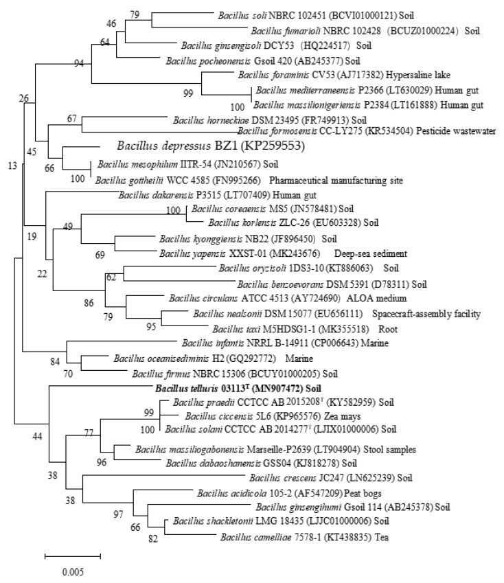
The complete 16S rRNA gene sequence (1347 bp) was discovered from the draft genome of the novel strain. Pairwise comparisons showed that strain 03113T was related most closely to B. solani CCTCC AB 2014277T (98.14% similarity), followed by B. praedii CCTCC AB 2015208T (98.07%), and B. dabaoshanensis CCTCC AB 2013260T (98.0%). Phylogenetic trees were reconstructed using the maximum-likelihood, neighbour-joining, and minimum-evolution methods. All three treeing methods yielded a similar phylogeny. Strain 03113T was located within the genus Bacillus and had a separated clade based on the phylogenetic trees of 16S rRNA genes (Figure 1, Figure S2, Figure S3), indicating that 03113T was a novel species of genus Bacillus.
Figure 1.
Neighbour-joining phylogenetic tree based on the 16S rRNA gene sequence of strain 03113T and other closely related Bacillus species. The significance of each branch is indicated by a bootstrap value (%) calculated for 1000 subsets. Genbank accession numbers are given in parentheses. Bar, 0.005 substitutions per nucleotide position. Isolating source label has been annotated in the back.
3.4. Whole-Genome Analysis
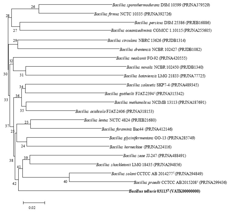
A total of 5,033,596 reads were obtained from draft genome sequencing of strain 03113T, which yielded a genome of 4,856,532 reads in length. N50 value was 190,698 bp, and the largest contig was 198,446 bp. The genome was predicted to contain a total of 4288 genes, which included 4241 protein-coding genes, 2 rRNA genes, and 45 tRNA genes. The genomic DNA G+C content of strain 03113T was 36.08 mol%. The phylogenomic tree based on the GGDC web also revealed the distinct phylogeny of strain 03113T and its close relationship with B. solani CCTCC AB 2014277T, B. praedii CCTCC AB 2015208T, and B. dabaoshanensis CCTCC AB 2013260T (Figure 2). ANIb and ANIm values of strain 03113T with the type strain of the most closely related species, B. solani CCTCC AB 2014277T, were 70.5% and 85.9%, respectively. All ANI values were much lower than the 96.0% cut-off value that was proposed previously for the genus Bacillus [38,39]. The DDH value of strain 03113T and B. solani CCTCC AB 2014277T was 22.6%, which was much lower than 70%. The ANI and DDH between strain 03113T and the other reference species B. praedii CCTCC AB 2015208T were 70.5% and 20.9%, respectively. This genome sequence, which was deposited in the GenBank/EMBL/DDBJ database under accession number VATK00000000, was used for further analysis. Thus, complete genome analysis combined with 16S rRNA phylogenetic, physiological, and biochemical properties all supported the conclusion that strain 03113T should be considered a novel species in the genus Bacillus.
Figure 2.
Phylogenomic tree generated with Genome-to-Genome Distance Calculator (GGDC), showing the phylogenomic position of strain 03113T and the type strains of related species of Bacillus. The numbers at the nodes indicate the gene support index. Bar, 0.02 substitutions per position.
3.5. Phylogenomic Comparative Analysis of Bacillus species
Based on the above database, we conducted a preliminary analysis of the pan-genome, which showed that 840 shared orthologous coding sequences were clustered into the core genome of Bacillus, 32,926 were represented in the accessory genome, and 22,024 were identified as strain-unique genes (Figure S4a). Therefore, a highly reliable mathematical extrapolation of the pan and core genome was constructed (Figure S4b). The total genes increase in the pan-genome of Bacillus with the rise in the analyzed genome number, suggesting that the pan-genome was open. The previous reports showed that the genes’ number of core genomes was highly conserved, while many strain-unique genomes and accessory genomes are thought to contribute to species diversity [40], which indicated that species in the genus Bacillus were also multifarious. A phylogenetic tree reconstructed based on the concatenated alignment of these 840 core orthologous proteins (Figure S4c) showed that strain 03113T clustered closely with known species, indicating that it was a member of the genus Bacillus. This is consistent with the previous results.
3.6. Plant Growth-Promoting Characteristics of Isolates
The qualitative determination indicated that strain 03113T secreted auxin IAA, and the colour reaction was pink, at a concentration of 175.94 μg/mL (Figure S5). Strain 03113T did not generate a color ring on the CAS flat plate, and no clear zone was observed around each of the colonies of strain 03113T on inorganic or organic phosphate media, which indicated that it did not produce siderophores or dissolve phosphate.
Strain 03113T carried the nitrogenase iron protein (nifH) gene, based on the genome annotation, which is a key enzyme for fixing nitrogen in bacteria. The nifH gene of 03113T had a very low similarity with published sequences based on Blast in Genbank (https://blast.ncbi.nlm.nih.gov/Blast.cgi). It was mostly related to B. alkalidiazotrophicus MS6 and B. arsenniciselenatis E1H with a similarity of 80%, which indicated that strain 03113T has the potential to fix nitrogen.
4. Conclusions
From the phenotypic and chemotaxonomic properties of strain 03113T, 16S rRNA gene sequence comparisons, and DNA–DNA hybridization, we concluded that strain 03113T (=ACCC 03113T=JCM 33017T) was distinguished from the known species in the genus Bacillus. Based on the present polyphasic analysis, strain 03113T is considered to represent a novel species within the genus Bacillus, for which we propose the name Bacillus telluris sp. nov. The description of Bacillus telluris sp. nov. is summarized in Appendix A.
Supplementary Materials
The following are available online at https://www.mdpi.com/2076-2607/8/5/702/s1, Figure S1: (a) The morphology and Gram-staining of strain 03113T of the genus Bacillus. (b) The morphology and Spore-staining of the strain 03113T in the genus Bacillus. Figure S2: Maximum-Likelihood phylogenetic tree based on the 16S rRNA gene sequence of strain 03113T and other closely related Bacillus species. The significance of each branch is indicated by a bootstrap value (%) calculated for 1000 subsets. Genbank accession numbers are given in parentheses. Bar, 0.005 substitutions per nucleotide position. Figure S3: Minimum-Evolution phylogenetic tree based on the 16S rRNA gene sequence of strains 03113T and other closely related Bacillus species. The significance of each branch is indicated by a bootstrap value (%) calculated for 1000 subsets. Genbank accession numbers are given in parentheses. Bar, 0.005 substitutions per nucleotide position. Figure S4: The Pan-genome analysis and the genome phylogenetic tree of strains belonging to the Bacillus genus. (a) Petal diagram of the pan-genome. Each strain is represented by a colored oval. The center is the number of orthologous coding sequences shared by all strains. Numbers in nonoverlapping portions of each oval show the numbers of CDSs unique to each strain. (b) Mathematical modeling of the pan-genome and core genome of Bacillus. (c) Tree constructed according to the maximum-likelihood method based on 840 core orthologous proteins of strain 03113T and closely related species of the genus Bacillus. Bootstrap values are expressed as percentages of 1000 replications, and those over 70% are shown at branch points. Bar, 0.05 substitutions per nucleotide position. Figure S5: Colour reaction of strain 03113T compared to CK (CK: Salkowski solution mixed with IAA). Figure S6: Polar lipids of strain 03113T after two-dimensional TLC and detection with (I) molybdophosphoric acid spray reagent and heating at 150 °C for 10 min, (II) molybdenum blue spray reagent, and (III) ninhydrin spray reagent and heating at 100 °C for 10 min. No spots were detected by α-naphthhol spray reagent and heating at 100 °C for 5 min. Chloroform/methanol/water (65:25:4, by vol.) was used in the first direction. (1), followed by chloroform/acetic acid/methanol/water (80:15:12:4, by vol.) in the second direction. (2), DPG, diphosphatidylglycerol; PG, phosphatidylglycerol; PE, phosphatidylethanolamine; PSer, phosphatidylserine; APL1-3, unknown aminophospholipid; PL1, unknown phospholipids.
Appendix A
Description of Bacillus telluris sp. nov.
Bacillus telluris (tel. lu′ris. L. gen. n. telluris from soil, the origin of the strain).
Cells are Gram-stain-positive, endospore-forming rods, motile, about 1−2 mm in diameter. Colonies are circular and smooth on TSA at 30 °C. Ellipsoidal endospores were observed at the terminal position. Growth occurred at 4 °C −50 °C (optimum 37 °C), at pH 3.0−12.0 (optimum pH 8.0), and with 0–10% (w/v) NaCl (optimum 5%). Catalase and oxidase activity were all positive. It produced the biological characteristics of IAA. Reactions of L-arabinose, ribose, D-xylose, galactose, sorbose, rhamnose, dulcitol, α-metyl-D-glucoside, amygdalin, arbutin, esculin, maltose, melibiose, sucrose, trehalose, D-turanose, and gluconate were positive. Positive reactions for β-galactosidase (ONPG), arginine, lysine, sodium citrate, urease, pyruvate, and kohn gelatin, but negative reactions for H2S production, indole production, ornithine, tryptophan, glucose, mannose, inositol, sorbitol, rhamnose, sucrose, melibiose, amygdalin, and arabinose. Dextrin, D-maltose, D-maltose, D-trehalose, sucrose, D-turanose, D-raffinose, α-D-glucose, stachyose, D-mannose, D-fructose, and sodium lactate were assimilated, but gentiobiose, D-melibiose, D-fructose, D-serine, myo-inositol, gelatin, and D-glucuronic acid were not. The main cellular fatty acids were anteiso-C15:0, iso-C15:0, and iso-C14:0. Major polar lipids were diphosphatidylglycerol, phosphatidylglycerol, phosphatidylethanolamine, and phosphatidylserine (Figure S6). The main isoprenoid quinone was MK-7.
The strain, 03113T (=ACCC 03113T=JCM 33017T), was isolated from greenhouse soil collected in Wangsiying, Chaoyang District, Beijing, China. The DNA G+C content of the genome of the strain was 36.08 mol%.
The GenBank/EMBL/DDBJ accession number for the 16S rRNA sequence of Bacillus telluris 03113T is MN907472, and the complete genome is deposited under the accession number VATK00000000.
Author Contributions
H.-B.G., S.-W.H., X.W., K.-K.T., H.-L.W., and X.-X.Z. conceived and supervised the study; H.-L.W. and X.-X.Z. designed the experiments; H.-B.G. performed the experiments; S.-W.H. took over genomic analysis; H.-B.G., X.W., K.-K.T. analyzed the data, prepared the figures and wrote the manuscript; H.-B.G., S.-W.H., X.W., K.-K.T., H.-L.W., and X.-X.Z. edited the manuscript and reviewed the literature. All authors have read and agreed to the published version of the manuscript.
Funding
This work was supported by Beijing Science and Technology Project (Z191100004019025), Fundamental Research Funds for Central Non-profit Scientific Institution (No. 1610132019015), and Central Public-interest Scientific Institution Basal Research Fund (No. Y2019XK07).
Conflicts of Interest
The authors declare no conflict of interest.
References
- 1.McKillip J.L. Prevalence and expression of enterotoxins in Bacillus cereus and other Bacillus spp., a literature review. Antonie Leeuwenhoek. 2000;77:393–399. doi: 10.1023/A:1002706906154. [DOI] [PubMed] [Google Scholar]
- 2.Claus D., Berkeley R.C. Bergey’s Manual of Systematic Bacteriology. Wiley; Hoboken, NJ, USA: 1986. Genus Bacillus; pp. 1105–1139. [Google Scholar]
- 3.Alcaraz L.D., Moreno-Hagelsieb G., Eguiarte L.E., Souza V., Herrera-Estrella L., Olmedo G. Understanding the evolutionary relationships and major traits of Bacillus through comparative genomics. BMC Genom. 2010;11:332. doi: 10.1186/1471-2164-11-332. [DOI] [PMC free article] [PubMed] [Google Scholar]
- 4.Logan N.A., de Vos P. Bacillus. In: De Vos P., Garrity G., Jones D., Krieg N.R., Ludwig W., Rainey F.A., Schleifer K.H., Whitman W.B., editors. Bergey’s Manual of Systematic Bacteriology: The Firmicutes. 2nd ed. Volume 3. Springer; New York, NY, USA: 2009. pp. 21–128. [Google Scholar]
- 5.Read T.D., Peterson S.N., Tourasse N., Baillie L.W., Fraser C.M. The genome sequence of Bacillus anthracis Ames and comparison to closely related bacteria. Nature. 2003;423:23–25. doi: 10.1038/nature01586. [DOI] [PubMed] [Google Scholar]
- 6.Rosovitz M.J., Leppla S.H. Medicine: Virus deals anthrax a killer blow. Nature. 2002;418:825–826. doi: 10.1038/418825a. [DOI] [PubMed] [Google Scholar]
- 7.Liu B., Liu G.H., Sengonca C., Schumann P., Ge C.B., Wang J.P., Cui W.D., Lin N.Q. Bacillus solani sp. nov. isolated from rhizosphere soil of potato field in Xinjiang of China. Int. J. Syst. Evol. Microbiol. 2015;65:4066–4071. doi: 10.1099/ijsem.0.000539. [DOI] [PubMed] [Google Scholar]
- 8.Liu B., Liu G.H., Sengonca C., Schumann P., Wang M.K. Bacillus praedii sp. nov. isolated from purplish paddy soil. Int. J. Syst. Evol. Microbiol. 2017;67:2823–2826. doi: 10.1099/ijsem.0.002030. [DOI] [PubMed] [Google Scholar]
- 9.Meierkolthoff J.P., Auch A.F., Klenk H.P., Markus G. Genome sequence-based species delimitation with confidence intervals and improved distance functions. BMC Bioinform. 2013;14:60. doi: 10.1186/1471-2105-14-60. [DOI] [PMC free article] [PubMed] [Google Scholar]
- 10.Murray R.G., Doetsch R.N., Robinow C.F. Determinative and cytological light microscopy. In: Gerhardt P., Murray R.G., Wood W.A., Krieg N.R., editors. Methods for General and Molecular Bacteriology. American Society for Microbiology (ASM); Washington, DC, USA: 1994. pp. 21–41. [Google Scholar]
- 11.Chen Y.G., Cui X.L., Pukall R., Li H.M., Yang Y.L., Xu L.H., Wen M.L., Peng Q., Jiang C.L. Salinicoccus kunmingensis sp. nov., a moderately halophilic bacterium isolated from a salt mine in Yunnan, south-west China. Int. J. Syst. Evol. Microbiol. 2007;57:2327–2332. doi: 10.1099/ijs.0.64783-0. [DOI] [PubMed] [Google Scholar]
- 12.Dong X.Z., Cai M.Y. Manual for the Systematic Identification of General Bacteria. Science Press; Beijing, China: 2001. Determination of Biochemical Properties; pp. 370–398. [Google Scholar]
- 13.Collins M.D., Pirouz T., Goodfellow M., Minnikin D.E. Distribution of Menaquinones in Actinomycetes and Corynebacteria. J Gen Microbiol. 1977;100:221–230. doi: 10.1099/00221287-100-2-221. [DOI] [PubMed] [Google Scholar]
- 14.Groth I., Schumann P., Weiss N., Martin K., Rainey F.A. Agrococcus jenensis gen. nov., sp. nov., a new genus of actinomycetes with diaminobutyric acid in the cell wall. Int. J. Syst. Bacteriol. 1996;46:234–239. doi: 10.1099/00207713-46-1-234. [DOI] [PubMed] [Google Scholar]
- 15.Minnikin D.E., Collins M.D., Goodfellow M. Fatty acid and polar lipid composition in the classification of Cellulomonas, Oerskovia and related taxa. J. Appl. Bacteriol. 1979;47:87–95. doi: 10.1111/j.1365-2672.1979.tb01172.x. [DOI] [PubMed] [Google Scholar]
- 16.Sasser M. Identification of bacteria by gas chromatography of cellular fatty acids. USFCC News. 1990;20:16. [Google Scholar]
- 17.Lane D.J. 16S/23S rRNA Sequencing. In: Stackebrandt E., Goodfellow M., editors. Nucleic Acid Techniques in Bacterial Systematics. John Wiley and Sons; New York, NY, USA: 1991. pp. 115–175. [Google Scholar]
- 18.Kim O.S., Cho Y.J., Lee K., Yoon S.H., Kim M., Na H., Park S.C., Jeon Y.S., Lee J.H., Yi H., et al. Introducing EzTaxon-e: A prokaryotic 16S rRNA gene sequence database with phylotypes that represent uncultured species. Int. J. Syst. Evol. Microbiol. 2012;62:716–721. doi: 10.1099/ijs.0.038075-0. [DOI] [PubMed] [Google Scholar]
- 19.Saitou N., Nei M. The neighbor-joining method: A new method for reconstructing phylogenetic trees. Mol. Biol. Evol. 1987;4:406–425. doi: 10.1093/oxfordjournals.molbev.a040454. [DOI] [PubMed] [Google Scholar]
- 20.Rzhetsky A., Nei M. A simple method for estimating and testing minimum evolution trees. Mol. Biol. Evol. 1992;9:945–967. [Google Scholar]
- 21.Felsenstein J. Evolutionary trees from DNA sequences: A maximum likelihood approach. J. Mol. Evol. 1981;17:368–376. doi: 10.1007/BF01734359. [DOI] [PubMed] [Google Scholar]
- 22.Tamura K., Dudley J., Nei M., Kumar S. MEGA 4: Molecular evolutionary genetics analysis (MEGA) software version 4.0. Mol. Biol. Evol. 2007;24:1596–1599. doi: 10.1093/molbev/msm092. [DOI] [PubMed] [Google Scholar]
- 23.Felsenstein J. Confidence limits on phylogenies: An approach using the bootstrap. Evolution. 1985;39:783–791. doi: 10.1111/j.1558-5646.1985.tb00420.x. [DOI] [PubMed] [Google Scholar]
- 24.Schubert M., Lindgreen S., Orlando L. AdapterRemoval v2: Rapid adapter trimming, identification, and read merging. BMC Res. Notes. 2016;9:88. doi: 10.1186/s13104-016-1900-2. [DOI] [PMC free article] [PubMed] [Google Scholar]
- 25.Coil D., Jospin G., Darling A.E. A5-miseq: An updated pipeline to assemble microbial genomes from Illumina MiSeq data. Bioinformatics. 2014;31:587–589. doi: 10.1093/bioinformatics/btu661. [DOI] [PubMed] [Google Scholar]
- 26.John B.E., Bass L. Usability and software architecture. Behav. Inform. Technol. 2001;20:329–338. doi: 10.1080/01449290110081686. [DOI] [Google Scholar]
- 27.Lowe T.M., Eddy S.R. tRNAscan-SE: A program for improved detection of transfer RNA genes in genomic sequence. Nucleic Acids Res. 1997;25:955–964. doi: 10.1093/nar/25.5.955. [DOI] [PMC free article] [PubMed] [Google Scholar]
- 28.Auch A.F., von Jan M., Klenk H.P., Göker M. Digital DNA-DNA hybridization for microbial species delineation by means of genome-to-genome sequence comparison. Stand Genom. Sci. 2010;2:117–134. doi: 10.4056/sigs.531120. [DOI] [PMC free article] [PubMed] [Google Scholar]
- 29.Chaudhari N.M., Gupta V.K., Dutta C. BPGA—An ultra-fast pan-genome analysis pipeline. Sci. Rep. 2016;6:24373. doi: 10.1038/srep24373. [DOI] [PMC free article] [PubMed] [Google Scholar]
- 30.Emms D.M., Kelly S. OrthoFinder: Solving fundamental biases in whole genome comparisons dramatically improves orthogroup inference accuracy. Genome Biol. 2015;16:157. doi: 10.1186/s13059-015-0721-2. [DOI] [PMC free article] [PubMed] [Google Scholar]
- 31.Emms D.M., Kelly S. OrthoFinder: Phylogenetic orthology inference for comparative genomics. Genome Biol. 2019;20:238. doi: 10.1186/s13059-019-1832-y. [DOI] [PMC free article] [PubMed] [Google Scholar]
- 32.Katoh K., Standley D.M. MAFFT multiple sequence alignment software version 7: Improvements in performance and usability. Mol. Biol. Evol. 2013;30:772–780. doi: 10.1093/molbev/mst010. [DOI] [PMC free article] [PubMed] [Google Scholar]
- 33.Glickmann E., Dessaux Y. Critical Examination of the Specificity of the Salkowski Reagent for Indolic Compounds Produced by Phytopathogenic Bacteria. Appl. Environ. Microbiol. 1995;61:793. doi: 10.1128/AEM.61.2.793-796.1995. [DOI] [PMC free article] [PubMed] [Google Scholar]
- 34.Machuca A., Milagres A.M.F. Use of CAS-agar plate modified to study the effect of different variables on the siderophore production by Aspergillus. Lett. Appl. Microbiol. 2003;36:177–181. doi: 10.1046/j.1472-765X.2003.01290.x. [DOI] [PubMed] [Google Scholar]
- 35.Fankem H., Nwaga D., Deubel A., Dieng L., Merbach W., Etoa F.X. Occurrence and functioning of phosphate solubilizing microorganisms from oil palm tree (Elaeis guineensis) rhizosphere in Cameroon. Afr. J. Biotechnol. 2007;5:2450–2460. [Google Scholar]
- 36.Kampfer P. Limits and Possibilities of Total Fatty Acid Analysis for Classification and Identification of Bacillus Species. Syst. Appl. Microbiol. 1994;17:86–98. doi: 10.1016/S0723-2020(11)80035-4. [DOI] [Google Scholar]
- 37.Cui X., Wang Y., Liu J., Chang M., Zhao Y., Zhou S., Zhuang L. Bacillus dabaoshanensis sp. nov., a Cr(VI)-tolerant bacterium isolated from heavy-metal-contaminated soil. Arch Microbiol. 2015;197:513–520. doi: 10.1007/s00203-015-1082-7. [DOI] [PubMed] [Google Scholar]
- 38.Goris J., Konstantinidis K.T., Klappenbach J.A., Coenye T., Vandamme P., Tiedje J.M. DNA-DNA hybridization values and their relationship to whole-genome sequence similarities. Int. J. Syst. Evol. Microbiol. 2007;57:81–91. doi: 10.1099/ijs.0.64483-0. [DOI] [PubMed] [Google Scholar]
- 39.Liu B., Hu G.P., Tang W.Q. Characteristic of average nucleotide identity (ANI) based on the whole genomes from Bacillus species in Bacillus-like genus. Fujian J. Agric. Sci. 2013;28:833–843. [Google Scholar]
- 40.Li J., Gao R., Chen Y., Xue D., Han J., Wang J., Dai Q., Lin M., Ke X., Zhang W. Isolation and Identification of Microvirga thermotolerans HR1, a Novel Thermo-Tolerant Bacterium, and Comparative Genomics among Microvirga Species. Microorganisms. 2020;8:101. doi: 10.3390/microorganisms8010101. [DOI] [PMC free article] [PubMed] [Google Scholar]
Associated Data
This section collects any data citations, data availability statements, or supplementary materials included in this article.


