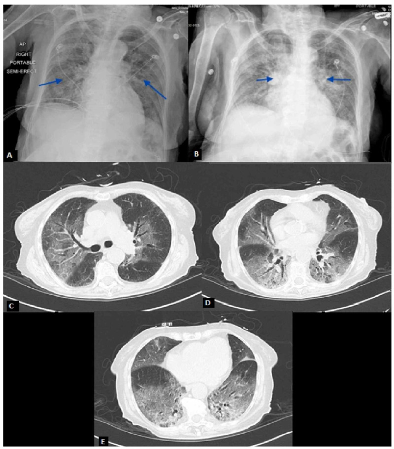
Figure 2. Chest X-Rays and CT Scan Demonstrating Bilateral Pulmonary Infiltrates.
(A) Hazy appearance of the lung fields with scattered airspace opacities. (B) Bilateral faint patchy airspace opacities slightly increased compared to initial imaging in Figure 2A. (C-E) Patchy ground-glass attenuation and smooth interlobular septal thickening involving all lobes, most pronounced within the right upper and bilateral lower lobes.

