Figure 2.
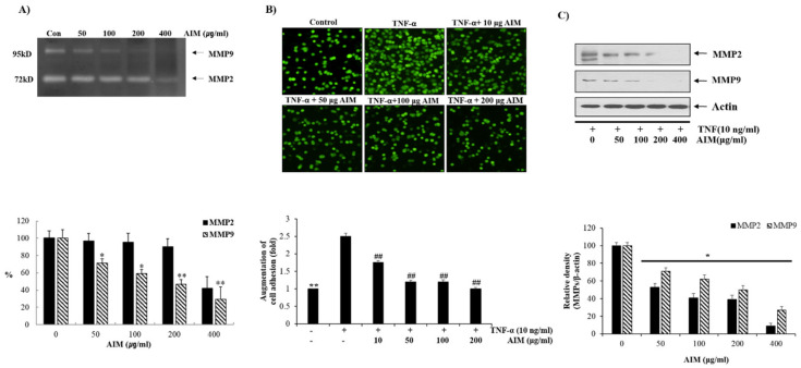
Inhibitory effects of AIM on cancer cell adhesion of MCF-7 Cells. (A) The cells were grown in 6-well plates with the seeding density of 5 × 10⁴ cells/well. MMP-2 and MMP-9 levels in gelatin zymography were assessed by densitometry. (Upper) The cells were treated with and without AIM for 24 h and the conditioned medium was taken to perform gelatin zymography. (Lower) MMP-2 and MMP-9 are expressed as a percentage of the activities against untreated cells. (B) The cells were seeded at the density of 5 × 10⁴ cells/mL. The cells were treated with indicated concentrations of AIM (0, 50, 100, and 200 μg/mL) for 24 h and the AIM effect on cancer cell invasion was assessed. For the group treated with AIM and TNF-α, cells were pre-treated with AIM (0, 10, 50, 100, and 200 μg/mL) for 1 h and then treated with TNF-α (10 ng/mL). ** p < 0.01 vs. the control group, ## p < 0.01 vs. the TNF-treated group. (C) Cells (5 × 10⁴ cells), either left untreated or pre-treated with AIM for 1 h, were exposed to TNF-α (10 ng/mL) for 24 h. (Upper) 30 μg of whole cell protein lysate were used for western blot analysis using antibodies of MMP-2 and MMP9. (Lower) Densitometry analysis of the data in western blot analysis by ImageJ software. The values were normalized against β-actin. ** p < 0.01 vs. the control group.

