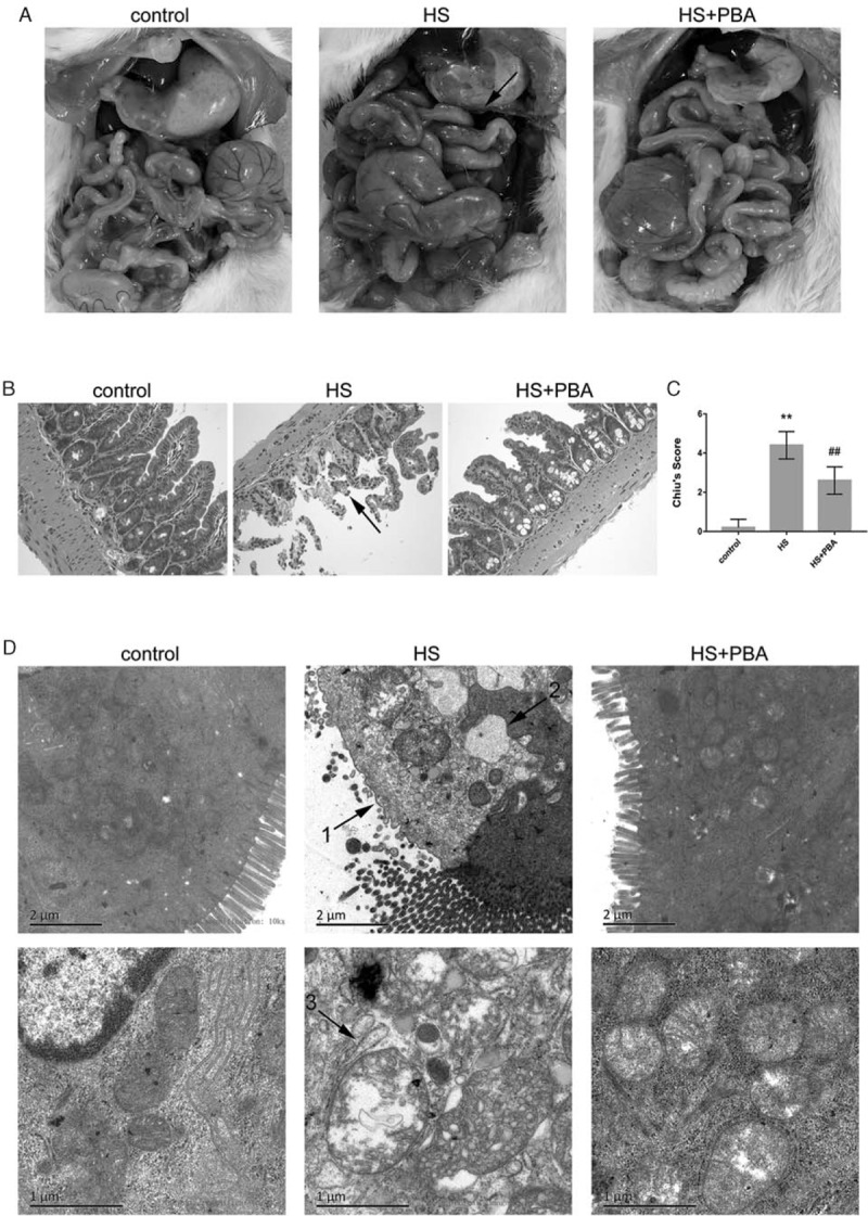
Fig. 1.
(A) Gross morphological changes in the ileum of mice.

Edema, hyperemia, and petechial hemorrhages in the mesentery are clearly seen after heatstroke. 4-PBA attenuated intestinal damage. (B) Pathological changes in the ileum of mice (magnification ×200). Samples were harvested 6 h after heatstroke and stained with H&E. The arrow indicates inflammatory cell infiltration and loss of villi. (C) Histological scores for the ileum are presented as mean ± SD. Significant differences are indicated as follows: ∗∗P < 0.01 and ∗P < 0.05 vs. the control group; ##P < 0.01 and #P < 0.05 vs. the heatstroke group. (D) Changes in the mucosal ultrastructure of the small intestine at 10,000× magnification and 25,000× magnification by transmission electron microscopy. The arrows indicate the following: 1, loss of intestinal microvilli; 2, extreme expansion of the endoplasmic reticulum; 3, vacuole-like structure and mitochondrial swelling.
