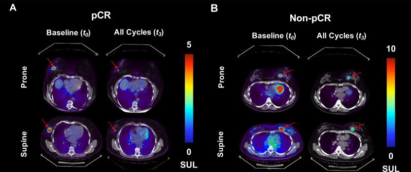
Figure 1.
Representative examples of prone and supine 18F-fluorodeoxyglucose (FDG) positron emission tomography (PET)/computed tomography (CT) images for a patient with pathologic complete response (pCR) [“responder”; (A)] and a patient with non-pCR patient [“nonresponder”; (B)] are presented at baseline and after all cycles of neoadjuvant therapy. The image corresponding to the central tumor slice is presented for each subject. FDG uptake decreased in both patients after therapy, although the responder had the largest decrease in radiotracer accumulation. In addition, the spatial distribution of radiotracer uptake within the lesion appears to be larger in the supine position than in the prone position, especially visible in the nonresponder (B). Also noted in (B) is the radiotracer accumulation within the myocardium of the heart in the prone images at baseline. Similar uptake is observed within the myocardium in the supine image at baseline, as well as both prone and supine image post-treatment. However the images that included the central tumor slice did not include the same anatomical section of the heart.

