PLOS ONE logoLink to PLOS ONE
. 2020 Jun 11;15(6):e0234441. doi: 10.1371/journal.pone.0234441

3D culture of functional human iPSC-derived hepatocytes using a core-shell microfiber

Shogo Nagata 1, Fumisato Ozawa 1, Minghao Nie 2, Shoji Takeuchi 1,2,3,*
Editor: Hiroaki Onoe4
PMCID: PMC7289419  PMID: 32525941

Abstract

Human iPSC-derived hepatocytes hold great promise as a cell source for cell therapy and drug screening. However, the culture method for highly-quantified hepatocytes has not yet been established. Herein, we have developed an encapsulation and 3D cultivation method for iPSC-hepatocytes in core-shell hydrogel microfibers (a.k.a. cell fiber). In the fiber-shaped 3D microenvironment consisting of abundant extracellular matrix (ECM), the iPSC-hepatocytes exhibited many hepatic characteristics, including the albumin secretion, and the expression of the hepatic marker genes (ALB, HNF4α, ASGPR1, CYP2C19, and CYP3A4). Furthermore, we found that the fibers were mechanically stable and can be applicable to hepatocyte transplantation. Three days after transplantation of the microfibers into the abdominal cavity of immunodeficient mice, human albumin was detected in the peripheral blood of the transplanted mice. These results indicate that the iPSC-hepatocyte fibers are promising either as in vitro models for drug screening or as implantation grafts to treat liver failure.

Introduction

Hepatocytes derived from human induced pluripotent stem cells (iPSC-hepatocytes) are promising cell sources in the fields of drug development, transplantation, and regenerative medicine [1]. For the culture of iPSC-hepatocytes, in comparison with the two-dimensional (2D) culture methods, three-dimensional (3D) culture methods have drawn much research attention recently; 3D microenvironments can promote the physiologically relevant hepatic functions of the hepatocytes [2,3]. For the 3D culture of hepatocytes, spheroid formation, in which dissociated hepatocytes are spontaneously aggregated by cell-cell interactions, is conventionally used [4,5]. However, there are problems associated with spheroid culture; cell aggregates are formed depending on their cell-cell junctions in suspension cultures which lack ECM. It is difficult to add the optimal amount and type of ECM to the 3D microenvironment in the conventional spheroid culture conditions. ECM is an important factor for the positive regulation of hepatocyte characteristics in various 3D hydrogel culture conditions [6,7]; the cell-ECM interaction promotes hepatic functions [8,9] and prevents cell death such as anoikis (loss of cell anchorage triggers apoptosis), which is induced in dissociated cells during the reconstruction of cell-cell and cell-ECM interactions in the suspension culture [10].

In this study, we establish the 3D culture of human iPSC-derived hepatocytes in Matrigel using a microfluidic fiber encapsulation technique called “cell fibers” [11]. The cell fiber is constructed using a 3D ECM-rich microenvironment as the core and mechanically stable alginate hydrogel as the shell. To construct the cell fibers based on iPSC-derived hepatocytes, we first mix commercially available human iPSC-hepatocytes with Matrigel and encapsulate the mixture into the core of the hydrogel microfibers; then the Matrigel is crosslinked and the cells are cultured in this 3D microenvironment. We demonstrate the advantage of our fiber culture conditions by examining the cell functions in the cell fibers both in vitro and in vivo. In vitro, we show that the hepatic function of hepatocytes can be enhanced using microfibers by assessing the secretion of albumin and hepatocyte-specific protein of the cell fibers, and by comparing the gene expression pattern of the hepatic cell markers, hepatic stem cell markers, and members of the CYP family among the fibers, spheroids, and 2D culture. In vivo, we show that the iPSC-hepatocyte fibers have good handleability and function as transplantable grafts by transplanting the iPSC-fibers into the abdominal cavity of mice.

Materials and methods

Cell culture

For conventional 2D culture, the commercially available iPSC-hepatocytes (REPROCELL) were seeded onto Matrigel-coated culture plate at a cell density of 2.4 × 10⁵ cells/cm2, following the manufacturer’s instructions. For the spheroid culture, the iPSC-hepatocytes were suspended in their culture medium at a cell density of 1.0 × 10⁵ cells/mL, and cell aggregates were formed by suspension culture in low-attachment 96-well V-bottom culture plates, a cell density of 1.0 × 10⁴ cells/well.

Formation and cultivation of core-shell hydrogel microfibers

Core-shell hydrogel fibers were prepared using a double-coaxial laminar-flow microfluidic device that was fabricated as described previously [11]. For the fiber formation, the iPSC-hepatocytes (REPROCELL) were recovered following the manufacturer’s instructions and suspended in Matrigel (CORNING) as the core solution. A pre-gel solution of 2.0% Na-alginate (ALG100, Mochida Pharmaceutical Co. Ltd.) in saline was used as the shell solution, and CaCl2 solution (100 mM CaCl2, 3% sucrose) or BaCl2 solution (20 mM BaCl2, 250 mM D-Mannitol, and 25 mM HEPES) was used as the sheath solution for alginate gelation. The typical flow rates of the core, shell, and We compared the morphology respectively. The fibers generated in the device were collected in a tube filled with CaCl2 (for in vitro assay) or BaCl2 solution (for in vivo assay) and were incubated in the collection bath for 10 min. Then, the fibers were washed with DMEM medium (Sigma-Aldrich) to remove the sheath solution and transferred to a culture dish filled with ReproHepato Culture Medium (REPROCELL) for 3D cultivation under conditions of 5% CO2 at 37°C. The medium was changed 1, 3, and 5 days after starting the 3D cultivation.

ELISA

For the measurement of human albumin in vitro, the supernatant of the culture medium was collected 3 and 6 days after the fiber formation and cultivation (n = 3, respectively). For the measurement of human albumin in vivo, peripheral blood was isolated from the transplanted and sham mice (n = 3, respectively). A human albumin-specific ELISA kit (Bethyl Laboratories) was used according to the manufacturer’s instructions.

Immunocytochemistry

Cultured fibers were fixed with 4% paraformaldehyde in saline for 10 min at room temperature, washed with saline solution, and pre-treated with blocking buffer [5% BSA in PBST (PBS with 0.1% Triton X-100)] at 4°C overnight. The cells were immunostained with primary antibodies (S1 Table) in the antibody buffer [1% BSA in PBST] overnight at 4°C and incubated with secondary antibodies in the antibody buffer for 1 h. After being mounted within a SlowFadeTM antifade mountant (Thermo Fisher) containing Hoechst 33342 dye (Lonza), the samples were observed using a confocal laser fluorescent microscope (LSM780, Carl Zeiss).

Gene expression analysis

Total RNA was isolated from iPSC-hepatocytes cultured in the conventional 2D condition and the cell fiber (n = 3, respectively) using the PureLink® RNA Mini Kit (Thermo Fisher) and was then treated with recombinant DNase I (Takara Bio) to eliminate genomic DNA contamination. For RT-PCR, the total RNA was used for the reverse transcription reaction with Superscript III™ reverse transcriptase (invitrogen), according to the manufacturer’s instructions. PCR was performed using the TB Green Premix Ex-TaqTM II system (Takara). GAPDH was used as an internal control for ΔΔCt quantitation method. The primer sequences are provided in S2 Table. For whole-transcriptome analysis, RNA sequencing was performed by Macrogen Inc. (Seoul, Korea). Briefly, library construction was performed using TruSeq RNA Sample Prep Kit v2 (Illumina) according to the manufacturer’s instructions. The sequencing was performed by Illumina NovaSeq 6000 platform. Expression profile was calculated for each sample and transcript/gene as read count and fragment per kilobase of transcript per million mapped reads (FPKM). Furthermore, differentially expressed genes (DEG) analysis was performed on a comparison pair as requested using FPKM. For significant lists, gene-set enrichment analysis was performed based on gene ontology (GO) (http://geneontology.org/). The result of the associated gene and the enrichment test was summarized by GO ID. The significance of specific GO ID in the enrichment test with the DEG set was calculated by modified fisher's exact test.

In vivo assay

For the functional evaluation of the iPSC-hepatocyte fibers in vivo, 1×10⁶ cells were encapsulated into a core-shell microfiber formed using Ba2+ as a crosslinker for the gelation of the alginate hydrogel. The cell fibers were cultured for 7 days before transplantation. After washing the cell fibers using saline solution, they were picked up and placed onto spatulas using tweezers. For the transplantation of the cell fibers into the abdominal cavity, celiotomy was performed on the eight-week-old male NOD/SCID mice (n = 3). The fibers were transplanted into their abdominal cavities using the spatulas, and the incision was closed using the non-absorbable surgical sutures. Three days after the transplantation, peripheral blood samples were collected from the cervical veins of the mice and evaluated by ELISA. This study was carried out in strict accordance with the recommendations in the Guidelines for Proper Conduct of Animal Experiments of Science Council of Japan. The protocol was approved by the University of Tokyo Institutional Animal Care and Use Committee (Permission number: 26–14). For anesthesia and euthanasia, Isoflurane was used in this study, and all efforts were made to minimize suffering.

Statistical analysis

Results are expressed as mean ± s.d. The data were analyzed using the two-tailed Student’s t-test.

Results and discussion

Hydrogel encapsulation and 3D cultivation of human iPSC-hepatocytes in a cell-laden core-shell microfiber

We used commercially available human iPSC-hepatocytes (cryopreserved) as a cell source. To demonstrate that the core-shell microfiber culture condition is useful for the encapsulation and 3D culture of iPSC-hepatocytes (Fig 1), we compared this system with the conventional 2D and spheroid culture conditions. For the fiber encapsulation, the iPSC-hepatocytes were thawed and suspended in Matrigel, and the Matrigel solution containing the iPSC-hepatocytes was then used as the core solution for the formation of core-shell microfibers (S1A Fig). In order to mimic the physiologically relevant 3D microenvironment, we chose Matrigel as the ECM for the fibers; the Matrigel contains basal membrane components and is known to be able to support the expression of hepatocyte function [8]. The initial cell density in the core of the fibers was fixed to 1.0 × 10⁸ cells/mL because the hepatocytes need not only cell-ECM interactions but also cell-cell interactions to survive and express their functions (Fig 2A).

Fig 1. Conceptual illustration of iPSC-hepatocyte fiber technology.

Fig 1

Human iPSC-hepatocytes are encapsulated in a 3D ECM-rich microenvironment using the cell fiber technology, and the iPSC-hepatocyte fibers are applicable to cell therapy for liver failure and drug development.

Fig 2. Cell encapsulation and 3D cultivation of human iPSC-hepatocytes in a core-shell hydrogel fiber.

Fig 2

(A) Human iPSC-hepatocytes singly dissociated and diluted into Matrigel were encapsulated into the core region of the fiber at a density of 1 × 10⁸ cells / mL, using the cell fiber technology. (B) The iPSC-hepatocytes were cultured for 7 days in the ECM-rich 3D microenvironment, and compacted cell aggregates were acquired in the fiber.

We compared the morphology of the iPSC-hepatocytes cultured for a week in the conventional 2D, spheroid, and the cell fiber culture conditions. In the 2D culture, the hepatocytes attached to the Matrigel-coated plate, and formed 2D cell-cell interactions and exhibited a cobblestone morphology, which is characteristic of typical hepatocytes. In the spheroid culture, loose spheroid in distorted shapes was generated (S2A Fig). In contrast, in the cell fiber culture, the iPSC-hepatocytes formed compact cell aggregates with various 3D structures (Fig 2B). We found that the iPSC-hepatocytes were alive in the aggregates in the fiber, but the cells that did not form aggregates were dead (S2B Fig); the aggregates maintained their shape even after the degradation of the alginate shell was degraded using alginate lyase (S2C Fig). These results suggest that both cell-ECM and cell-cell interactions are needed for cell survival and the formation of 3D compacted cell aggregates in the fibers (S1B Fig).

Hepatic characterization of iPSC-hepatocytes in the cell fibers compared to those in the 2D and spheroid culture

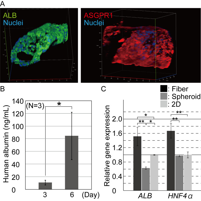
For the iPSC-hepatocytes in the cell fibers, we analyzed our samples by both immunostaining and ELISA. First, the iPSC-hepatocyte fibers on day 7 after the encapsulation were fixed and immunostained for functional mature hepatocyte marker proteins, albumin, asialoglycoprotein receptor 1 (ASGPR1) [12], and the hepatic cell marker HNF4α [13]. Cells comprising the aggregates in the fibers were highly positive for these protein markers (Fig 3A and S3 Fig), indicating that the encapsulated iPSC-hepatocytes maintained their hepatic characteristics during the 3D culture. The hepatic-marker-positive cells formed cell aggregates in the core region of the fibers. Second, using the ELISA, we evaluated whether the iPSC-hepatocytes were potent to secrete albumin in vitro. On both day 3 and day 6 of 3D culture, albumin protein was detected from culture medium at concentrations of 10.9 ± 3.8 ng/ml and 84.6 ± 37.2 ng/ml (mean ± s.d.), respectively (Fig 3B). These results indicate that the encapsulated iPSC-hepatocytes secreted albumin, expressed gradually enhanced hepatic characteristics with increasing culture time and that the shell of the fiber does not prevent the permeation of the secreted albumin protein into the culture medium. These results suggest that the iPSC-hepatocytes progress their maturation in the ECM-rich microenvironment of the fibers. Thus, the fiber culture can be useful for constructing functional 3D hepatic tissue in vitro.

Fig 3. Characterization of the hepatic function of the encapsulated hepatocytes.

Fig 3

(A) Immunocytochemistry was performed for hepatic marker proteins 7 days after hepatocyte cultivation. The iPSC-hepatocytes were positive for albumin and ASGPR1. (B) ELISA was performed for quantifying the albumin secreted into the culture medium. Secreted albumin was detected and its contents increased in the fibers during the culturing process (n = 3, P < 0.07). The error bars represent the standard deviation (s.d.) of triplicate samples. (C.) Quantitative RT-PCR was performed for the hepatic marker genes, ALB, and HNF4α. The expression of the marker genes in the hepatocytes from the cell fiber culture was significantly upregulated, compared to those from the conventional spheroid and 2D culture conditions (n = 3, *; P<0.05), **; P<0.01). The error bars represent the s.d. of triplicate samples.

We then compared the quality of the iPSC-hepatocytes in the cell fibers, spheroids, and 2D culture conditions. The expression level of the hepatic-specific genes ALB, and HNF4α, were evaluated by quantitative reverse transcription-polymerase chain reaction (qRT-PCR). The iPSC-hepatocytes in the fibers showed significantly high expression level of both ALB and HNF4α, compared to those in the spheroids and 2D culture conditions (Fig 3C). In addition, in our 2D culture, the hepatocytes exhibited a higher hepatic property than those in the spheroid culture, although it is generally known that 3D cultures—including spheroids—greatly enhance the cell characteristics of primary and stem cell-derived hepatocytes [29]. The purchased iPSC-hepatocytes used in this study were found to be unsuitable for the spheroid culture (S2A Fig); in literature, to form spheroids from the commercially available iPSC-hepatocytes, magnetic beads were used to modify the conventional suspension culture in order to support the efficient generation of cell-cell interactions [14]. In the cell fibers, the iPSC-hepatocytes were compacted to form cell aggregates, unlike those in the spheroids (Fig 2B), suggesting that the Matrigel functions as an ECM-rich microenvironment and supports the 3D culture of hepatocytes, with the upregulated expression of hepatic cell characteristics and prevented anoikis (S1B Fig).

Comparison of gene expression patterns of the iPSC-hepatocytes in the fiber, spheroid, and 2D culture

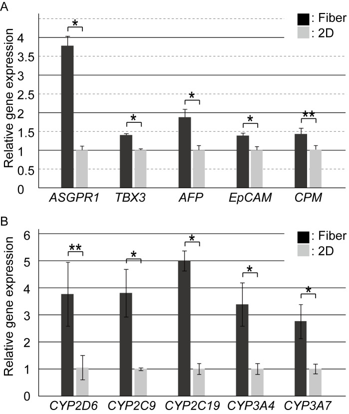
To precisely evaluate the characteristics of the iPSC-hepatocytes cultured in the fibers, we performed qRT-PCR for the quantification of hepatic genes. Gene expression levels of the hepatocytes in the fiber were compared to those in the 2D culture condition (the gold standard of hepatocyte culture). Compared to the iPSC-hepatocytes in the 2D culture, those in the cell fibers showed high expression of ALB and HNF4α (Fig 3C); the mature hepatocyte marker ASGPR1, early hepatic-lineage marker TBX3, the immature hepatocyte marker AFP, and the hepatic stem cell markers EpCAM [15] and CPM [16] were also significantly highly expressed in the fibers (Fig 4A). These results indicate that the iPSC-hepatocytes cultured in the fibers were "younger" hepatocytes than those in the 2D culture, and that the hepatic characteristics were promoted after their encapsulation into the ECM-rich 3D microenvironment and culturing using the core-shell fibers. In addition, the expressions of another marker of hepatic function, i.e. the CYP family genes which encode the CYP enzymes for the metabolism of chemicals, was also evaluated. Compared to the 2D culture, in our cell fiber culture, the iPSC-hepatocytes showed highly upregulated expression of the CYP family genes including CYP2D6, CYP2C9, CYP2C19, CYP3A4, and CYP3A7 (Fig 4B), suggesting that drug metabolism capacity of the iPSC-hepatocytes in the fiber culture was higher than those in the 2D culture. To precisely investigate effects of the encapsulation of the iPSC-hepatocytes into the fiber, RNA-sequencing was performed, and the 2D and the cell fiber culture condition was compared on global gene expression. In S4A Fig, the heat map of the one-way hierarchical clustering (1,053 genes satisfying with fold change 2 (FC2)), and top 30 genes which were upregulated or downregulated in the cell fiber were shown based on DEG analysis, indicating that global gene expression of the iPSC-hepatocytes was changed in the cell fiber. In addition, gene ontology analysis indicated that not only hepatocyte-specific pathways but also wide biological processes were changed (S4B Fig). Moreover, to evaluate their hepatic developmental stage in the ECM-rich 3D microenvironment, the gene expression of hepatocyte-related integrin was assessed. The expression level of ITGA1, ITGA5, ITGA6, and ITGB1 were also assessed since they are known to be highly expressed in hepatoblasts and fetal hepatocytes [17]. As a result, hepatocytes in the fibers and the 2D cultured were unchanged. The expression of ITGB3, which is associated with cellular senescence [18], was not also changed (S4C Fig), indicating that the cell fiber culture did not affect the expression profile of the hepatocyte-related integrin genes.

Fig 4. Evaluation of the hepatic gene expression profile of the iPSC-hepatocytes cultured in the cell fibers.

Fig 4

(A) qRT-PCR was performed for marker genes of the developmental stage of hepatocytes (n = 3, *; P<0.05), **; P<0.01). The error bars represent the s.d. of triplicate samples. (B) qRT-PCR was performed for quantifying the CYP family genes, which encode the CYP enzymes for drug metabolism (n = 3, *; P<0.05), **; P<0.01). The error bars represent the s.d. of triplicate samples.

Although the iPSC-hepatocytes are expected to be used for chemical screening of efficacy and toxicity for drug development, it has been known that the hepatocytes show low drug metabolism functions in conventional 2D culture condition; hence, various culture methods and conditions have been developed and investigated to promote the functions of cultured iPSC-hepatocytes [24,19,20]. Our results suggest that the iPSC-hepatocyte fibers can significantly promote hepatic function and therefore be one of the candidates to replace 2D cultures for drug screening.

Application of the iPSC-hepatocyte cell fibers for cell transplantation

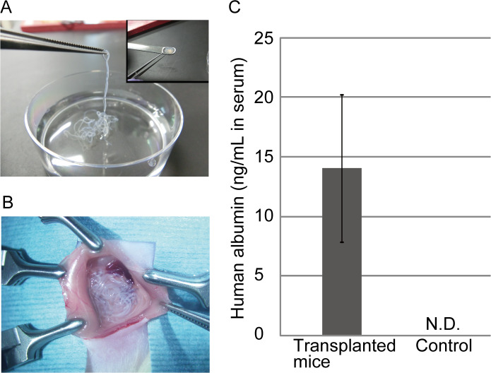
To evaluate the applicability of the iPSC-hepatocyte fibers as functional transplantable grafts, we performed an in vivo assay for albumin secretion after the transplantation of the microfibers into the abdominal cavity of immunodeficient NOD/SCID mice. For transplantation, our cell fibers have two advantages over hydrogel microbeads, which are widely used for cell encapsulation and transplantation research: first, the fiber shape is continuous in comparison to the separated droplets/beads, and therefore, the fibers can be easily picked up by tweezers and be efficiently implanted and retrieved as a whole; second, the fiber shell consists of alginate hydrogel, which makes the fibers mechanically strong to prevent the unwanted broad dispersion of the transplanted cells away from the injection site (Fig 5A). In the experiment, we transplanted the fibers that initially contained 1 × 10⁶ cells into the abdominal cavity of immunodeficient NOD/SCID mice using a spatula (Fig 5B). Three days after the transplantation, we assessed the human albumin concentration in the blood plasma of the mice by ELISA. Human albumin was detected at a concentration of 14.1 ± 6.2 ng/ml (mean ± s.d.) in the transplanted mice and was not detected in the blood of non-transplanted mice, which were used as controls (Fig 5C). The dosage of the secreted human albumin is enough to improve the survival ratio of acute liver failure (ALF) model mice as previously reported [2123]. These results indicate that the cell fiber technology is useful not only for the 3D culture of human iPSC-hepatocytes with a high expression of hepatic functions but also for cell therapy, due to their mechanical strength and handleability as transplantable grafts.

Fig 5. Application of the iPSC-hepatocytes cell fibers as transplantable grafts.

Fig 5

(A) The fibers were easily picked up and collected in optimal shapes for transplantation. (B) The fibers were transplanted into the mouse abdominal cavity. (C) Human albumin was detected in the peripheral blood samples from the transplanted mice by ELISA (n = 3). The error bars represent the s.d. of triplicate samples. N.D.; not-detected.

In general, hepatocyte transplantation has been used as a symptomatic treatment for ALF, fulminant liver failure, and metabolic disorders to bridge the gap period when liver transplantation is not available due to the shortage of liver donors [24,25]. Furthermore, hepatocyte transplantation is expected to be applied especially for the treatment of congenital liver failure in the field of pediatric care [25,26]. Although there are many reports about clinical trials involving hepatocyte transplantation, these trials are not yet accepted as conventional treatment, compared to liver transplantation because the methods for hepatocyte preparation and transplantation are not yet well-established [27]. Therefore, the development and establishment of techniques for hepatocyte transplantation are highly desired. Our results suggest that transplantation of the iPSC-hepatocyte fibers can be a powerful tool for efficient and safe cell therapy for treating liver failure and also be an alternative option for enzyme replacement therapy.

Conclusions

We developed a method for cell encapsulation and the 3D cultivation of human iPSC-hepatocytes in core-shell microfibers, with Matrigel as the ECM and alginate hydrogel. In the fibers, the iPSC-hepatocytes formed compacted cell aggregates, secreting albumin and highly expressing hepatic marker genes (including the CYP family genes). Furthermore, we demonstrate that the iPSC-hepatocyte fibers are mechanically stable and functional implantation grafts through the transplantation of the microfibers into the abdominal cavity of immunodeficient mice. In summary, the iPSC-hepatocyte fibers are suitable for the in vitro formation of 3D hepatic tissues, which can be applied for drug screening and cell therapy for liver disorders.

Supporting information

S1 Fig. Schematic illustration of the core-shell microfiber formation and cell culture in the ECM-rich 3D microenvironment.

(A) A double co-axial laminar flow microfluidic device was used for the formation of the iPSC-hepatocyte-laden core-shell hydrogel microfiber (iPSC-hepatocyte fiber). (B) The cells interacted with both other cells and the ECM, and were mechanically stimulated and regulated to spread and migrate three-dimensionally. Gradients of soluble factors, nutrients, and oxygen are also generated by diffusion through the ECM gel.

(TIF)

S2 Fig. 3D and 2D cultivation of human iPSC-hepatocytes.

(A) Human iPSC-hepatocytes formed loose and distorted spheroid in the suspension culture and exhibited a cobblestone morphology in the 2D culture on Matrigel-coated plate. (B) Live/dead staining was performed 7 days after the 3D culture of the hepatocyte fibers. The iPSC-hepatocytes in the cell aggregates were alive. (C) The shell of the fibers was degraded by alginate lyase after the 3D culture. The iPSC-hepatocytes maintained their compacted cell aggregates (arrowheads).

(TIF)

S3 Fig. Hepatic function characterization of the encapsulated hepatocytes 7 days after 3D cultivation.

Immunocytochemistry was performed for the hepatic stem/progenitor marker EpCAM, and the hepatic marker HNF4α. Some of the iPSC-hepatocytes were positive for EpCAM merging with albumin, and almost of HNF4α-positive cells were also positive for ASGPR1.

(TIF)

S4 Fig. Evaluation of the gene expression profile of the iPSC-hepatocytes in the cell fibers.

(A) RNA-sequencing was performed for hierarchical clustering analysis. Gene expression level is shown in normalized value (log2 based) using z-score, and color-coded with the color range shown in the top. Top 30 genes upregulated and downregulated in the fiber were selected and indicated. (B) Gene-set enrichment analysis which based on GO was conducted with the significant gene list and progressed about 3 categories of GO (biological processes, cellular component, and molecular function). The bar plot shown here is the top 10 terms of GO functional analysis in biological processes (*; P<0.05), **; P<0.01, ***; P<0.001). (C) qRT-PCR was performed for quantifying the expression of the integrin genes (n = 3). The error bars represent the s.d. of triplicate samples.

(TIF)

S1 Table. Antibodies used for immunocytochemistry.

(TIF)

S2 Table. Primers used for qRT-PCR.

(TIF)

Acknowledgments

We thank Dr. T. Kido and Dr. A. Miyajima for their constructive comments and help with discussions regarding the experimental design. We also thank H. Aoyagi for assistance on the in-vivo assay, T. Saito for assistance with fiber formation, and Dr. T. Kobayashi for help with statistical analysis.

Data Availability

All relevant data are within the manuscript and its Supporting Information files.

Funding Statement

S.N. is partly supported by JSPS KAKENHI (https://www.jsps.go.jp/j-grantsinaid/12_kiban/index.html, Grant Number 16H06329), and the Japan Agency for Medical Research and Development (AMED), Research Center Network for Realization of Regenerative Medicine (https://www.amed.go.jp/program/list/01/02/001.html, 16bm0304005h0004 and 18bm0404021h0001).

References

  • 1.Goldman O, Gouon-Evans V. Human Pluripotent Stem Cells: Myths and Future Realities for Liver Cell Therapy. Cell Stem Cell. 2016;18: 703–706. 10.1016/j.stem.2016.05.019 [DOI] [PubMed] [Google Scholar]
  • 2.Wang B, Jakus AE, Baptista PM, Soker S, Soto-Gutierrez A, Abecassis MM, et al. Functional Maturation of Induced Pluripotent Stem Cell Hepatocytes in Extracellular Matrix-A Comparative Analysis of Bioartificial Liver Microenvironments. Stem Cells Transl Med. 2016;5: 1257–1267. 10.5966/sctm.2015-0235 [DOI] [PMC free article] [PubMed] [Google Scholar]
  • 3.Takebe T, Sekine K, Enomura M, Koike H, Kimura M, Ogaeri T, et al. Vascularized and functional human liver from an iPSC-derived organ bud transplant. Nature. 2013;499: 481–484. 10.1038/nature12271 [DOI] [PubMed] [Google Scholar]
  • 4.Song W, Lu YC, Frankel AS, An D, Schwartz RE, Ma M. Engraftment of human induced pluripotent stem cell-derived hepatocytes in immunocompetent mice via 3D co-aggregation and encapsulation. Sci Rep. 2015;23: 16884. [DOI] [PMC free article] [PubMed] [Google Scholar]
  • 5.Bell CC, Hendriks DF, Moro SM, Ellis E, Walsh J, Renblom A, et al. Characterization of primary human hepatocyte spheroids as a model system for drug-induced liver injury, liver function and disease. Sci Rep. 2016;4: 25187. [DOI] [PMC free article] [PubMed] [Google Scholar]
  • 6.Pampaloni F, Reynaud EG, Stelzer EH. The third dimension bridges the gap between cell culture and live tissue. Nat Rev Mol Cell Biol. 2007;8: 839–845. 10.1038/nrm2236 [DOI] [PubMed] [Google Scholar]
  • 7.Toivonen S, Malinen MM, Küblbeck J, Petsalo A, Urtti A, Honkakoski P, et al. Regulation of Human Pluripotent Stem Cell-Derived Hepatic Cell Phenotype by Three-Dimensional Hydrogel Models. Tissue Eng Part A. 2016;22: 971–984. 10.1089/ten.TEA.2016.0127 [DOI] [PubMed] [Google Scholar]
  • 8.Borlak J, Singh PK, Rittelmeyer I. Regulation of Liver Enriched Transcription Factors in Rat Hepatocytes Cultures on Collagen and EHS Sarcoma Matrices. PLoS One. 2015;10(4): e0124867 10.1371/journal.pone.0124867 [DOI] [PMC free article] [PubMed] [Google Scholar]
  • 9.Wang Y, Kim MK, Shirahama H, Lee JH, Ng SS, Glenn JS, et al. ECM proteins in a microporous scaffold influence hepatocyte morphology, function, and gene expression. Sci Rep. 2016;6: 37427 10.1038/srep37427 [DOI] [PMC free article] [PubMed] [Google Scholar]
  • 10.Smets FN, Chen Y, Wang LJ, Soriano HE. Loss of cell anchorage triggers apoptosis (anoikis) in primary mouse hepatocytes. Mol. Genet. Metab. 2002;75: 344–352. 10.1016/S1096-7192(02)00004-5 [DOI] [PubMed] [Google Scholar]
  • 11.Onoe H, Okitsu T, Itou A, Kato-Negishi M, Gojo R, Kiriya D, et al. Metre-long cell-laden microfibres exhibit tissue morphologies and functions. Nat. Mater. 2013;12: 584–590. 10.1038/nmat3606 [DOI] [PubMed] [Google Scholar]
  • 12.Peters DT, Henderson CA, Warren CR, Friesen M, Xia F, Becker CF, et al. Asialoglycoprotein receptor 1 is a specific cell-surface marker for isolating hepatocytes derived from human pluripotent stem cells. Development. 2016;143: 1475–1481. 10.1242/dev.132209 [DOI] [PMC free article] [PubMed] [Google Scholar]
  • 13.Odom DT, Zizlsperger N, Gordon DB, Bell GW, Rinaldi NJ, Murray HL, et al. Control of pancreas and liver gene expression by HNF transcription factors. Science. 2004;303: 1378–1381. 10.1126/science.1089769 [DOI] [PMC free article] [PubMed] [Google Scholar]
  • 14.Pujan DK, Hubert T, Glauco SR. Assembly of Hepatocyte Spheroids Using Magnetic 3D Cell Culture for CYP450 Inhibition/Induction. Int J Mol Sci. 2017;18: 1085. [DOI] [PMC free article] [PubMed] [Google Scholar]
  • 15.Yoon SM, Gerasimidou D, Kuwahara R, Hytiroglou P, Yoo JE, Park YN, et al. Epithelial cell adhesion molecule (EpCAM) marks hepatocytes newly derived from stem/progenitor cells in humans. Hepatology. 2011;53: 964–973. 10.1002/hep.24122 [DOI] [PubMed] [Google Scholar]
  • 16.Kido T, Koui Y, Suzuki K, Kobayashi A, Miura Y, Chern EY, et al. CPM Is a Useful Cell Surface Marker to Isolate Expandable Bi-Potential Liver Progenitor Cells Derived from Human iPS Cells. Stem Cell Reports. 2015;5: 508–515. 10.1016/j.stemcr.2015.08.008 [DOI] [PMC free article] [PubMed] [Google Scholar]
  • 17.Couvelard A, Bringuier AF, Dauge MC, Nejjari M, Darai E, Benifla JL, et al. Expression of integrins during liver organogenesis in humans. Hepatology, 1998;27: 839–847. 10.1002/hep.510270328 [DOI] [PubMed] [Google Scholar]
  • 18.Rapisarda V, Borghesan M, Miguela V, Encheva V, Snijders AP, Lujambio A, et al. Integrin Beta 3 Regulates Cellular Senescence by Activating the TGF-β Pathway. Cell Rep. 2017,18: 2480–2493. 10.1016/j.celrep.2017.02.012 [DOI] [PMC free article] [PubMed] [Google Scholar]
  • 19.Takayama K, Kawabata K, Nagamoto Y, Kishimoto K, Tashiro K, Sakurai F, et al. 3D spheroid culture of hESC/hiPSC-derived hepatocyte-like cells for drug toxicity testing. Biomaterials. 2013;34: 1781–1789. 10.1016/j.biomaterials.2012.11.029 [DOI] [PubMed] [Google Scholar]
  • 20.Kim JH, Jang YJ, An SY, Son J, Lee J, Lee G, et al. Enhanced Metabolizing Activity of Human ES Cell-Derived Hepatocytes Using a 3D Culture System With Repeated Exposures to Xenobiotics. Toxicol Sci. 2016;149: 269 10.1093/toxsci/kfv248 [DOI] [PubMed] [Google Scholar]
  • 21.Sgroi A, Mai G, Morel P, Baertschiger RM, Gonelle-Gispert C, Serre-Beinier V, et al. Transplantation of encapsulated hepatocytes during acute liver failure improves survival without stimulating native liver regeneration. Cell Transplant. 2011;20: 1791–1803. 10.3727/096368911X564976 [DOI] [PubMed] [Google Scholar]
  • 22.Liu H, Kim Y, Sharkis S, Marchionni L, Jang YY. In vivo liver regeneration potential of human induced pluripotent stem cells from diverse origins. Sci Transl Med. 2011;3: 82ra39 10.1126/scitranslmed.3002376 [DOI] [PMC free article] [PubMed] [Google Scholar]
  • 23.Duan Y, Catana A, Meng Y, Yamamoto N, He S, Gupta S, et al. Differentiation and enrichment of hepatocyte-like cells from human embryonic stem cells in vitro and in vivo. Stem Cells. 2007;25: 3058–3068. 10.1634/stemcells.2007-0291 [DOI] [PubMed] [Google Scholar]
  • 24.Fitzpatrick E, Mitry RR, Dhawan A. Human hepatocyte transplantation: state of the art. J Intern Med. 2009;266: 339–357. 10.1111/j.1365-2796.2009.02152.x [DOI] [PubMed] [Google Scholar]
  • 25.Iansante V, Mitry RR, Filippi C, Fitzpatrick E, Dhawan A. Human hepatocyte transplantation for liver disease: current status and future perspectives. Pediatr Res. 2018;83: 232–240. 10.1038/pr.2017.284 [DOI] [PubMed] [Google Scholar]
  • 26.Meyburg J, Das AM, Hoerster F, Lindner M, Kriegbaum H, Engelmann G, et al. One liver for four children: first clinical series of liver cell transplantation for severe neonatal urea cycle defects. Transplantation. 2009;87, 636–641. 10.1097/TP.0b013e318199936a [DOI] [PubMed] [Google Scholar]
  • 27.Fisher RA, Strom SC, Human hepatocyte transplantation: worldwide results. Human hepatocyte transplantation: worldwide results. Transplantation. 2006;82: 441–449. 10.1097/01.tp.0000231689.44266.ac [DOI] [PubMed] [Google Scholar]

Decision Letter 0

Hiroaki Onoe

31 Mar 2020

PONE-D-20-04950

3D culture of functional human iPSC-derived hepatocytes using a core-shell microfiber

PLOS ONE

Dear Prof. Takeuchi,

Thank you for submitting your manuscript to PLOS ONE. After careful consideration, we feel that it has merit but does not fully meet PLOS ONE’s publication criteria as it currently stands. Therefore, we invite you to submit a revised version of the manuscript that addresses the points raised during the review process.

We would appreciate receiving your revised manuscript by May 15 2020 11:59PM. When you are ready to submit your revision, log on to https://www.editorialmanager.com/pone/ and select the 'Submissions Needing Revision' folder to locate your manuscript file.

If you would like to make changes to your financial disclosure, please include your updated statement in your cover letter.

To enhance the reproducibility of your results, we recommend that if applicable you deposit your laboratory protocols in protocols.io, where a protocol can be assigned its own identifier (DOI) such that it can be cited independently in the future. For instructions see: http://journals.plos.org/plosone/s/submission-guidelines#loc-laboratory-protocols

Please include the following items when submitting your revised manuscript:

  • A rebuttal letter that responds to each point raised by the academic editor and reviewer(s). This letter should be uploaded as separate file and labeled 'Response to Reviewers'.

  • A marked-up copy of your manuscript that highlights changes made to the original version. This file should be uploaded as separate file and labeled 'Revised Manuscript with Track Changes'.

  • An unmarked version of your revised paper without tracked changes. This file should be uploaded as separate file and labeled 'Manuscript'.

Please note while forming your response, if your article is accepted, you may have the opportunity to make the peer review history publicly available. The record will include editor decision letters (with reviews) and your responses to reviewer comments. If eligible, we will contact you to opt in or out.

We look forward to receiving your revised manuscript.

Kind regards,

Hiroaki Onoe

Academic Editor

PLOS ONE

Journal Requirements:

When submitting your revision, we need you to address these additional requirements:

1. Please ensure that your manuscript meets PLOS ONE's style requirements, including those for file naming. The PLOS ONE style templates can be found at http://www.plosone.org/attachments/PLOSOne_formatting_sample_main_body.pdf and http://www.plosone.org/attachments/PLOSOne_formatting_sample_title_authors_affiliations.pdf

2. Thank you for stating the following in the Competing Interests section:

"The authors declare competing financial interests: ST is a inventor on intellectual property rights related to the cell fibre technology, and stockholders of Cellfiber Inc, a start-up company based on the cell fibre technology."

We note that one or more of the authors are employed by a commercial company: Cellfiber Inc.

1.     Please provide an amended Funding Statement declaring this commercial affiliation, as well as a statement regarding the Role of Funders in your study. If the funding organization did not play a role in the study design, data collection and analysis, decision to publish, or preparation of the manuscript and only provided financial support in the form of authors' salaries and/or research materials, please review your statements relating to the author contributions, and ensure you have specifically and accurately indicated the role(s) that these authors had in your study. You can update author roles in the Author Contributions section of the online submission form.

Please also include the following statement within your amended Funding Statement.

“The funder provided support in the form of salaries for authors [insert relevant initials], but did not have any additional role in the study design, data collection and analysis, decision to publish, or preparation of the manuscript. The specific roles of these authors are articulated in the ‘author contributions’ section.”

If your commercial affiliation did play a role in your study, please state and explain this role within your updated Funding Statement.

2. Please also provide an updated Competing Interests Statement declaring this commercial affiliation along with any other relevant declarations relating to employment, consultancy, patents, products in development, or marketed products, etc.  

Within your Competing Interests Statement, please confirm that this commercial affiliation does not alter your adherence to all PLOS ONE policies on sharing data and materials by including the following statement: "This does not alter our adherence to  PLOS ONE policies on sharing data and materials.” (as detailed online in our guide for authors http://journals.plos.org/plosone/s/competing-interests) . If this adherence statement is not accurate and  there are restrictions on sharing of data and/or materials, please state these. Please note that we cannot proceed with consideration of your article until this information has been declared.

Please include both an updated Funding Statement and Competing Interests Statement in your cover letter. We will change the online submission form on your behalf.

Please know it is PLOS ONE policy for corresponding authors to declare, on behalf of all authors, all potential competing interests for the purposes of transparency. PLOS defines a competing interest as anything that interferes with, or could reasonably be perceived as interfering with, the full and objective presentation, peer review, editorial decision-making, or publication of research or non-research articles submitted to one of the journals. Competing interests can be financial or non-financial, professional, or personal. Competing interests can arise in relationship to an organization or another person. Please follow this link to our website for more details on competing interests: http://journals.plos.org/plosone/s/competing-interests

3. We note that you have a patent relating to material pertinent to this article. Please provide an amended statement of Competing Interests to declare this patent (with details including name and number), along with any other relevant declarations relating to employment, consultancy, patents, products in development or modified products etc. Please confirm that this does not alter your adherence to all PLOS ONE policies on sharing data and materials, as detailed online in our guide for authors http://journals.plos.org/plosone/s/competing-interests by including the following statement: "This does not alter our adherence to  PLOS ONE policies on sharing data and materials.” If there are restrictions on sharing of data and/or materials, please state these. Please note that we cannot proceed with consideration of your article until this information has been declared.

This information should be included in your cover letter; we will change the online submission form on your behalf.

Please know it is PLOS ONE policy for corresponding authors to declare, on behalf of all authors, all potential competing interests for the purposes of transparency. PLOS defines a competing interest as anything that interferes with, or could reasonably be perceived as interfering with, the full and objective presentation, peer review, editorial decision-making, or publication of research or non-research articles submitted to one of the journals. Competing interests can be financial or non-financial, professional, or personal. Competing interests can arise in relationship to an organization or another person. Please follow this link to our website for more details on competing interests: http://journals.plos.org/plosone/s/competing-interests

[Note: HTML markup is below. Please do not edit.]

Reviewers' comments:

Reviewer's Responses to Questions

Comments to the Author

1. Is the manuscript technically sound, and do the data support the conclusions?

The manuscript must describe a technically sound piece of scientific research with data that supports the conclusions. Experiments must have been conducted rigorously, with appropriate controls, replication, and sample sizes. The conclusions must be drawn appropriately based on the data presented.

Reviewer #1: Yes

Reviewer #2: Partly

Reviewer #3: Yes

Reviewer #4: Yes

**********

2. Has the statistical analysis been performed appropriately and rigorously?

Reviewer #1: I Don't Know

Reviewer #2: Yes

Reviewer #3: Yes

Reviewer #4: Yes

**********

3. Have the authors made all data underlying the findings in their manuscript fully available?

The PLOS Data policy requires authors to make all data underlying the findings described in their manuscript fully available without restriction, with rare exception (please refer to the Data Availability Statement in the manuscript PDF file). The data should be provided as part of the manuscript or its supporting information, or deposited to a public repository. For example, in addition to summary statistics, the data points behind means, medians and variance measures should be available. If there are restrictions on publicly sharing data—e.g. participant privacy or use of data from a third party—those must be specified.

Reviewer #1: Yes

Reviewer #2: Yes

Reviewer #3: Yes

Reviewer #4: Yes

**********

4. Is the manuscript presented in an intelligible fashion and written in standard English?

PLOS ONE does not copyedit accepted manuscripts, so the language in submitted articles must be clear, correct, and unambiguous. Any typographical or grammatical errors should be corrected at revision, so please note any specific errors here.

Reviewer #1: Yes

Reviewer #2: Yes

Reviewer #3: Yes

Reviewer #4: Yes

**********

5. Review Comments to the Author

Please use the space provided to explain your answers to the questions above. You may also include additional comments for the author, including concerns about dual publication, research ethics, or publication ethics. (Please upload your review as an attachment if it exceeds 20,000 characters)

Reviewer #1: The authors reported a novel cultivation method of Human iPSC-hepatocytes with a fiber-shaped 3D scaffold. They encapsulated hepatocytes into the core of core-shell hydrogel fibers to achieve both a 3D ECM-rich environment and easy manipulation. The approach is interesting and they showed that the protein secretion and gene expression increase compare to 2D or spheroid culture. However, the results are a bit unclear because the manuscript lacks some information or experiments as follows:

1. For line 185 and Fig. 3B, there is a contradiction in the culture time until evaluating the albumin secretion (The manuscript is written as day 3 & 7, while the figure is shown as day 3 & 6). They need to be corrected.

2. In Fig. 3C, 4A, 4B, and S4, the authors compared the gene expression of hepatocytes. However, there is no description of the normalization of cell numbers. Since the number of cells affects to the amount of expression, the results should be normalized for comparison. The description should be added. If the results are not the normalized one, they should be revised.

3. In Fig. 5C and lines 276-280, the authors indicated that the concentration of albumin increases by transplantation of fibers compared to the non-transplanted case. However, since the purpose of the authors is to increase the cell function by their culture method, the effect should be compared not with the non-transplanted case but with the previous culture methods (e.g. spheroids).

Reviewer #2: Takeuchi and co-workers reported a culture system for human iPSC-derived hepatocyte-like cells using the core-shell microfibers. The authors compared the cell functions between the fiber and conventional 2D culture conditions, and claimed that the presented approach was effective. In addition, the authors perform in vivo experiments to show the applicability of the fiber to transplantation therapy. In general, organization of individual cells into 3D platforms is a good strategy to maximize the cell functions, and the presented paper is interesting as one of new approaches. However, following points should be properly explained and/or reflected before publication of this paper in this journal.

(1) It was unclear why the authors obtained good results when they encapsulated cells in the fiber, because there is no rational explanation for this point. There should be some important cell-cell and cell-matrix interactions in 3D formats that potentially enhanced cell functions, with proper signal transductions. These points, especially from the viewpoint of the molecular signalings, should be properly explained.

(2) The results of the animal study were not so convincing, because the authors did not mention the effectiveness of the fiber-based cell encapsulation and transplantation. Many researchers have transplanted cells (hepatocytes) that were encapsulated in hydrogel matrices, but the comparison with conventional strategies was not described at all in this paper. The reason for choosing the abdominal cavity as the transplantation site is also unclear; the liver itself, the kidney capsule, or subcutaneous site would be superior because of the presence of the blood flow. The blood concentration of human albumin might not be sufficiently high considering the number of the transplanted cells; I am wondering the cell viability. It was unclear what types of liver diseases could be improved by this approach, especially for humans. These points should be clarified.

(3) It was unclear why the authors used Matrigel as the matrix, instead of collagen, the gold-standard for hepatocyte culture.

(4) Some of the experimental conditions were not properly described. For example, following points are unclear: (i) the housekeeping gene used for qPCR, (ii) the age of the animal, and (iii) how many times the authors repeated the experiments, especially for the figures.

(5) The manuscript contains many typos and unnatural expressions. The entire manuscript should be thoroughly checked once again before submitting the revision.

Reviewer #3: This manuscript presents the 3D culture technique of human iPSC-derived hepatocytes using a core-shell microfiber. The core-shell microfiber is a unique, superior 3D culture platform that provides an ECM-abundant core and a mechanically-strong shell. The authors proved the potential of their original core-shell fibers for cell transplantation applications. The manuscript would seem of considerable interest to those working in tissue engineering and regenerative medicines. However, the authors should describe the differences with their previous publication in IEEE MEMS 2020 titled “3D hepatic tissue formed by iPSC-derived hepatocytes using a cell fiber technology.” Figure 1 includes the exactly same figures in Figure 3 of the previous publication. After polished based on the aforementioned critiques, this manuscript may be able to be published in PLOS ONE. I would recommend that this paper needs minor revision to be published in PLOS ONE.

Reviewer #4: Nagata et al. apply cell fiber biofabrication techniques to create core-shell fibers of human iPSC-derived hepatocytes and perform in vitro and in vivo characterizations, including transplantation in a mouse model. Overall, the work is thorough; however, there are several aspects that should be expanded before publication. First, the introduction is missing a lot of prior work on cell fibers. The state of the art should be discussed in more detail (e.g., which cells have been demonstrated as compatible with the cell fiber approach, why haven't human iPSC-derived hepatocytes been done before, etc.). Such discussion on how this work is different from prior developments is important. I think a figure and supplementary movie featuring the biofabrication process is needed. Fabrication results are included in Fig. 2, but that figure should be expanded with the fabrication setup and process, which will be helpful for the readership. Also, the conclusion reads like a quick summary, but conclusions should really be used to provide some deeper insights into the study. There are some other minor notes, like the format of randomly including the figure captions in the main text being difficult for the reviewers and some weird callouts to the suppl. figures (S2A Fig). Overall though, should the aforementioned changes be made, my recommendation is for the manuscript to be accepted.

**********

6. PLOS authors have the option to publish the peer review history of their article (what does this mean?). If published, this will include your full peer review and any attached files.

If you choose “no”, your identity will remain anonymous but your review may still be made public.

Do you want your identity to be public for this peer review? For information about this choice, including consent withdrawal, please see our Privacy Policy.

Reviewer #1: No

Reviewer #2: No

Reviewer #3: No

Reviewer #4: No

[NOTE: If reviewer comments were submitted as an attachment file, they will be attached to this email and accessible via the submission site. Please log into your account, locate the manuscript record, and check for the action link "View Attachments". If this link does not appear, there are no attachment files to be viewed.]

While revising your submission, please upload your figure files to the Preflight Analysis and Conversion Engine (PACE) digital diagnostic tool, https://pacev2.apexcovantage.com/. PACE helps ensure that figures meet PLOS requirements. To use PACE, you must first register as a user. Registration is free. Then, login and navigate to the UPLOAD tab, where you will find detailed instructions on how to use the tool. If you encounter any issues or have any questions when using PACE, please email us at figures@plos.org. Please note that Supporting Information files do not need this step.

PLoS One. 2020 Jun 11;15(6):e0234441. doi: 10.1371/journal.pone.0234441.r002

Author response to Decision Letter 0


5 May 2020

Response to Reviewer# 1

Comment 1: For line 185 and Fig. 3B, there is a contradiction in the culture time until evaluating the albumin secretion (The manuscript is written as day 3 & 7, while the figure is shown as day 3 & 6). They need to be corrected.

Response 1:

We apologize for the incorrect information and have corrected the statement in the manuscript.

Comment 2: In Fig. 3C, 4A, 4B, and S4, the authors compared the gene expression of hepatocytes. However, there is no description of the normalization of cell numbers. Since the number of cells affects to the amount of expression, the results should be normalized for comparison. The description should be added. If the results are not the normalized one, they should be revised.

Response 2:

We apologize for causing this misunderstanding. For analyzing gene expression, we used RT-PCR technique. In the assay, the gene expression level of each sample can be normalized by the expression level of the housekeeping gene instead of cell number. Here we choose GAPDH, which is widely used as the housekeeping gene, for the normalization, so it is not necessary to mention cell number of the samples. We have revised the sentence on quantitative RT-PCR in the materials and methods section.

Comment 3: In Fig. 5C and lines 276-280, the authors indicated that the concentration of albumin increases by transplantation of fibers compared to the non-transplanted case. However, since the purpose of the authors is to increase the cell function by their culture method, the effect should be compared not with the non-transplanted case but with the previous culture methods (e.g. spheroids).

Response 3:

We appreciate the reviewer for this comment. In this study, we demonstrate that the cell fiber technique improve hepatocyte functionality in vitro, and we have not compared the function of the fibers as a transplant with conventional hepatocyte transplantation methods in the animal experiment. Here, we focus on showing the possibility of applying the hepatocyte fibers to cell transplantation therapy. This is because the transplantation of the hepatocyte fibers into the abdominal cavity performed in this study is a completely novel transplantation method, and thus it is difficult to compare with the conventional transplantation methods, in which hepatocytes are transplanted into the liver by vein administration of hepatocytes.

Response to Reviewer# 2

Comment 1: It was unclear why the authors obtained good results when they encapsulated cells in the fiber, because there is no rational explanation for this point. There should be some important cell-cell and cell-matrix interactions in 3D formats that potentially enhanced cell functions, with proper signal transductions. These points, especially from the viewpoint of the molecular signalings, should be properly explained.

Response 1:

We appreciate the reviewer for this comment. We are also focusing on the molecular mechanism on the enhancement of hepatocyte characteristics of the encapsulated iPSC-hepatocytes in the fiber. This is still challenging and many researchers are investigating the issue. Here we evaluated global gene expression profiles and compared the whole-transcriptome between the hepatocytes in the fiber and them in the 2D culture condition using RNA-sequencing technique. We have found some genes as candidates for enhancing hepatocyte characteristics in the fiber, but the details of their molecular functions are still under analysis and further research is needed in the future. In this revision, we do not mention the molecular mechanism but show the data of the RNA-sequencing analysis in S4 Fig. Furthermore, we have revised the sentences as shown below.

[Revised manuscript] (Results and Discussion section “To precisely investigate…” line 263)

To precisely investigate effects of the encapsulation of the iPSC-hepatocytes into the fiber, RNA-sequencing was performed, and the 2D and the cell fiber culture condition was compared on global gene expression. In S4A Fig, the heat map of the one-way hierarchical clustering (1,053 genes satisfying with fold change 2 (FC2)), and top 30 genes which were upregulated or downregulated in the cell fiber were shown based on DEG analysis, indicating that global gene expression of the iPSC-hepatocytes was changed in the cell fiber. In addition, gene ontology analysis indicated that not only hepatocyte-specific pathways but also wide biological processes were changed (S4B Fig).

Comment 2: The results of the animal study were not so convincing, because the authors did not mention the effectiveness of the fiber-based cell encapsulation and transplantation. Many researchers have transplanted cells (hepatocytes) that were encapsulated in hydrogel matrices, but the comparison with conventional strategies was not described at all in this paper. The reason for choosing the abdominal cavity as the transplantation site is also unclear; the liver itself, the kidney capsule, or subcutaneous site would be superior because of the presence of the blood flow. The blood concentration of human albumin might not be sufficiently high considering the number of the transplanted cells; I am wondering the cell viability. It was unclear what types of liver diseases could be improved by this approach, especially for humans. These points should be clarified.

Response 2:

We appreciate the reviewer for this comment. As the reviewer pointed out, there are reports on transplantation of hepatocytes encapsulated in the hydrogel, but, in most of the studies, hydrogel microbeads are used for the cell encapsulation as we mentioned in the manuscript. The advantage of our cell fiber technology compared to the microbeads is that, by using a hydrogel microfiber having core-shall structure, the core retained the encapsulated hepatocytes in the ECM-rich 3D microenvironment, while the outer shell composed of alginate hydrogel supported the physical strength and stability in vivo of the cell fiber. Furthermore, since the fiber is possible to be handled as a single transplant due to its shape and strength, the transplant operation can be simple. In addition, when a malignant event occurs in the transplant or when it is no longer needed, the transplanted fiber can be more efficiently retrieved from the host than the microbeads.

In this study, we chose intraperitoneal transplantation of the fiber because it is generally known that transplantation of hydrogel encapsulated hepatocytes into the peritoneal cavity is an attractive option for the management of acute liver failure providing short-term support to allow native liver regeneration [PLoS One. 2014 Dec 1;9(12):e113609.]. As the reviewer commented, transplantation into regions rich in blood vessels (e.g. subcutaneous site) is especially required for organ transplantation that requires permanent expression of cellular function, but, in this study, hepatocyte fiber transplantation is thought as one of novel cell therapy techniques and required to express the function in the short term as mentioned above. To clearly show that, we have revised the sentence as shown below.

[Revised manuscript] (Results and Discussion section “For transplantation,…” line 300)

For transplantation, our cell fibers have two advantages over hydrogel microbeads, which are widely used for cell encapsulation and transplantation research: first, the fiber shape is continuous in comparison to the separated droplets/beads, and therefore, the fibers can be easily picked up by tweezers and be efficiently implanted and retrieved as a whole; second, the fiber shell consists of alginate hydrogel, which makes the fibers mechanically strong to prevent the unwanted broad dispersion of the transplanted cells away from the injection site (Fig 5A).

Comment 3: It was unclear why the authors used Matrigel as the matrix, instead of collagen, the gold-standard for hepatocyte culture.

Response 3:

We appreciate the reviewer for this comment. Type I collagen has been known as a suitable ECM for hepatocytes, but Matrigel is also known to be one of the most useful matrices for static, traditional hepatocyte culture [Tissue Eng Part A. 2010 Mar; 16(3): 1075–1082.]. Here we chose Matrigel for constructing the cell fiber due to basal membrane components for supporting the iPSC-hepatocytes in the core region of the fiber. Since various gels can be used for the cell fiber formation, it is possible to use an arbitrary gel (e.g. collagen) depending on the application.

Comment 4: Some of the experimental conditions were not properly described. For example, following points are unclear: (i) the housekeeping gene used for qPCR, (ii) the age of the animal, and (iii) how many times the authors repeated the experiments, especially for the figures.

Response 4:

We appreciate the reviewer for this comment and have revised the sentence in the materials and methods section.

Comment 5: The manuscript contains many typos and unnatural expressions. The entire manuscript should be thoroughly checked once again before submitting the revision.

Response 5:

We have checked again and revised the manuscript according to advice from experts on English.

Response to Reviewer# 3

Comment :This manuscript presents the 3D culture technique of human iPSC-derived hepatocytes using a core-shell microfiber. The core-shell microfiber is a unique, superior 3D culture platform that provides an ECM-abundant core and a mechanically-strong shell. The authors proved the potential of their original core-shell fibers for cell transplantation applications. The manuscript would seem of considerable interest to those working in tissue engineering and regenerative medicines. However, the authors should describe the differences with their previous publication in IEEE MEMS 2020 titled “3D hepatic tissue formed by iPSC-derived hepatocytes using a cell fiber technology.” Figure 1 includes the exactly same figures in Figure 3 of the previous publication. After polished based on the aforementioned critiques, this manuscript may be able to be published in PLOS ONE. I would recommend that this paper needs minor revision to be published in PLOS ONE.

Response :

We appreciate the reviewer for this comment. As pointed out by the reviewer, a related but preliminary work was reported in the MEMS conference. Comparing to the conference proceedings, we have significantly polished the initial ideas based on the molecular biological insight which was gained by new experiments that precisely evaluated the hepatic function and the cell characteristics and showed that the cell fiber was useful for efficient 3D culture system of the iPSC-hepatocytes and cell therapy for liver failure treatment.

On the comment for figure 1, we modified and revised parts of the figure.

Response to Reviewer# 4

Comment : Nagata et al. apply cell fiber biofabrication techniques to create core-shell fibers of human iPSC-derived hepatocytes and perform in vitro and in vivo characterizations, including transplantation in a mouse model. Overall, the work is thorough; however, there are several aspects that should be expanded before publication. First, the introduction is missing a lot of prior work on cell fibers. The state of the art should be discussed in more detail (e.g., which cells have been demonstrated ascompatible with the cell fiber approach, why haven't human iPSC-derived hepatocytes been done before, etc.). Such discussion on how this work is different from prior developments is important. I think a figure and supplementary movie featuring the biofabrication process is needed. Fabrication results are included in Fig.2, but that figure should be expanded with the fabrication setup and process, which will be helpful for the readership. Also, the conclusion reads like a quick summary, but conclusions should really be used to provide some deeper insights into the study. There are some other minor notes, like the format of randomly including the figure captions in the main text being difficult for the reviewers and some weird callouts to the suppl. figures (S2A Fig). Overall though, should the aforementioned changes be made, my recommendation is for the manuscript to be accepted.

Response :

We appreciate the reviewer for this comment. We considered adding a video of the fabrication process, but due to the limited activity of our research facility caused by the spread of the coronavirus infection (COVID-19), it is difficult to do it within the revise period. However, the biofabrication technique used in this study was basically the same as in the previous report [Nat Mater. 2013 Jun;12(6):584-90.], so the details can be omitted in this manuscript. The modification in the fiber fabrication is that, for the transplantation experiment, barium ions was used as the crosslinker of alginate gel for the fiber strength and in vivo stability. To clearly show that, we have revised the sentence as shown below.

[Revised manuscript] (Materials and methods section “The fibers generated in the …” line 91)

The fibers generated in the device were collected in a tube filled with CaCl2 (for in vitro assay) or BaCl2 solution (for in vivo assay), and were incubated in the collection bath for 10 min.

As the reviewer pointed out, there have been reports on the cell fibers. The cell fiber technology is thought to be applicable to all cell types in principle, but previous reports mainly focused on the fabrication technology using various cell lines, which are relatively stable in cultivation. The difference between them and this study is that here we developed the fibers using the iPSC-derived functional cells, which can be directly applied to medical applications (e.g. cell therapy). This is the first report of the fiber composed of iPSC-hepatocytes, which required to preciously evaluate the cell characteristics and transplantation experiments as performed in this study.

The paper format follows the PloS ONE submission rules. Therefore, the figure captions are inserted in the main text immediately after the paragraph that cites the figure.

We appreciate the comment on S2A Fig and have revised the sentence as shown below.

[Revised manuscript] (Results and Discussion section “We compared the…” line 183)

We compared the morphology of the iPSC-hepatocytes cultured for a week in the conventional 2D, spheroid, and the cell fiber culture conditions. In the 2D culture, the hepatocytes attached onto the Matrigel-coated plate, and formed 2D cell-cell interactions and exhibited a cobblestone morphology, which is characteristic of typical hepatocytes. In the spheroid culture, loose spheroid in distorted shapes was generated (S2A Fig). In contrast, in the cell fiber culture, the iPSC-hepatocytes formed compact cell aggregates with various 3D structures (Fig 2B).

Attachment

Submitted filename: Response to reviewers.docx

Decision Letter 1

Hiroaki Onoe

27 May 2020

3D culture of functional human iPSC-derived hepatocytes using a core-shell microfiber

PONE-D-20-04950R1

Dear Dr. Takeuchi,

We are pleased to inform you that your manuscript has been judged scientifically suitable for publication and will be formally accepted for publication once it complies with all outstanding technical requirements.

Within one week, you will receive an e-mail containing information on the amendments required prior to publication. When all required modifications have been addressed, you will receive a formal acceptance letter and your manuscript will proceed to our production department and be scheduled for publication.

Shortly after the formal acceptance letter is sent, an invoice for payment will follow. To ensure an efficient production and billing process, please log into Editorial Manager at https://www.editorialmanager.com/pone/, click the "Update My Information" link at the top of the page, and update your user information. If you have any billing related questions, please contact our Author Billing department directly at authorbilling@plos.org.

If your institution or institutions have a press office, please notify them about your upcoming paper to enable them to help maximize its impact. If they will be preparing press materials for this manuscript, you must inform our press team as soon as possible and no later than 48 hours after receiving the formal acceptance. Your manuscript will remain under strict press embargo until 2 pm Eastern Time on the date of publication. For more information, please contact onepress@plos.org.

With kind regards,

Hiroaki Onoe

Academic Editor

PLOS ONE

Additional Editor Comments (optional):

Reviewers' comments:

Reviewer's Responses to Questions

Comments to the Author

1. If the authors have adequately addressed your comments raised in a previous round of review and you feel that this manuscript is now acceptable for publication, you may indicate that here to bypass the “Comments to the Author” section, enter your conflict of interest statement in the “Confidential to Editor” section, and submit your "Accept" recommendation.

Reviewer #1: All comments have been addressed

Reviewer #2: All comments have been addressed

Reviewer #3: All comments have been addressed

Reviewer #4: All comments have been addressed

**********

2. Is the manuscript technically sound, and do the data support the conclusions?

The manuscript must describe a technically sound piece of scientific research with data that supports the conclusions. Experiments must have been conducted rigorously, with appropriate controls, replication, and sample sizes. The conclusions must be drawn appropriately based on the data presented.

Reviewer #1: Yes

Reviewer #2: Yes

Reviewer #3: Yes

Reviewer #4: (No Response)

**********

3. Has the statistical analysis been performed appropriately and rigorously?

Reviewer #1: Yes

Reviewer #2: Yes

Reviewer #3: Yes

Reviewer #4: (No Response)

**********

4. Have the authors made all data underlying the findings in their manuscript fully available?

The PLOS Data policy requires authors to make all data underlying the findings described in their manuscript fully available without restriction, with rare exception (please refer to the Data Availability Statement in the manuscript PDF file). The data should be provided as part of the manuscript or its supporting information, or deposited to a public repository. For example, in addition to summary statistics, the data points behind means, medians and variance measures should be available. If there are restrictions on publicly sharing data—e.g. participant privacy or use of data from a third party—those must be specified.

Reviewer #1: Yes

Reviewer #2: Yes

Reviewer #3: Yes

Reviewer #4: (No Response)

**********

5. Is the manuscript presented in an intelligible fashion and written in standard English?

PLOS ONE does not copyedit accepted manuscripts, so the language in submitted articles must be clear, correct, and unambiguous. Any typographical or grammatical errors should be corrected at revision, so please note any specific errors here.

Reviewer #1: Yes

Reviewer #2: Yes

Reviewer #3: Yes

Reviewer #4: (No Response)

**********

6. Review Comments to the Author

Please use the space provided to explain your answers to the questions above. You may also include additional comments for the author, including concerns about dual publication, research ethics, or publication ethics. (Please upload your review as an attachment if it exceeds 20,000 characters)

Reviewer #1: Authors have properly addressed my concerns in the revision, and I agree to their explanations.

I have no more concerns on this article.

Reviewer #2: The revised manuscript by Takeuchi and co-workers properly reflected most of my previous concerns, and now the paper was improved. This paper is a nice example of the hydrogel fiber-based cell culture technique, and hence, I recommned accepting this paper for publication.

Reviewer #3: The authors replied to the reviewers' comments well and improved the manuscript. I recommend the manuscript for publication.

Reviewer #4: (No Response)

**********

7. PLOS authors have the option to publish the peer review history of their article (what does this mean?). If published, this will include your full peer review and any attached files.

If you choose “no”, your identity will remain anonymous but your review may still be made public.

Do you want your identity to be public for this peer review? For information about this choice, including consent withdrawal, please see our Privacy Policy.

Reviewer #1: No

Reviewer #2: No

Reviewer #3: No

Reviewer #4: No

Acceptance letter

Hiroaki Onoe

2 Jun 2020

PONE-D-20-04950R1

3D culture of functional human iPSC-derived hepatocytes using a core-shell microfiber

Dear Dr. Takeuchi:

I'm pleased to inform you that your manuscript has been deemed suitable for publication in PLOS ONE. Congratulations! Your manuscript is now with our production department.

If your institution or institutions have a press office, please let them know about your upcoming paper now to help maximize its impact. If they'll be preparing press materials, please inform our press team within the next 48 hours. Your manuscript will remain under strict press embargo until 2 pm Eastern Time on the date of publication. For more information please contact onepress@plos.org.

If we can help with anything else, please email us at plosone@plos.org.

Thank you for submitting your work to PLOS ONE and supporting open access.

Kind regards,

PLOS ONE Editorial Office Staff

on behalf of

Dr. Hiroaki Onoe

Academic Editor

PLOS ONE

Associated Data

    This section collects any data citations, data availability statements, or supplementary materials included in this article.

    Supplementary Materials

    S1 Fig. Schematic illustration of the core-shell microfiber formation and cell culture in the ECM-rich 3D microenvironment.

    (A) A double co-axial laminar flow microfluidic device was used for the formation of the iPSC-hepatocyte-laden core-shell hydrogel microfiber (iPSC-hepatocyte fiber). (B) The cells interacted with both other cells and the ECM, and were mechanically stimulated and regulated to spread and migrate three-dimensionally. Gradients of soluble factors, nutrients, and oxygen are also generated by diffusion through the ECM gel.

    (TIF)

    S2 Fig. 3D and 2D cultivation of human iPSC-hepatocytes.

    (A) Human iPSC-hepatocytes formed loose and distorted spheroid in the suspension culture and exhibited a cobblestone morphology in the 2D culture on Matrigel-coated plate. (B) Live/dead staining was performed 7 days after the 3D culture of the hepatocyte fibers. The iPSC-hepatocytes in the cell aggregates were alive. (C) The shell of the fibers was degraded by alginate lyase after the 3D culture. The iPSC-hepatocytes maintained their compacted cell aggregates (arrowheads).

    (TIF)

    S3 Fig. Hepatic function characterization of the encapsulated hepatocytes 7 days after 3D cultivation.

    Immunocytochemistry was performed for the hepatic stem/progenitor marker EpCAM, and the hepatic marker HNF4α. Some of the iPSC-hepatocytes were positive for EpCAM merging with albumin, and almost of HNF4α-positive cells were also positive for ASGPR1.

    (TIF)

    S4 Fig. Evaluation of the gene expression profile of the iPSC-hepatocytes in the cell fibers.

    (A) RNA-sequencing was performed for hierarchical clustering analysis. Gene expression level is shown in normalized value (log2 based) using z-score, and color-coded with the color range shown in the top. Top 30 genes upregulated and downregulated in the fiber were selected and indicated. (B) Gene-set enrichment analysis which based on GO was conducted with the significant gene list and progressed about 3 categories of GO (biological processes, cellular component, and molecular function). The bar plot shown here is the top 10 terms of GO functional analysis in biological processes (*; P<0.05), **; P<0.01, ***; P<0.001). (C) qRT-PCR was performed for quantifying the expression of the integrin genes (n = 3). The error bars represent the s.d. of triplicate samples.

    (TIF)

    S1 Table. Antibodies used for immunocytochemistry.

    (TIF)

    S2 Table. Primers used for qRT-PCR.

    (TIF)

    Attachment

    Submitted filename: Response to reviewers.docx

    Data Availability Statement

    All relevant data are within the manuscript and its Supporting Information files.


    Articles from PLoS ONE are provided here courtesy of PLOS

    RESOURCES