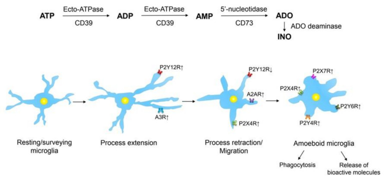
Figure 2.
Purinergic receptors at microglial cells exemplifying their different activation states. ATP is sequentially dephosphorylated by an enzymatic cascade to AMP by ecto-ATPase (NPDase-1; CD39) through the intermediary product ADP. AMP is further degraded to adenosine (ADO) by 5’-nucleotidase (CD73). Finally, adenosine is almost inactivated by adenosine deaminase to inosine (INO). Resting/ramified microglia extend and retrieve processes, thereby scanning their territories, non-overlapping with those of the neighboring microglial cells. When ATP is released/outpoured into the extracellular space from damaged CNS cells, as a first step of microglial activation, these cells extend their processes towards the site of injury triggered by stimulation of P2Y12 receptors (Rs). Both P2Y12 and A3Rs are upregulated in consequence of CNS damage, and they co-operate in steering the microglial process extension. Subsequently, these processes retract due to the downregulation of P2Y12Rs and the upregulation A2ARs; the migratory activity of this microglia is controlled by the interaction of P2Y12 and P2X4Rs. After the complete retraction of the microglial processes, an amoeboid phenotype is evolving. On this microglia, phagocytosis and pinocytosis are induced by P2Y6R and P2Y4R activation, respectively. P2X4Rs mediate the secretion of brain-derived neurotrophic factor (BDNF) in spinal cord microglia. P2X7Rs may initiate multiple secretory processes such as the release of pro-inflammatory cytokines, chemokines, growth factors, proteases, reactive oxygen/nitrogen species, cannabinoids, and probably also the excitotoxic ATP and glutamate. Upwardly directed arrows beside receptors indicate their upregulation or increased activation by agonists, while downwardly directed arrows indicate their downregulation or decreased activation by agonists. For further details, see [19,79,80]. Artwork by Dr. Haiyan Yin.

