Figure 1.
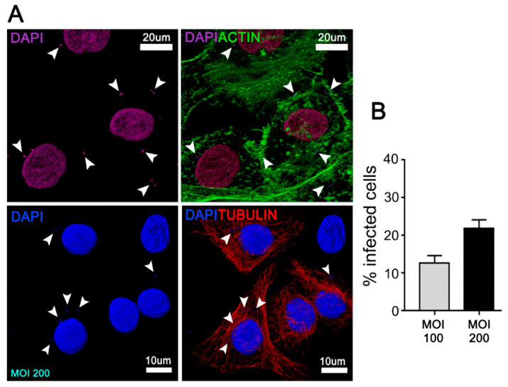
Junctional epithelium keratinocytes (JEKs)-invasion by Aggregatibacter actinomycetemcomitans (Aa). OKF6/TERT-2 cells were incubated with A.a (serotype b) at MOI = 100 and 200 for 3 h at 37 °C. After the corresponding washes, JEKs were fixed and subjected to confocal microscopy analysis for the quantification of infected cells. (A) Immunofluorescence staining using actin (upper panel in green) and tubulin (bottom panel in red) to delineate cell contour, as well as DAPI (purple, upper panel and blue, bottom panel) for visualization of cell nuclei and bacteria. White arrowheads indicate intracellular bacteria. (B) The graph shows the average of 15 images acquired by confocal microscopy using a 63× objective in five independent experiments (≥1000 cells). Scale bar 10 and 20 µm.

