Abstract
Breast cancer (BC) is frequent among women in worldwide as well as in India. Several studies have reported a wide variation (1.6–86.2%) in the frequency of incidence of human papillomavirus (HPV) infection in BC with high prevalence of high risk HPV16 subtype. HPV infection in breast can occur through different routes like body fluid or by micro-lesion of breast skin from genital/agential sites, though the actual mode of HPV transmission is not yet known in details. Frequent integration and sequence variation with low copy number of HPV16 were seen in this tumour. In addition, high frequencies of methylation in p97 promoter region of HPV16 were evident in this tumour. Novel splice variants of E6/E7 along with other common variants and their protein expression were seen in the tumour. This indicates the importance of HPV in this tumor, its early diagnosis and prognosis. Thus, HPV may be targeted through vaccination to control the disease. However, detailed analysis of HPV associated molecular pathogenesis of BC is warranted for proper therapeutic intervention.
Keywords: Breast cancer (BC), human papillomavirus (HPV), HPV transmission, management
Introduction
Globally, breast cancer (BC) is the most common cancer among the women registering a total of 2.08 million new cases (11.6% of all new cases among females) in the year 2018 alone (1). Accounting for 15% of the total cancer-related deaths, it is the first most common cause of cancer deaths among women, worldwide (1). In Indian context, BC remains the most frequent (27.7%) cancer among women with the urban and metropolitan regions reporting high rates of incidence than rural region (1,2). Going by the numbers, in 2018 about 87,090 women died due to BC in India (11.1% of total women cancer) (1).
The BC has several etiological factors like prolonged or elevated exposure to estrogen due to early age of menarche (younger than 12 years), nulliparity, late age of menopause (over 55 years), exposure to high doses of ionizing radiation, regular alcohol consumption and high fat diet (3). Among the different etiological factors, infection with several viruses has also been reported in BC (4). However, these etiological factors were involved in only 20–50% of BC cases (5). Recently, different studies suggested association of human papillomavirus (HPV) with BC (6). But, frequency of HPV infection in BC varied widely (1.6–86%) among different studies (7,8). Inconsistent HPV infection was also reported in different molecular subtypes of BC (9,10). The possible mode of HPV transmission in breast and its role in breast carcinogenesis are not well studied. In this review our aim is to discuss the role of HPV infection in breast carcinogenesis and its future management.
Association of HPV infection with BC
Prevalence of HPV infection in breast
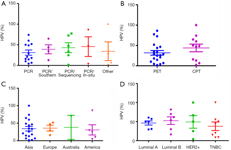
Recently, HPV infection in BC in different population around the world was reported by several authors (Table 1). However, many of them have not identified any HPV DNA in breast tumour. The prevalence of HPV in BC varied widely from 1.6–86.2% among the different continents of the world (7,8). According to screening methods, comparatively high frequency of HPV was detected in polymerase chain reaction (PCR) with sequencing or in-situ hybridization than only PCR method alone (Figure 1A). While a comparatively lower frequency of HPV DNA was found when the tissue source was formalin fixed paraffin-embedded tissue (PET) than the cryo-preserved tissue (CPT), the reason can be attributed towards the fact that the total DNA is severely degraded during the whole process of formalin fixation and paraffin embedding (47). So, this detection based difference in results might account partly for the wide range of frequency of HPV infection in BC, as reported by several studies (Figure 1B). On the other hand, HPV infection did not show significant variation among the different continents of the world (Figure 1C). To date, nine HPV types (HPV6, 11, 16, 18, 31, 33, 35, 45 and 52) are evident in BC across different population of the world. The prevalence of these HPV types showed variation among different population. The HPV16 was prevalent in American BC patients, whereas HPV18 and HPV33 were frequent in Australian and Chinese BC patients (Table 1). Apart from the above mentioned three subtypes, prevalence of other subtypes in BC patients among different population are as follows: HPV6/HPV11 in 5–12.6% patients of Iran and Spain (39,40), HPV31 in 1.5–11.5% patients of Brazil and UK (37,48), HPV35 in 16–19.2% of patients of Thailand and UK (37,49), HPV45 in 23% of UK BC patients (37) and HPV52 in 1.5–11% of Brazil, UK and Thailand patients (37,48,49).
Table 1. Worldwide HPV prevalence in breast tumour and adjacent normal breast tissue.
| Country | Breast tumour | Adjacent normal breast | Tissue preservation type | Methods of detection | |||||||
|---|---|---|---|---|---|---|---|---|---|---|---|
| Benign | Malignant | ||||||||||
| HPV (%) | HPV (%) | HPV16 (%) | HPV18 (%) | HPV33 (%) | Other HPV (%) | HPV (%) | |||||
| China [Yu et al. 1999] (11) | 1/20, 5.0 | 18/52, 34.6 | 1/52, 1.9 | 0/52, 0.0 | – | – | – | PET | PCR/Southern | ||
| China [Yu et al. 2000] (12) | 4/72, 5.0 | 14/32, 43.8 | – | – | 14/32, 43.8 | – | – | PET | PCR/Southern | ||
| USA [de Villiers et al. 2004] (8) | 25/29, 86.2 | 3/29, 10.3 | 0/29. 0.0 | 0/29,0.0 | 12/25, 48.0 | – | PET | PCR/In-situ | |||
| Brazil [Damin et al. 2004] (13) | 0/41, 0.0 | 25/101, 24.7 | 14/101, 13.8 | 10/101, 9.9 | – | – | – | PET | PCR/Seq | ||
| Turkey [Gumus et al. 2006] (14) | 37/50, 74.0 | – | 20/50, 40.0 | 35/50, 70.0 | – | 16/50, 32.0 | CPT | PCR | |||
| Greece [Kroupis et al. 2006] (15) | 17/107, 15.9 | 14/17, 67.0 | – | – | 7/17, 41.1 | – | CPT | PCR | |||
| Korea [Choi et al. 2007] (16) | 8/123, 6.5 | – | – | – | – | 0/31, 0.0 | PET | PCR/Chip | |||
| China [Tsai et al. 2005] (17) | 8/62, 12.9 | – | – | – | – | 8\62 12.9 | CPT | PCR/Southern | |||
| Japan [Khan et al. 2008] (18) | 26/124, 20.9 | 24/26, 92.3 | 3/124, 2.4 | 1/124, 0.8 | – | 0/11. 0.0 | PET | PCR | |||
| Mexico [de León DC et al. 2009] (19) | 15/51, 29.4 | 10/51, 19.6 | 3/51, 5.8 | – | – | 0/43. 0.0 | PET | PCR | |||
| Australia [Heng et al. 2009] (20) | 1/26, 3.8 | – | – | – | – | – | PET | PCR/In-situ | |||
| China [He et al. 2009] (21) | 24/40, 60.0 | – | – | – | – | 1/20. 5.0 | CPT | PCR | |||
| Mexico [Mendizabal-Ruiz et al. 2009] (22) | 3/67, 4.4 | – | – | – | – | 0/40, 0.0 | PET | PCR | |||
| Mexico [Herrera-Goepfert et al. 2011] (23) | 6/60, 10.0 | 6/60, 10.0 | – | – | – | 7/60, 11.6 | PET | PCR | |||
| China [Mou et al. 2011] (24) | 4/62, 6.4 | 3/62, 4.8 | 1/62, 1.6 | – | – | 0/46, 0.0 | CPT | PCR | |||
| Italy [Frega et al. 2012] (25) | 9/31, 29.0 | – | – | – | – | 0/12 | PET | INNO-Lipa HPV | |||
| Australia [Glenn et al. 2012] (26) | 25/50, 50.0 | 25/50, 50.0 | – | – | – | 8/40, 20.0 | CPT | PCR | |||
| Iran [Sigaroodi et al. 2012] (27) | 15/58, 25.8 | 4/79, 5.0 | 4/79, 5.0 | – | – | 1/41, 2.4 | PET | PCR/Seq | |||
| China [Liang et al. 2013] (28) | 48/224, 21.4 | – | – | – | – | 6/37, 16.2 | Lump | HC2 | |||
| China [Wang et al. 2014] (29) | 2/2, 100.0 | 7/7,100.0 | – | – | – | – | – | CPT | HC/seq | ||
| Iraq [Ali et al. 2014] (30) | 60/129, 46.5 | 33/129, 25.5 | 35/129, 27.1 | 16/129, 12.4 | – | 3/44, 6.8 | PET | In-situ | |||
| Iran [Ahangar-Oskouee et al. 2014] (31) | 22/65, 33.8 | 1/65, 1.5 | – | – | – | 0/65, 0.0 | PET | PCR/Seq | |||
| Iran [Manzouri et al. 2014] (32) | 10/55, 18.1 | 2/55, 3.6 | 1/55, 1.8 | 1/55, 1.8 | – | 7/51, 13.7 | PET | PCR | |||
| China [Peng et al. 2014] (33) | 2/100, 2.0 | 2/100, 2.0 | – | – | – | 0/50, 0.0 | CPT | MS-PCR | |||
| China [Fu et al. 2015] (34) | 25/169, 14.7 | – | – | – | – | 1/83, 1.2 | PET | PCR | |||
| China [Li et al. 2015] (7) | 3/187, 1.6 | – | – | – | – | 0/92, 0.0 | PET | PCR/Seq | |||
| Australia [Lawson et al. 2015] (35) | 29/40, 72.5 | 29/40, 72.5 | 4/40, 10.0 | 22/40, 55.0 | 8/40, 20.0 | – | 6/20, 30.0 | PET | PCR/Seq | ||
| Australia [Ngan et al. 2015] (36) | 23/31, 74.1 | 24/31, 77.4 | 3/31, 9.6 | 21/31, 67.7 | 4/31, 12.9 | – | – | PET | PCR/Seq | ||
| UK [Salman et al. 2017] (37) | 6/36, 16.6 | 35/74, 47.2 | 7/35, 20.0 | 8/35, 22.8 | 3/35, 8.5 | 25/35, 71.4 | – | CPT | PCR/Seq | ||
| India [Islam et al. 2017] (38) | 5/7, 71 | 203/213, 64.8 | 120/174, 69 | 61/174, 35.0 | 5/174, 2.9 | – | 2/21, 9.5 | CPT | PCR/Southern | ||
| Spain [Delgado-García et al. 2017] (39) | 130/251, 51.8 | – | – | – | – | 49/186. 26.3 | PET | PCR | |||
| Iran [Khodabandehlou et al. 2019] (40) | 35/72, 48.6 | – – | – | – | – | 5/36, 16.1 | CPT | PCR | |||
| UK [Wrede et al. 1992] (41) | – | 0/80, 0.0 | 0/80, 0.0 | 0/80, 0.0 | 0/80, 0.0 | – | – | PET | PCR | ||
| USA [Bratthauer et al. 1992] (42) | – | 0/13, 0.0 | 0/13, 0.0 | 0/13, 0.0 | 0/13, 0.0 | 0/13, 0.0 | 0/15, 0.0 | PET | PCR | ||
| India [Gopalkrishna et al. 1996] (43) | – | 0/25, 0.0 | 0/25, 0.0 | 0/25, 0.0 | – | – | 0/5, 0.0 | FNAC | PCR | ||
| Switzerland [Lindel et al. 2007] (44) | – | 0/81, 0.0 | 0/81, 0.0 | 0/81, 0.0 | 0/81, 0.0 | 0/81, 0.0 | – | PET | PCR | ||
| France [de Cremoux et al. 2008] (45) | – | 0/50, 0.0 | 0/50, 0.0 | 0/50, 0.0 | 0/50, 0.0 | 0/50, 0.0 | – | CPT | PCR | ||
| China [Chang et al. 2012] (46) | – | 0/48, 0.0 | – | – | – | – | 3/30, 10.0 | PET | PCR | ||
PCR, polymerase chain reaction; PCR/Seq, polymerase chain reaction followed by sequencing; PCR/southern, polymerase chain reaction followed by Southern blot; PCR/in-situ, PCR followed by in-situ hybridisation; MS-PCR, mutagenically separated PCR; HC2, hybrid capture 2; PET, paraffin-embedment tissue; CPT, cryo preserve tissue.
Figure 1.
HPV prevalence in breast cancer in worldwide. (A) Frequency of HPV among the methods of detection. (B) Frequency of HPV among the preservation type of tissue samples. (C) Distribution of HPV among different continents of the world. (D) Frequency of HPV among different subtypes of breast cancer (BC). PET, paraffin-embedded tissue; CPT, cryo preserved tissue.
HPV infection was also evident among the different subtypes of BC (Table 2). Among these subtypes, comparative high HPV infection was observed in Luminal B than other BC subtypes indicating that these cells might be favourable for HPV survival or may serve as an initial target of HPV infection due to the cooperative interaction with HER2 as well as ER (Figure 1D) (55,56). HPV infection in Triple Negative Breast Cancer (TNBC) varied from 15–50% in different studies, in which HPV16 was the most prevalent subtype (Table 2). In addition, HPV infection was also reported in adjacent normal and benign breast tissue (Table 1) (57) as well as in BC cell lines MDA-MB-175-VII, SK-BR-3 and MCF7 (20,38). HPV infection was also reported in nipple tissue, breast ductal lavage, nipple discharge and even from breast milk (8,58-62). Interestingly, presence of HPV was also observed in the serum-derived extracellular vesicles (58). In many studies, the presence of HPV genome in Indian, Italian and Australian BC patients was confirmed by sequencing analysis apart from PCR based methods (35,38,58).
Table 2. Worldwide prevalence of HPV infection in different subtypes of breast cancer.
| Country | TNBC | Her2+ | Luminal B | Luminal A | |||||||||||
|---|---|---|---|---|---|---|---|---|---|---|---|---|---|---|---|
| HPV (%) | HPV16/18/33 (%) | Other HPV (%) |
HPV (%) | HPV16/18/33 (%) | Other HPV (%) | HPV (%) | HPV16/18/33 (%) | Other HPV (%) | HPV (%) | HPV16/18/33 (%) | Other HPV (%) | ||||
| Algeria [Corbex et al. 2014] (50) | 5/25, 20.0 | 4/25, 16.0 | 1/25, 4.0 | – | – | – | – | – | – | – | – | – | |||
| Italy [Piana et al. 2014] (51) | 6/40, 15.0 | 28.6 | 14.3 | 0/2, 0.0 | – | – | – | – | – | 0/38, 0.0 | – | – | |||
| Australia [Lawson et al. 2015] (35) | 1/2, 50.0 | 1/2, 50.0 | – | 2/2, 100.0 | 2/2, 100.0 | – | 18/22, 81.8 | 14/22, 81.8 | 4/22, 18.1 | 3/6, 50.0 | 3/6, 50.0 | – | |||
| Spain [Vernet-Tomas et al. 2015] (52) | 0/16, 0.0 | – | – | – | – | – | – | – | – | – | |||||
| Venezuela [Fernandes et al. 2015] (53) | 2/2, 100 | – | – | 0 | – | – | 4/7, 54.1 | – | – | 4/13, 30.7 | – | – | |||
| India [Islam et al. 2017] (38) | 37/67, 55.2 | – | – | 56/84, 66.6 | – | – | 58/83, 69.9 | – | – | 23/38, 60.5 | – | – | |||
| Spain [Delgado-García et al. 2017] (39) | 11/24, 8.7 | – | – | 5/12, 4.0 | – | – | 73/118, 61.8 | – | – | 37/88, 29.4 | – | – | |||
| Morocco [Habyarimana et al. 2018] (54) | 4\9, 44.4 | 2\2, 100.0 | 2\2, 100.0 | 3\6, 50.0 | 2\3, 66.6 | 1\3, 33.3 | 3\10, 30.0 | 2\3, 66.6 | 1\3, 33.3 | 12\21, 57.1 | 11\12, 91.6 | 1\12, 8.3 | |||
TNBC, triple negative breast cancer.
Significant association between HPV infection, clinical grade, young age of the patients and histology were reported by different investigators worldwide (38,53,56), which further establish the clinical implication of HPV infection in BC. In addition, HPV associated poor prognosis of BC patients was also reported by our group and Ohba et al. (38,56).
Possible route of HPV infection in breast:
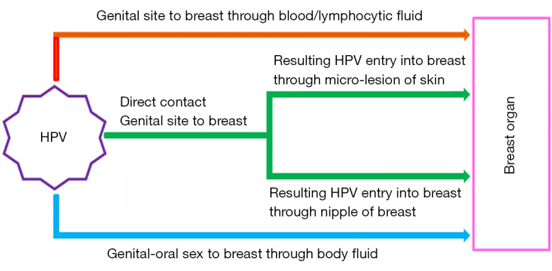
HPV infection can be transmitted through both sexual and nonsexual contacts. The genital HPV is mostly transmitted by direct skin-to-skin contact during sexual intercourse with an infected person (63). Generally, HPVs enter into the body through the skin and epidermal injuries, mucous membranes, skin abrasions and infects the cells of the basal layer of the stratified epithelium (64). The internalization of virions occurs slowly by endocytosis of clathrin coated vesicles in the presence of heparin sulphate. This ultimately leads to the transport of viral DNA to the nucleus and in the process disruption of the intracapsomeric disulphide bonds of the viral capsid occurs in the reducing environment of the cell (65-70). However, there can be three possible mode of HPV infection in breast tissue (Figure 2). According to the first one, HPV may be transmitted to breast from the genital region of the patients having a previous history of HPV-positive uterine cervical cancer (CACX) through blood, lymphatic systems or any other body fluid (71). It may be the case where a secondary malignant transformation of breast tissue could occur by an HPV infected malignant cell, which is derived from the primary tumour of any other site (72,73). It may also be due to spill over of HPV virion to the circulation system from HPV infected primary tumour site (74). As per the second mechanism, transmission of HPV can occur to breast from any oral site due to oral sexual practices (46). Third one suggests that the transmission of HPV may occur to breast by nipple or micro-lesion of breast skin due to genital-breast sexual activity (75,76).
Figure 2.

Representative diagram showing possible route of HPV transmission to breast tissue. There are mainly three possible mechanisms: (I) infected genital site to breast through blood/body fluid, (II) direct contact between genital and breast due abnormal sexual activity and (III) oral to breast due to oral sex activity.
Molecular profiles of HPV in BC
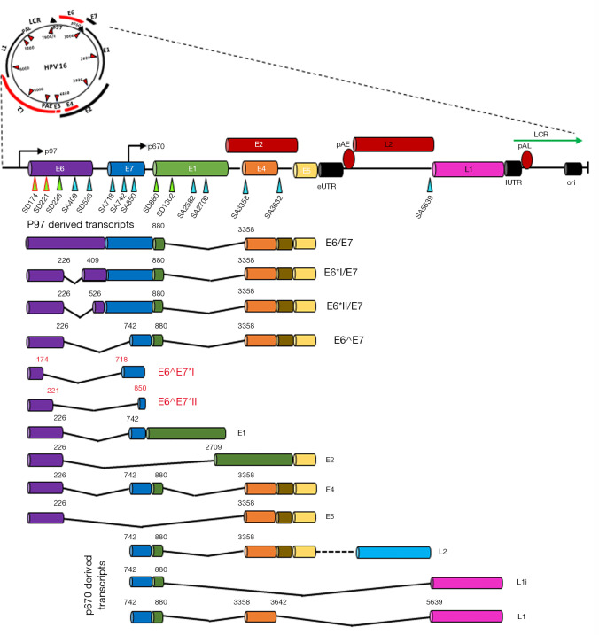
The persistent high-risk (hr) HPV infection are well known prerequisite factor for clinical progression and the development of Cervical intraepithelial neoplasia III (CIN III) and CACX (77-79). The persistent infections with hrHPVs have been identified as an essential but not sufficient factor in the pathogenesis of anogenital and other epithelial carcinomas (80). It was evident that sequential changes in the molecular profiles (genetic/epigenetic expression) of HPV occurred during development of tumour. Recent studies have shown that the majority (86–100%) of HPV genome present in breast tissue in an integrated form, an important step of HPV induced normal epithelial cell transformation as well as carcinogenesis (Table 3) (85). On the other hand, low copy number of HPV genome with range 0.00054–9.3 copies/cell in breast tumor was reported by different investigators including our group (Table 3). Based on sequence variation of the HPV genome, four naturally occurring lineages have been characterized like European-Asian (A), African-1(Af-1) (B) African-2(Af-2) (C) and Asian-American-North American (D) (86). Among these, American-North American (D) lineage was associated with the virulence property (87). Our previous sequence variation analysis of E6-E7 and LCR regions of HPV16 genome revealed that “A” lineage was frequent in BC (64.2%, 36/56) followed by D (33.9%) and B (1.78%) (38). Among these, frequent variants such as 7521 G > A at LCR and 350T > G at E6 regions indicated their importance in the process of carcinogenesis (88). HPV genome is functionally subdivided into three regions: early, late and the regulatory-long control region (LCR) or non-coding region (NCR), each are separated by two polyadenylation (pA) sites: early pA (pAE) and late pA (pAL) sites (Figure 3) (89). After HPV infection and capsid uncoating, P97 promoter derived early poly-cistronic mRNA transcript is responsible for production of early response proteins i.e., E1, E2, E4, E5, E6 and E7 by differential splicing (90). On the other hand, the poly-cistronic mRNA transcript from the late promoter P670 through differential splicing could produce E1, E2, E4, L1 and L2 proteins. Our previous study showed high methylation in p97 promoter (97%) and enhancer (51%) at LCR region of HPV16 genome, indicating the importance of this epigenetic modification in regulation of the viral genome expression (38) (Table 3).
Table 3. Molecular profiles of HPV in breast cancer.
| References | Molecular profiles | Description | |||
|---|---|---|---|---|---|
| Physical Status | Integrated (%) | Mix (%) | Episomal (%) | ||
| Khodabandehlou et al. 2019 (40) | 86 (30/35) | 14 (5/35) | -- | ||
| Khan et al. 2008 (18) | 96 (25/26) | -- | 4 (1/26) | ||
| Islam et al. 2017 (38) | 87.5 (105/120) | 8.3 (9/120) | 4.2 (5/120) | ||
| Aguayo et al. 2011 (81) | 100.0 (4/4) | -- | -- | ||
| Herrera-Goepfert et al. 2013 (82) | Viral Load | 0.20892 copies/cell | |||
| Lawson et al. 2016 (71,83) | 0.00054–0.0021 copies/cell | ||||
| Khan et al. 2008 (18) | 5.4 copies/cell | ||||
| Islam et al. 2017 (38) | 9.3 copies/50 ng gDNA | ||||
| Islam et al. 2017 (38) | Sequence variants | 70.8% (34/48) | |||
| Islam et al. 2017 (38) | Methylation status | P97 promoter: 96.7%, (30/31), Enhancer: 51.6%, (16/31) | |||
| Lawson et al. 2015 (35) | E6 expression (mRNA/protein) | 76% (16/21) | |||
| Islam et al. 2017 (38) | 53.3% (16/30) | ||||
| Suarez et al. 2013 (84) | 56.2 (9/16) | ||||
| Lawson et al. 2015 (35) | E7 expression (mRNA/protein) | 24% (5/21) | |||
| Islam et al. 2017 (38) | 53.3% (16/30) | ||||
| Suarez et al. 2013 (84) | 56.2 (9/16) | ||||
| Ngan et al. 2015 (36) | 62.5% (20/32) | ||||
Figure 3.
Schematic representation of molecular portrait of human papillomavirus 16 (HPV16) genome. The ~8 kb human papillomavirus genome may be found as an episomal or linear integrated form in the nucleus of the infected cell. The viral genome harbours two polyadenylation signals such as early polyadenylation signal (pAE) and late polyadenylation signal (pAL). The pAE signal terminates the transcription of early (E) genes such as E1–E7, whereas pAL signal terminate transcription of late (L) genes L1 and L2. The LCR of the genome contains the origin of DNA replication (ori) and the early viral promoter, p97 while the late promoter, p670, is located in the E7 coding region. eUTR and lUTR represent the early and late 3'UTR respectively. Known 5' splice donor site (SD) like SD226, SD880, SD1302 and SD3632 are shown as green circle with black border whereas 3' splice acceptors (SA) SA409, SA526, SA742, SA2582, SA2709, SA3358 and SA5639 are shown as blue circle with black border. Apart from these, two novel splice donor sites SD174 & SD221 and accepter sites SA718 & SA850 are depicted as green circle with red border and blue circle with red border respectively. Alternative splicing among these splice sites are produce two sets of mRNA transcripts from respective promoter p97 and p670. Red colour E6^E7*I & E6^E7*II represent the novel transcripts. Each transcript represents the most likely candidate mRNA for production of the corresponding proteins.
The expression of E6 and E7 oncogenes have their significant biological implications in HPV induced carcinogenesis. The E6/E7 transcripts were detected in 24–100% of BC samples by different researchers including our group (Table 3). Apart from the existing transcripts of E6/E7, two novel fusion transcripts of E6/E7 (E6^E7*I, E6^E7*II) in breast tumour were detected by us suggesting the underlying differences in molecular pathogenesis of HPV in BC compared to other cancers (Figure 3) (38). Going further, different investigators including our group detected the E6/E7 protein expression in 24–76% breast samples indicating functional relevance of HPV in breast tumour tissue (Table 3) (35). In addition, E6 and E7 expression was also evident in adjacent normal tissue, nipple tissue and epithelial layer of normal breast skin (8,38,71).
Molecular pathogenesis of HPV associated BC
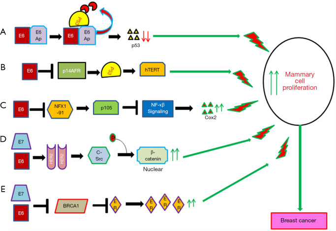
The molecular mechanism of HPV infection in promoting cervical cancer development and progression has been studied comprehensively (91). However, the exact mechanism by which HPV induces or promotes breast carcinogenesis is not well defined yet. It was evident that the E6 and E7 oncoproteins of HPV16 could immortalize human mammary epithelial cells through inactivation of p53 and RB respectively indicating their importance in cellular transformation (55,92). Different in-vitro studies showed association of E6/E7 with multiple cellular pathways in transformation of mammary epithelial cells (Figure 4) (5). Among these pathways, E6/E7 could down regulate P53, NFX1 and BRCA1 resulting up regulation of CoX2, NF-κβ and ER associated pathways (72,93-97). On the other hand, E6/E7 could stabilize HER2 receptor resulting in the activation of beta-catenin and thus enhance cellular proliferation (Figure 3) (55,98). Al Moustafa et al. observed co-over expression of E6/E7 and HER-2 in 40% of HPV16 positive BC (99). Ohba et al. showed association of the APOBEC3B pathway with the ER-positive breast tumors in presence of HPV (56). The association of E6 with these pathways in breast carcinogenesis has been validated in murine model systems (100).
Figure 4.
Schematic diagram represent the Putative mechanism of HPV in breast carcinogenesis. (A) Interaction of E6 with E6-AP leads to the degradation of p53 resulting in increased cellular proliferation eventually transforming into immortalized mammary epithelial cells (MEC). (B) E6 linked with hTERT can mediate immortalization of MEC through inactivation of p14ARF-p53 pathway (V) E6 could increase the mammary cell proliferation through up regulation of Cox2. This occurs due to E6 mediated degradation of NFX1 resulting in p105 down regulation and stabilizing NF-κβ which can now activate transcription of COX2. (D) E6/E7 interaction with HER2 results in its activation. HER2 in-turn activates c-Src which leads to the phoshorylation of beta-catenin at its C-terminal end as a result of which beta-catenin translocates to nucleus and activates different proliferation associated genes. (E) E6/E7 inhibits the function of BRCA1 resulting in restoration of expression of ER. High expression of ER leads to increased proliferation of mammary cell due to modulation of different proliferation associated genes.
Future management of HPV associated BC
In this review, it is evident that HPV is associated with a sub set of BC irrespective of different molecular subtypes. As HPV infects the breast through nipple and micro-lesions on the breast skin due to genetial-breast sex activity, hygienic sexual practice could prevent HPV infection to the breast. In conventional cervical cancer screening, cervical swab is used for HPV test followed by Pap test leading to early diagnosis of cervical cancer (101). Likewise, it is pertinent to detect HPV in breast ductal lavage, breast nipple discharge and breast milk which will be useful for determination of risk of BC as well as early diagnosis of BC. Apart from these, detection of HPV in breast tissue will be powerful biomarker for specific treatment protocol of the HPV infected BC. Moreover, the presence of HPV in blood plasma of BC patients can be the indicator of dissemination of tumour cells from the primary site which can serve as a useful prognostic tool of the disease. The prevalence of HPV in BC indicates that prophylactic vaccination against HPV is needed to restrict the disease in women (102).
Conclusions
In this review, we suggest that HPV is an important etiological factor in the development of a sub-set of BC and also HPV associated BC has some distinct molecular profile than other HPV associated cancers like cervical cancer (CACX), head and neck squamous cell carcinoma (HNSCC). Thus an in-depth understanding and analysis of the molecular profile of BC in the light of HPV is essentially needed for the proper management of the disease.
Supplementary
The article’s supplementary files as
Acknowledgments
The authors thank the Director, Chittaranjan National Cancer Institute, Kolkata, India for kind interest in the work. We would like to thank Mr. Aniban Roychowdhury for his language editing help and valuable suggestions.
Funding: The financial support for this work was provided by UGC-NET Fellowship grant Sr. No. 2121430433, Ref. No.: 21/12/2014(ii) EU-V dated 08.06.2015 to Mr. BC.
Ethical Statement: The authors are accountable for all aspects of the work in ensuring that questions related to the accuracy or integrity of any part of the work are appropriately investigated and resolved.
Footnotes
Conflict of Interest: All authors have completed the ICMJE uniform disclosure form (available at http://dx.doi.org/10.21037/atm-19-2756). The authors have no conflicts of interest to declare.
References
- 1.Bray F, Ferlay J, Soerjomataram I, et al. Global cancer statistics 2018: GLOBOCAN estimates of incidence and mortality worldwide for 36 cancers in 185 countries. CA Cancer J Clin 2018;68:394-424. 10.3322/caac.21492 [DOI] [PubMed] [Google Scholar]
- 2.Malvia S, Bagadi SA, Dubey US, et al. Epidemiology of breast cancer in Indian women. Asia Pac J Clin Oncol 2017;13:289-95. 10.1111/ajco.12661 [DOI] [PubMed] [Google Scholar]
- 3.Hankinson SE, Colditz GA, Willett WC. Towards an integrated model for breast cancer etiology: the lifelong interplay of genes, lifestyle, and hormones. Breast Cancer Res 2004;6:213-8. 10.1186/bcr921 [DOI] [PMC free article] [PubMed] [Google Scholar]
- 4.Alibek K, Kakpenova A, Mussabekova A, et al. Role of viruses in the development of breast cancer. Infect Agent Cancer 2013;8:32. 10.1186/1750-9378-8-32 [DOI] [PMC free article] [PubMed] [Google Scholar]
- 5.de Lima EG, do Amaral CM, Peixe FC, et al. Putative Mechanisms of Viral Transmission and Molecular Dysregulation of Mammary Epithelial Cells by Human Papillomavirus: Implications for Breast Cancer. Curr Mol Med 2016. [Epub ahead of print]. [PubMed] [Google Scholar]
- 6.Wang T, Chang P, Wang L, et al. The role of human papillomavirus infection in breast cancer. Med Oncol 2012;29:48-55. 10.1007/s12032-010-9812-9 [DOI] [PubMed] [Google Scholar]
- 7.Li J, Ding J, Zhai K. Detection of Human Papillomavirus DNA in Patients with Breast Tumor in China. PLoS One 2015;10:e0136050. 10.1371/journal.pone.0136050 [DOI] [PMC free article] [PubMed] [Google Scholar]
- 8.de Villiers EM, Sandstrom RE, zur Hausen H, et al. Presence of papillomavirus sequences in condylomatous lesions of the mamillae and in invasive carcinoma of the breast. Breast Cancer Res 2005;7:R1-11. 10.1186/bcr940 [DOI] [PMC free article] [PubMed] [Google Scholar]
- 9.Polyak K. Breast cancer: origins and evolution. J Clin Invest 2007;117:3155-63. 10.1172/JCI33295 [DOI] [PMC free article] [PubMed] [Google Scholar]
- 10.Ma H, Wang Y, Sullivan-Halley J, et al. Use of four biomarkers to evaluate the risk of breast cancer subtypes in the women's contraceptive and reproductive experiences study. Cancer Res 2010;70:575-87. 10.1158/0008-5472.CAN-09-3460 [DOI] [PMC free article] [PubMed] [Google Scholar]
- 11.Yu Y, Morimoto T, Sasa M, et al. HPV33 DNA in premalignant and malignant breast lesions in Chinese and Japanese populations. Anticancer Res 1999;19:5057-61. [PubMed] [Google Scholar]
- 12.Yu Y, Morimoto T, Sasa M, et al. Human papillomavirus type 33 DNA in breast cancer in Chinese. Breast Cancer 2000;7:33-6. 10.1007/BF02967185 [DOI] [PubMed] [Google Scholar]
- 13.Damin AP, Karam R, Zettler CG, et al. Evidence for an association of human papillomavirus and breast carcinomas. Breast Cancer Res Treat 2004;84:131-7. 10.1023/B:BREA.0000018411.89667.0d [DOI] [PubMed] [Google Scholar]
- 14.Gumus M, Yumuk PF, Salepci T, et al. HPV DNA frequency and subset analysis in human breast cancer patients' normal and tumoral tissue samples. J Exp Clin Cancer Res 2006;25:515-21. [PubMed] [Google Scholar]
- 15.Kroupis C, Markou A, Vourlidis N, et al. Presence of high-risk human papillomavirus sequences in breast cancer tissues and association with histopathological characteristics. Clin Biochem 2006;39:727-31. 10.1016/j.clinbiochem.2006.03.005 [DOI] [PubMed] [Google Scholar]
- 16.Choi YL, Cho EY, Kim JH, et al. Detection of human papillomavirus DNA by DNA chip in breast carcinomas of Korean women. Tumour Biol 2007;28:327-32. 10.1159/000124238 [DOI] [PubMed] [Google Scholar]
- 17.Tsai JH, Tsai CH, Cheng MH, et al. Association of viral factors with non-familial breast cancer in Taiwan by comparison with non-cancerous, fibroadenoma, and thyroid tumor tissues. J Med Virol 2005;75:276-81. 10.1002/jmv.20267 [DOI] [PubMed] [Google Scholar]
- 18.Khan NA, Castillo A, Koriyama C, et al. Human papillomavirus detected in female breast carcinomas in Japan. Br J Cancer 2008;99:408-14. 10.1038/sj.bjc.6604502 [DOI] [PMC free article] [PubMed] [Google Scholar]
- 19.de León DC, Montiel DP, Nemcova J, et al. Human papillomavirus (HPV) in breast tumors: prevalence in a group of Mexican patients. BMC Cancer 2009;9:26. 10.1186/1471-2407-9-26 [DOI] [PMC free article] [PubMed] [Google Scholar]
- 20.Heng B, Glenn WK, Ye Y, et al. Human papilloma virus is associated with breast cancer. Br J Cancer 2009;101:1345-50. 10.1038/sj.bjc.6605282 [DOI] [PMC free article] [PubMed] [Google Scholar]
- 21.He Q, Zhang SQ, Chu YL, et al. The correlations between HPV16 infection and expressions of c-erbB-2 and bcl-2 in breast carcinoma. Mol Biol Rep 2009;36:807-12. 10.1007/s11033-008-9249-9 [DOI] [PubMed] [Google Scholar]
- 22.Mendizabal-Ruiz AP, Morales JA, Ramirez-Jirano LJ, et al. Low frequency of human papillomavirus DNA in breast cancer tissue. Breast Cancer Res Treat 2009;114:189-94. 10.1007/s10549-008-9989-1 [DOI] [PubMed] [Google Scholar]
- 23.Herrera-Goepfert R, Khan NA, Koriyama C, et al. High-risk human papillomavirus in mammary gland carcinomas and non-neoplastic tissues of Mexican women: no evidence supporting a cause and effect relationship. Breast 2011;20:184-9. 10.1016/j.breast.2010.11.006 [DOI] [PubMed] [Google Scholar]
- 24.Mou X, Chen L, Liu F, et al. Low prevalence of human papillomavirus (HPV) in Chinese patients with breast cancer. J Int Med Res 2011;39:1636-44. 10.1177/147323001103900506 [DOI] [PubMed] [Google Scholar]
- 25.Frega A, Lorenzon L, Bononi M, et al. Evaluation of E6 and E7 mRNA expression in HPV DNA positive breast cancer. Eur J Gynaecol Oncol 2012;33:164-7. [PubMed] [Google Scholar]
- 26.Glenn WK, Heng B, Delprado W, et al. Epstein-Barr virus, human papillomavirus and mouse mammary tumour virus as multiple viruses in breast cancer. PLoS One 2012;7:e48788. 10.1371/journal.pone.0048788 [DOI] [PMC free article] [PubMed] [Google Scholar]
- 27.Sigaroodi A, Nadji SA, Naghshvar F, et al. Human papillomavirus is associated with breast cancer in the north part of Iran. ScientificWorldJournal 2012;2012:837191. 10.1100/2012/837191 [DOI] [PMC free article] [PubMed] [Google Scholar]
- 28.Liang W, Wang J, Wang C, et al. Detection of high-risk human papillomaviruses in fresh breast cancer samples using the hybrid capture 2 assay. J Med Virol 2013;85:2087-92. 10.1002/jmv.23703 [DOI] [PubMed] [Google Scholar]
- 29.Wang T, Zeng X, Li W, et al. Detection and analysis of human papillomavirus (HPV) DNA in breast cancer patients by an effective method of HPV capture. PLoS One 2014;9:e90343. 10.1371/journal.pone.0090343 [DOI] [PMC free article] [PubMed] [Google Scholar]
- 30.Ali SH, Al-Alwan NA, Al-Alwany SH. Detection and genotyping of human papillomavirus in breast cancer tissues from Iraqi patients. East Mediterr Health J 2014;20:372-7. 10.26719/2014.20.6.372 [DOI] [PubMed] [Google Scholar]
- 31.Ahangar-Oskouee M, Shahmahmoodi S, Jalilvand S, et al. No detection of 'high-risk' human papillomaviruses in a group of Iranian women with breast cancer. Asian Pac J Cancer Prev 2014;15:4061-5. 10.7314/APJCP.2014.15.9.4061 [DOI] [PubMed] [Google Scholar]
- 32.Manzouri L, Salehi R, Shariatpanahi S, et al. Prevalence of human papilloma virus among women with breast cancer since 2005-2009 in Isfahan. Adv Biomed Res 2014;3:75. 10.4103/2277-9175.125873 [DOI] [PMC free article] [PubMed] [Google Scholar]
- 33.Peng J, Wang T, Zhu H, et al. Multiplex PCR/mass spectrometry screening of biological carcinogenic agents in human mammary tumors. J Clin Virol 2014;61:255-9. 10.1016/j.jcv.2014.07.010 [DOI] [PubMed] [Google Scholar]
- 34.Fu L, Wang D, Shah W, et al. Association of human papillomavirus type 58 with breast cancer in Shaanxi province of China. J Med Virol 2015;87:1034-40. 10.1002/jmv.24142 [DOI] [PubMed] [Google Scholar]
- 35.Lawson JS, Glenn WK, Salyakina D, et al. Human Papilloma Viruses and Breast Cancer. Front Oncol 2015;5:277. 10.3389/fonc.2015.00277 [DOI] [PMC free article] [PubMed] [Google Scholar]
- 36.Ngan C, Lawson JS, Clay R, et al. Early Human Papilloma Virus (HPV) Oncogenic Influences in Breast Cancer. Breast Cancer (Auckl) 2015;9:93-7. 10.4137/BCBCR.S35692 [DOI] [PMC free article] [PubMed] [Google Scholar]
- 37.Salman NA, Davies G, Majidy F, et al. Association of High Risk Human Papillomavirus and Breast cancer: A UK based Study. Sci Rep 2017;7:43591. 10.1038/srep43591 [DOI] [PMC free article] [PubMed] [Google Scholar]
- 38.Islam S, Dasgupta H, Roychowdhury A, et al. Study of association and molecular analysis of human papillomavirus in breast cancer of Indian patients: Clinical and prognostic implication. PLoS One 2017;12:e0172760. 10.1371/journal.pone.0172760 [DOI] [PMC free article] [PubMed] [Google Scholar]
- 39.Delgado-García S, Martinez-Escoriza JC, Alba A, et al. Presence of human papillomavirus DNA in breast cancer: a Spanish case-control study. BMC Cancer 2017;17:320. 10.1186/s12885-017-3308-3 [DOI] [PMC free article] [PubMed] [Google Scholar]
- 40.Khodabandehlou N, Mostafaei S, Etemadi A, et al. Human papilloma virus and breast cancer: the role of inflammation and viral expressed proteins. BMC Cancer 2019;19:61. 10.1186/s12885-019-5286-0 [DOI] [PMC free article] [PubMed] [Google Scholar]
- 41.Wrede D, Luqmani YA, Coombes RC, et al. Absence of HPV 16 and 18 DNA in breast cancer. Br J Cancer 1992;65:891-4. 10.1038/bjc.1992.186 [DOI] [PMC free article] [PubMed] [Google Scholar]
- 42.Bratthauer GL, Tavassoli FA, O'Leary TJ. Etiology of breast carcinoma: no apparent role for papillomavirus types 6/11/16/18. Pathol Res Pract 1992;188:384-6. 10.1016/S0344-0338(11)81229-X [DOI] [PubMed] [Google Scholar]
- 43.Gopalkrishna V, Singh UR, Sodhani P, et al. Absence of human papillomavirus DNA in breast cancer as revealed by polymerase chain reaction. Breast Cancer Res Treat 1996;39:197-202. 10.1007/BF01806186 [DOI] [PubMed] [Google Scholar]
- 44.Lindel K, Forster A, Altermatt HJ, et al. Breast cancer and human papillomavirus (HPV) infection: no evidence of a viral etiology in a group of Swiss women. Breast 2007;16:172-7. 10.1016/j.breast.2006.09.001 [DOI] [PubMed] [Google Scholar]
- 45.de Cremoux P, Thioux M, Lebigot I, et al. No evidence of human papillomavirus DNA sequences in invasive breast carcinoma. Breast Cancer Res Treat 2008;109:55-8. 10.1007/s10549-007-9626-4 [DOI] [PubMed] [Google Scholar]
- 46.Chang P, Wang T, Yao Q, et al. Absence of human papillomavirus in patients with breast cancer in north-west China. Med Oncol 2012;29:521-5. 10.1007/s12032-011-9945-5 [DOI] [PubMed] [Google Scholar]
- 47.Lüder Ripoli F, Mohr A, Conradine Hammer S, et al. A Comparison of Fresh Frozen vs. Formalin-Fixed, Paraffin-Embedded Specimens of Canine Mammary Tumors via Branched-DNA Assay. Int J Mol Sci 2016;17:724. 10.3390/ijms17050724 [DOI] [PMC free article] [PubMed] [Google Scholar]
- 48.Cavalcante JR, Pinheiro LGP, Almeida PRC, et al. Association of breast cancer with human papillomavirus (HPV) infection in Northeast Brazil: molecular evidence. Clinics (Sao Paulo) 2018;73:e465. 10.6061/clinics/2018/e465 [DOI] [PMC free article] [PubMed] [Google Scholar]
- 49.Ngamkham J, Karalak A, Chaiwerawattana A, et al. Prevalence of Human Papillomavirus Infection in Breast Cancer Cells from Thai Women. Asian Pac J Cancer Prev 2017;18:1839-45. [DOI] [PMC free article] [PubMed] [Google Scholar]
- 50.Corbex M, Bouzbid S, Traverse-Glehen A, et al. Prevalence of papillomaviruses, polyomaviruses, and herpesviruses in triple-negative and inflammatory breast tumors from algeria compared with other types of breast cancer tumors. PLoS One 2014;9:e114559. 10.1371/journal.pone.0114559 [DOI] [PMC free article] [PubMed] [Google Scholar]
- 51.Piana AF, Sotgiu G, Muroni MR, et al. HPV infection and triple-negative breast cancers: an Italian case-control study. Virol J 2014;11:190. 10.1186/s12985-014-0190-3 [DOI] [PMC free article] [PubMed] [Google Scholar]
- 52.Vernet-Tomas M, Mena M, Alemany L, et al. Human papillomavirus and breast cancer: no evidence of association in a Spanish set of cases. Anticancer Res 2015;35:851-6. [PubMed] [Google Scholar]
- 53.Fernandes A, Bianchi G, Feltri AP, et al. Presence of human papillomavirus in breast cancer and its association with prognostic factors. Ecancermedicalscience 2015;9:548. 10.3332/ecancer.2015.548 [DOI] [PMC free article] [PubMed] [Google Scholar]
- 54.Habyarimana T, Attaleb M, Mazarati JB, et al. Detection of human papillomavirus DNA in tumors from Rwandese breast cancer patients. Breast Cancer 2018;25:127-33. 10.1007/s12282-018-0831-2 [DOI] [PubMed] [Google Scholar]
- 55.Woods Ignatoski KM, Dziubinski ML, Ammerman C, et al. Cooperative interactions of HER-2 and HPV-16 oncoproteins in the malignant transformation of human mammary epithelial cells. Neoplasia 2005;7:788-98. 10.1593/neo.05106 [DOI] [PMC free article] [PubMed] [Google Scholar]
- 56.Ohba K, Ichiyama K, Yajima M, et al. In vivo and in vitro studies suggest a possible involvement of HPV infection in the early stage of breast carcinogenesis via APOBEC3B induction. PLoS One 2014;9:e97787. 10.1371/journal.pone.0097787 [DOI] [PMC free article] [PubMed] [Google Scholar]
- 57.Bae JM, Kim EH. Human papillomavirus infection and risk of breast cancer: a meta-analysis of case-control studies. Infect Agent Cancer 2016;11:14. 10.1186/s13027-016-0058-9 [DOI] [PMC free article] [PubMed] [Google Scholar]
- 58.Carolis S, Pellegrini A, Santini D, et al. Liquid biopsy in the diagnosis of HPV DNA in breast lesions. Future Microbiol 2018;13:187-94. 10.2217/fmb-2017-0145 [DOI] [PubMed] [Google Scholar]
- 59.Balci FL, Uras C, Feldman SM. Is human papillomavirus associated with breast cancer or papilloma presenting with pathologic nipple discharge? Cancer Treat Res Commun 2019;19:100122. 10.1016/j.ctarc.2019.100122 [DOI] [PubMed] [Google Scholar]
- 60.Louvanto K, Sarkola M, Rintala M, et al. Breast Milk Is a Potential Vehicle for Human Papillomavirus Transmission to Oral Mucosa of the Spouse. Pediatr Infect Dis J 2017;36:627-30. 10.1097/INF.0000000000001546 [DOI] [PubMed] [Google Scholar]
- 61.Tuominen H, Rautava S, Collado MC, et al. HPV infection and bacterial microbiota in breast milk and infant oral mucosa. PLoS One 2018;13:e0207016. 10.1371/journal.pone.0207016 [DOI] [PMC free article] [PubMed] [Google Scholar]
- 62.Diaz S, Boulle N, Moles JP, et al. Human papillomavirus (HPV) shedding in breast milk from African women living with HIV. J Clin Virol 2018;106:41-3. 10.1016/j.jcv.2018.07.004 [DOI] [PubMed] [Google Scholar]
- 63.Stevens-Simon C, Nelligan D, Breese P, et al. The prevalence of genital human papillomavirus infections in abused and nonabused preadolescent girls. Pediatrics 2000;106:645-9. 10.1542/peds.106.4.645 [DOI] [PubMed] [Google Scholar]
- 64.Beutner KR, Wiley DJ, Douglas JM, et al. Genital warts and their treatment. Clin Infect Dis 1999;28 Suppl 1:S37-56. 10.1086/514722 [DOI] [PubMed] [Google Scholar]
- 65.Giroglou T, Florin L, Schafer F, et al. Human papillomavirus infection requires cell surface heparan sulfate. J Virol 2001;75:1565-70 10.1128/JVI.75.3.1565-1570.2001 [DOI] [PMC free article] [PubMed] [Google Scholar]
- 66.Joyce JG, Tung JS, Przysiecki CT, et al. The L1 major capsid protein of human papillomavirus type 11 recombinant virus-like particles interacts with heparin and cell-surface glycosaminoglycans on human keratinocytes. J Biol Chem 1999;274:5810-22. 10.1074/jbc.274.9.5810 [DOI] [PubMed] [Google Scholar]
- 67.Culp TD, Christensen ND. Kinetics of in vitro adsorption and entry of papillomavirus virions. Virology 2004;319:152-61. 10.1016/j.virol.2003.11.004 [DOI] [PubMed] [Google Scholar]
- 68.Day PM, Lowy DR, Schiller JT. Papillomaviruses infect cells via a clathrin-dependent pathway. Virology 2003;307:1-11. 10.1016/S0042-6822(02)00143-5 [DOI] [PubMed] [Google Scholar]
- 69.Selinka HC, Giroglou T, Sapp M. Analysis of the infectious entry pathway of human papillomavirus type 33 pseudovirions. Virology 2002;299:279-87. 10.1006/viro.2001.1493 [DOI] [PubMed] [Google Scholar]
- 70.Li M, Beard P, Estes PA, et al. Intercapsomeric disulfide bonds in papillomavirus assembly and disassembly. J Virol 1998;72:2160-7. 10.1128/JVI.72.3.2160-2167.1998 [DOI] [PMC free article] [PubMed] [Google Scholar]
- 71.Lawson JS, Glenn WK, Salyakina D, et al. Human Papilloma Virus Identification in Breast Cancer Patients with Previous Cervical Neoplasia. Front Oncol 2016;5:298. 10.3389/fonc.2015.00298 [DOI] [PMC free article] [PubMed] [Google Scholar]
- 72.Widschwendter A, Brunhuber T, Wiedemair A, et al. Detection of human papillomavirus DNA in breast cancer of patients with cervical cancer history. J Clin Virol 2004;31:292-7. 10.1016/j.jcv.2004.06.009 [DOI] [PubMed] [Google Scholar]
- 73.Hennig EM, Suo Z, Thoresen S, et al. Human papillomavirus 16 in breast cancer of women treated for high grade cervical intraepithelial neoplasia (CIN III). Breast Cancer Res Treat 1999;53:121-35. 10.1023/A:1006162609420 [DOI] [PubMed] [Google Scholar]
- 74.Bodaghi S, Wood LV, Roby G, et al. Could human papillomaviruses be spread through blood? J Clin Microbiol 2005;43:5428-34. 10.1128/JCM.43.11.5428-5434.2005 [DOI] [PMC free article] [PubMed] [Google Scholar]
- 75.Islam S, Dasgupta H, Basu M, et al. Skin mediates Human Papilloma Virus (HPV) infection in breast: A report of four cases. Available online: https://www.researchgate.net/publication/324008020_Skin_mediated_human_papillomavirus_infection_in_breast_A_report_of_four_cases
- 76.Breast cancer may be sexually transmitted. 2006. Available online: www.abc.net.au/science/news/stories/2006/1808903.htm. Accessed 12 December.
- 77.Wallin KL, Wiklund F, Angstrom T, et al. Type-specific persistence of human papillomavirus DNA before the development of invasive cervical cancer. N Engl J Med 1999;341:1633-8. 10.1056/NEJM199911253412201 [DOI] [PubMed] [Google Scholar]
- 78.Zielinski GD, Snijders PJ, Rozendaal L, et al. HPV presence precedes abnormal cytology in women developing cervical cancer and signals false negative smears. Br J Cancer 2001;85:398-404. 10.1054/bjoc.2001.1926 [DOI] [PMC free article] [PubMed] [Google Scholar]
- 79.zur Hausen H. Papillomavirus infections--a major cause of human cancers. Biochim Biophys Acta 1996;1288:F55-78. [DOI] [PubMed] [Google Scholar]
- 80.zur Hausen H. Papillomaviruses and cancer: from basic studies to clinical application. Nat Rev Cancer 2002;2:342-50. 10.1038/nrc798 [DOI] [PubMed] [Google Scholar]
- 81.Aguayo F, Khan N, Koriyama C, et al. Human papillomavirus and Epstein-Barr virus infections in breast cancer from chile. Infect Agent Cancer 2011;6:7. 10.1186/1750-9378-6-7 [DOI] [PMC free article] [PubMed] [Google Scholar]
- 82.Herrera-Goepfert R, Vela-Chavez T, Carrillo-Garcia A, et al. High-risk human papillomavirus (HPV) DNA sequences in metaplastic breast carcinomas of Mexican women. BMC Cancer 2013;13:445. 10.1186/1471-2407-13-445 [DOI] [PMC free article] [PubMed] [Google Scholar]
- 83.Lawson JS, Glenn WK, Whitaker NJ. Human Papilloma Viruses and Breast Cancer - Assessment of Causality. Front Oncol 2016;6:207. 10.3389/fonc.2016.00207 [DOI] [PMC free article] [PubMed] [Google Scholar]
- 84.Pereira Suarez AL, Lorenzetti MA, Gonzalez Lucano R, et al. Presence of human papilloma virus in a series of breast carcinoma from Argentina. PLoS One 2013;8:e61613. 10.1371/journal.pone.0061613 [DOI] [PMC free article] [PubMed] [Google Scholar]
- 85.McBride AA, Warburton A. The role of integration in oncogenic progression of HPV-associated cancers. PLoS Pathog 2017;13:e1006211. 10.1371/journal.ppat.1006211 [DOI] [PMC free article] [PubMed] [Google Scholar]
- 86.Burk RD, Harari A, Chen Z. Human papillomavirus genome variants. Virology 2013;445:232-43. 10.1016/j.virol.2013.07.018 [DOI] [PMC free article] [PubMed] [Google Scholar]
- 87.Mirabello L, Yeager M, Cullen M, et al. HPV16 Sublineage Associations With Histology-Specific Cancer Risk Using HPV Whole-Genome Sequences in 3200 Women. J Natl Cancer Inst 2016;108:djw100. 10.1093/jnci/djw100 [DOI] [PMC free article] [PubMed] [Google Scholar]
- 88.DeFilippis VR, Ayala FJ, Villarreal LP. Evidence of diversifying selection in human papillomavirus type 16 E6 but not E7 oncogenes. J Mol Evol 2002;55:491-9. 10.1007/s00239-002-2344-y [DOI] [PubMed] [Google Scholar]
- 89.Doorbar J, Egawa N, Griffin H, et al. Human papillomavirus molecular biology and disease association. Rev Med Virol 2015;25 Suppl 1:2-23. 10.1002/rmv.1822 [DOI] [PMC free article] [PubMed] [Google Scholar]
- 90.Johansson C, Schwartz S. Regulation of human papillomavirus gene expression by splicing and polyadenylation. Nat Rev Microbiol 2013;11:239-51. 10.1038/nrmicro2984 [DOI] [PubMed] [Google Scholar]
- 91.Balasubramaniam SD, Balakrishnan V, Oon CE, et al. Key Molecular Events in Cervical Cancer Development. Medicina (Kaunas) 2019;55:384. 10.3390/medicina55070384 [DOI] [PMC free article] [PubMed] [Google Scholar]
- 92.Wazer DE, Liu XL, Chu Q, et al. Immortalization of distinct human mammary epithelial cell types by human papilloma virus 16 E6 or E7. Proc Natl Acad Sci U S A 1995;92:3687-91. 10.1073/pnas.92.9.3687 [DOI] [PMC free article] [PubMed] [Google Scholar]
- 93.Liu Y, Chen JJ, Gao Q, et al. Multiple functions of human papillomavirus type 16 E6 contribute to the immortalization of mammary epithelial cells. J Virol 1999;73:7297-307. 10.1128/JVI.73.9.7297-7307.1999 [DOI] [PMC free article] [PubMed] [Google Scholar]
- 94.Wang YX, Zhang ZY, Wang JQ, et al. HPV16 E7 increases COX-2 expression and promotes the proliferation of breast cancer. Oncol Lett 2018;16:317-25. [DOI] [PMC free article] [PubMed] [Google Scholar]
- 95.Zhang Y, Fan S, Meng Q, et al. BRCA1 interaction with human papillomavirus oncoproteins. J Biol Chem 2005;280:33165-77. 10.1074/jbc.M505124200 [DOI] [PubMed] [Google Scholar]
- 96.Rosen EM, Fan S, Isaacs C. BRCA1 in hormonal carcinogenesis: basic and clinical research. Endocr Relat Cancer 2005;12:533-48. 10.1677/erc.1.00972 [DOI] [PubMed] [Google Scholar]
- 97.Hilakivi-Clarke L. Estrogens, BRCA1, and breast cancer. Cancer Res 2000;60:4993-5001. [PubMed] [Google Scholar]
- 98.Yasmeen A, Bismar TA, Kandouz M, et al. E6/E7 of HPV type 16 promotes cell invasion and metastasis of human breast cancer cells. Cell Cycle 2007;6:2038-42. 10.4161/cc.6.16.4555 [DOI] [PubMed] [Google Scholar]
- 99.Al Moustafa AE, Kassab A, Darnel A, et al. High-risk HPV/ErbB-2 interaction on E-cadherin/catenin regulation in human carcinogenesis. Curr Pharm Des 2008;14:2159-72. 10.2174/138161208785740216 [DOI] [PubMed] [Google Scholar]
- 100.Shai A, Pitot HC, Lambert PF. p53 Loss synergizes with estrogen and papillomaviral oncogenes to induce cervical and breast cancers. Cancer Res 2008;68:2622-31. 10.1158/0008-5472.CAN-07-5266 [DOI] [PMC free article] [PubMed] [Google Scholar]
- 101.Koliopoulos G, Nyaga VN, Santesso N, et al. Cytology versus HPV testing for cervical cancer screening in the general population. Cochrane Database Syst Rev 2017;8:CD008587. [DOI] [PMC free article] [PubMed] [Google Scholar]
- 102.Purdie J. Can Human Papillomavirus (HPV) Cause Breast Cancer? healthline. 2018. Available online: https://www.healthline.com/health/breast-cancer/breast-cancer-and-hpv. Accessed December 14 2018.
Associated Data
This section collects any data citations, data availability statements, or supplementary materials included in this article.
Supplementary Materials
The article’s supplementary files as


