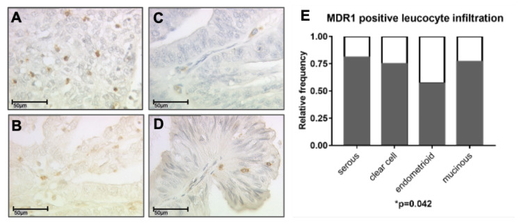
Figure 2.
A MDR1+ leucocyte infiltrate was detected by immunohistochemistry in all subtypes: serous (A), clear cell (B), endometrioid (C) and mucinous carcinoma (D). (A–D) are shown in 40× magnification (scale bar = 50 µm), 25× magnification is provided in the supplementary Figure S4. The highest relative frequency of cases with MDR1+ leucocyte infiltration was found for serous histology (E, p = 0.042) followed by mucinous, clear cell and endometrioid.

