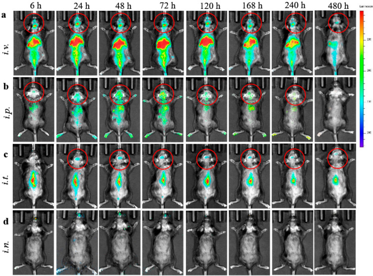
Figure 1.
Biodistribution of 1,1’-Dioctadecyl-3,3,3’,3’-tetramethylindo-carbocyanine iodide (DIR)- extracellular vesicles (EVs) in CLN2 KO mice by IVIS. DIR-labeled EVs were administered in BD mice (1 month old, N = 4), though: (a) i.v. (6 × 10¹¹ particles/200 µL), (b) i.p. (6 × 10¹¹ particles/200 µL), (c) i.t. (1.5 × 10¹¹ particles/50 µL), or (d) i.n. (6 × 10¹⁰ particles/20 µL) routes, and imaged up to 480 h. Prone representative images show prolonged DIR signal accumulation in the brain for i.v., i.p., and i.t. administration routs, especially at 24–120 h. Little, if any, DIR signal was observed in live animals after i.n. administration (d). Accumulation of labeled EVs was also observed in the main peripheral organs for i.v. and i.p. injections.

