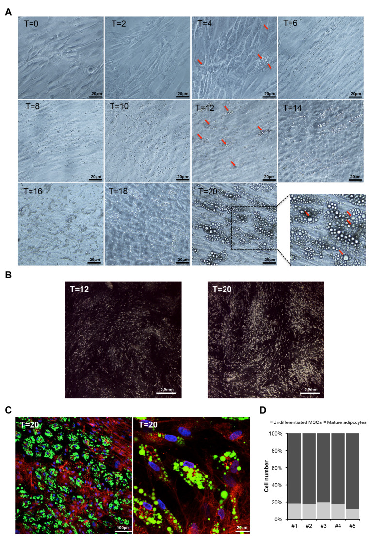
Figure 2.
Human mesenchymal stem cells (hMSCs) are a reliable in vitro model of human adipocyte differentiation. (A) Representative phase-contrast images showing morphological changes of hMSCs along adipocyte differentiation, i.e., at starting point (T = 0 h), at different time points upon adipogenesis induction (T = 2, 4, 6, 8, 10, 12, 14, 16, 18 days) and at terminal differentiation (T = 20 days). Red arrows indicate some lipid droplets visible by optical microscopy during adipocyte differentiation (scale bar, 20 µm). (B) Representative images of hMSCs at 12 days and 20 days upon induction fixed with osmium tetraoxide and observed in dark field microscopy. The LDs are showed as white dots (scale bar, 1 mm). (C) Representative confocal microscopy images of hMSCs differentiated in mature adipocytes (T = 20 days): nuclei in blue (DAPI), lipid droplets in green (Bodipy 495/503) and cell membranes in red (WGA 632/647). Clusters of “bunch of grapes”–like droplets are evident (scale bar, 100 µm left panel; scale bar, 20 µm right panel). (D) Bar graph indicating the percentage of undifferentiated and differentiated cells in five independent experiments measured analyzing confocal images of hMSCs at terminal adipogenic differentiation (T = 20 days). Total number of cells was calculated counting nuclei stained with DAPI, differentiated cells were identified by positive staining of lipid droplets (Bodipy 495/503), and the number of undifferentiated hMSCs was calculated as the difference between total and differentiated cells. A total of ~6000 cells were analyzed.

