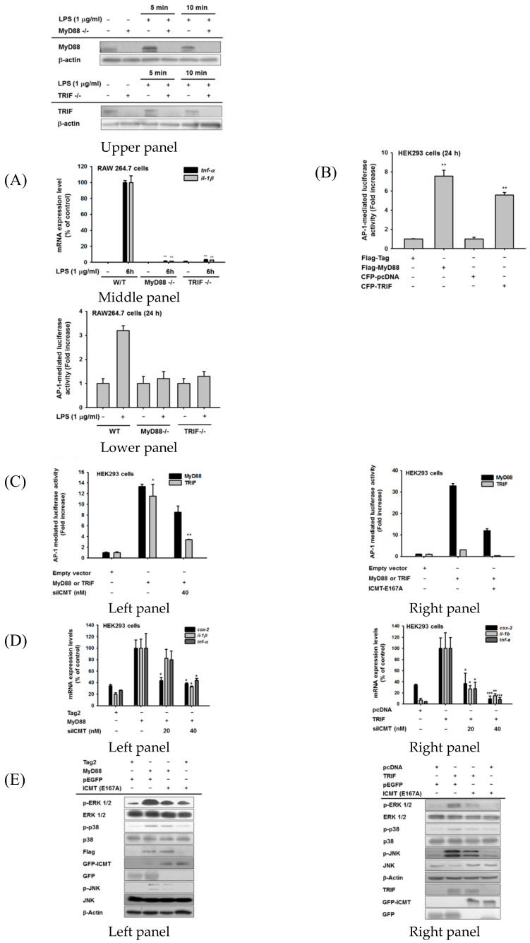
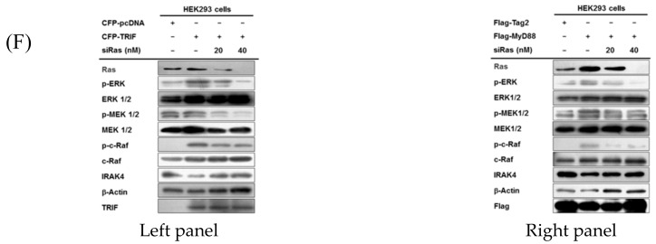
Figure 5.
ICMT/Ras expression is both MyD88- and TRIF-dependent. ((A), upper panel) Protein levels of MyD88 and TRIF in RAW264.7 cells under their knockout conditions in the presence or absence of LPS (1 µg/mL). (A, middle panel) Inhibitory effects of MyD88 and TRIF knockout on the expression of inflammatory genes (TNF-α and IL-1β) in RAW264.7 cells stimulated with LPS (1 µg/mL). ((A), lower panel, (B,D)) AP-1-mediated luciferase activity was analyzed by reporter gene assay in MyD88- and TRIF-knockout RAW264.7 cells ((A), lower panel), HEK293 cells transfected with Flag-MyD88 or CFP-TRIF (B), or HEK293 cells transfected with Flag-MyD88 or CFP-TRIF in the presence or absence of siICMT or ICMT-E167A ((C), left and right panels). Luminescence levels were determined with a luminometer. (D) mRNA expression levels of COX-2, TNF-α, and IL-1β were determined using real-time PCR in HEK293 cells transfected with MyD88 ((D), left panel) or TRIF ((D), right panel). (E) Total and phospho-protein levels of MAPK in HEK293 cells during transfection with MyD88, TRIF, and the dominant negative form of ICMT (ICMT-E167A). (F) Total and phospho-protein levels of ERK1/2 and their upstream enzymes (MEK1/2, c-Raf, and IRAK4) in HEK293 cells transfected with MyD88 or TRIF in the presence or absence of siRas were analyzed using immunoblotting analysis.


