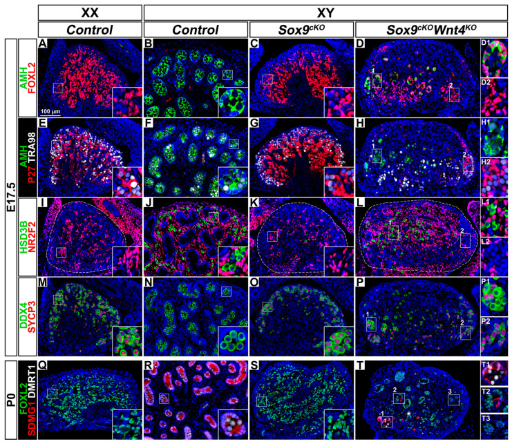
Figure 6.
Ovotestis development in XY Sox9; Wnt4 double mutant gonads at E17.5 and P0. (A–D) Immunodetection of the granulosa differentiation and Sertoli cell marker AMH (green) and the pre-granulosa cell marker FOXL2 (red) in E17.5 gonads of XX Control (A), XY Control (B), XY Sox9cKO (C), and XY Sox9cKO Wnt4KO (D) genotypes. (E–H) Immunodetection of AMH (green), the quiescent cell marker P27 (red) and the germ cell marker TRA98 (white) in E17.5 gonads of XX Control (E), XY Control (F), XY Sox9cKO (G), and XY Sox9cKO Wnt4KO (H) genotypes. (I–L) Immunodetection of the steroidogenic cell marker HSD3ß (green) and the interstitial and stromal cell marker NR2F2 (red) in E17.5 gonads of XX Control (I), XY Control (J), XY Sox9cKO (K), and XY Sox9cKO Wnt4KO (L) genotypes. (M–P) Immunodetection of the germ cell marker DDX4 (green) and the meiosis marker SYCP3 (red) in E17.5 gonads of XX Control (M), XY Control (N), XY Sox9cKO (O), and XY Sox9cKO Wnt4KO (P) genotypes. (Q–T) Immunodetection of the pre-granulosa cell marker FOXL2 (green), the Sertoli cell marker SDMG1 (red) and the Sertoli cell and male germ cell marker DMRT1 (white) in newborn gonads of XX Control (Q), XY Control (R), XY Sox9cKO (S), and XY Sox9cKO Wnt4KO (T) genotypes. Nuclei labeled with DAPI are shown in blue. Magnification is the same in all panels. Scale bar = 100 μm. Regions marked by numbers in panels D, H, L, P, and T are shown in the corresponding insets. Gonads are outlined with broken white lines in I, K, and L.

