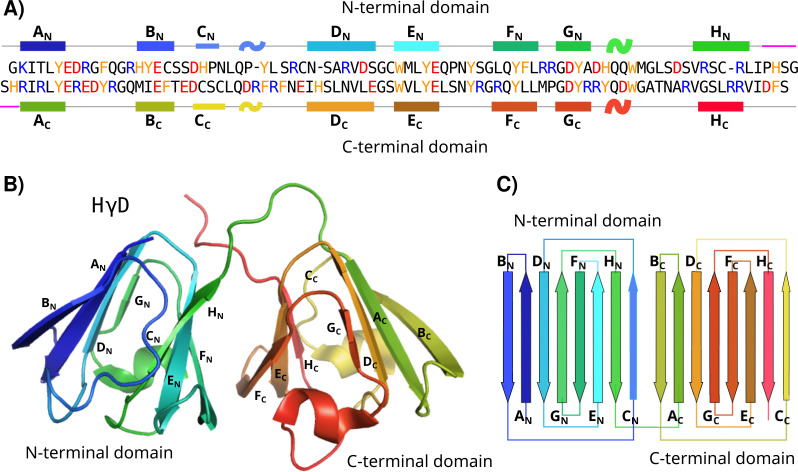
Figure 1. HγD structure.
(A) Sequence of HγD crystallin showing the N-terminal (top) and the C-terminal (bottom) domains. Positive-charged residues are shown in blue, negative-charged residues in red, and aromatic residues in yellow. Secondary structure elements are shown. Each strand is named from A to H for each domain and the linker region is shown in purple. (B) Three-dimensional structure of HγD crystallin labeled by strands (PDB id 1HK0). (C) Topology of the N-terminal and C-terminal domain describing the four Greek motifs.

