Abstract
Right-sided aortic arch with aberrant left subclavian artery is a rare variant of vascular anatomy. Three types of right-sided aortic arches are described and classified based on the arrangement of the aortic arch vessels, the presence or absence of congenital heart abnormalities, the relationship of the aortic arch to the trachea and esophagus, as well as the presence or absence of a complete or incomplete vascular ring. On review of the existing literature, 31 case reports were found with a spectrum of clinical presentation sand outcomes. In this case report, we highlight a case of a young female in her early 20’swho presented with choking spells, shortness of breath along with intermittent dysphagia since childhood. She was otherwise healthy and pregnant at 26 weeks gestational age. A Computed tomography scan with angiography (CTA) of the thorax was performed to rule out a pulmonary embolism (PE) however surprisingly, a right-sided aortic arch with aberrant left subclavian artery was revealed instead. Subsequently, an extensive literature review was carried out to better understand clinical presentation sand treatment strategies for this rather rare disorder.
Keywords: right-sided aortic arch with aberrant left subclavian artery, right aortic arch, congenital abnormalities, aberrant left subclavian artery, vascular ring
1. Introduction
Vascular rings can present with non-specific respiratory and or esophageal symptoms, most commonly identified in children. Several reports has also documented symptomatic vascular rings in adults. This case report will discuss aortic arch anomalies and will emphasize the necessity of maintaining a broad differential when facing shortness of breath. Right-sided aortic arch is a rare anatomical variant present in about 0.1% of the adult population [1,2,3]. It is usually asymptomatic and diagnosed incidentally in adulthood. A right-sided aortic arch is an anatomic variant resulting from persistence of the right fourth embryologic aortic arch and involution of the left aortic arch. Normal anatomy occurs when the left aortic arch persists and the right involutes. Normally, the left subclavian artery (LSA) arises directly from the aortic arch; however, an aberrant left subclavian artery arises from the diverticulum of Kommerell, a vascular structure derived from a remnant of the fourth aortic arch. When the diverticulum of Kommerell compresses the adjacent structures, apatient may become symptomatic. To date, approximately 31 cases from the year of 2011 of right-sided aortic arch and aberrant LSA have been published in the literature [4]. The most common symptoms in this group were dysphagia, dyspnea, and cough [4]. No case reports involving pregnant females were identified by our literature review.
2. Materials and Methods
An electronic search was performed using the PUBMED, Medline, and Cochrane Library databases at the library of the University Hospital of Brooklyn. Five sets of search terms were used to ensure an adequate and comprehensive literature review. These included search queries ‘right-sided aortic arch’, ‘right-sided aorta’, ‘aberrant left subclavian artery’, ‘vascular ring.’
3. Case Report
This is a case of a 23-year-oldpregnant female at 26 weeks gestational age who presented with shortness of breath. The patient reported brief, self-resolving episodes of shortness of breath and chest tightness every other day for the past 2-3 weeks. The symptoms were worse when laying supine. She remembered having similar episodes when she was a child between the ages of 9 to 12 and several more episodes throughout her adult life. She also reported symptoms of heartburn and mild intermittent dysphagia to solidsthat have persisted since childhood. The symptoms progressively worsened with the progression of the patient’s pregnancy. The patient reported a family history of hypertension and diabetes in her mother and heart disease in her father. She denied any history of smoking, alcohol intake or illicit substance use. She reported that she lives with her parents and doesn’t communicate with the father of her child. She had poor follow up with her obstetrician and missed multiple appointments. Her only medication were Ranitidine 150 mg once daily for occasional dysphagia and prenatal vitamins.
Physical examination revealed a blood pressure of 120/75 mm Hg with a pulse rate of 75. Neck exam revealed no signs of jugular venous distension (JVD). Pulmonary and cardiovascular exam were unremarkable. Laboratory work-up was unremarkable as well.
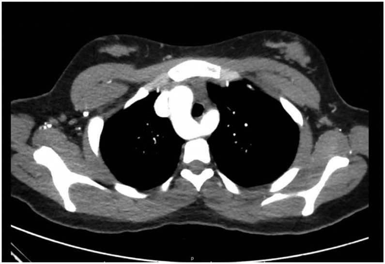
A Computed tomography with angiography (CTA) exam ruled out pulmonary embolism (PE). On close observation, a right aortic arch with aberrant left subclavian artery was incidentally discovered [Figure 1]. There was mild right-sided tracheal compression by Kommerell's diverticulum.
Figure 1.

Right-sided aortic arch (RAA) with aberrant left subclavian artery with Kommerell's diverticulum (KD).
Over the next few days the patient’s symptoms improved, and she was discharged with close follow up with vascular surgery, gastroenterology and pulmonary clinics. Given these findings, the patient was instructed to avoid exertion, follow up with gastroenterology for a barium contrast esophagogram to detect any esophageal compression, and follow up with vascular surgery for a possible surgical intervention after the delivery. Unfortunately following this admission, the patient was lost to follow-up.
4. Discussion
A review of the literature published between 2011 to 2019, revealed that thirty-one adult cases of right-sided aortic arch with aberrant left subclavian artery have been reported [Table 1]. Of these, 11patients presented with dysphagia, 6 patients were asymptomatic, 2 patients presented with an aortic dissection, 2 with aneurysms, 1 with respiratory symptoms, 1 with cerebrovascular insufficiency, 1 with left upper extremity pain and numbness on exercise, 1 with left cerebral infarction, 1 with a right nonrecurrent laryngeal nerve, 1 with acute superior vena cava syndrome, 1 with pseudo-occlusion of the left internal carotid artery, 1 with subclavian steal syndrome, and 2 with rupture. The patients’ ages ranged from 23 to 80 years.
Table 1.
Patient presentation in the studies reported in the systematic review
| Case number |
Year author | Reported/observed symptom/diagnostic finding |
|---|---|---|
| 1 | 2019, Arakoni R [5] | Dysphagia |
| 2 | 2019, Morosetti D [6] | Asymptomatic, incidental finding |
| 3 | 2016, Masuoka H [7] | Right nonrecurrent laryngeal nerve |
| 4 | 2019, Raymond S [8] | Dysphagia |
| 5 | 2019, Morishita A [9] | Dysphagia |
| 6 | 2019, Zhao C [10] | Right-sidedaortic arch aneurysm |
| 7 | 2018, Tanaka Y [11] | Aneurysm arising fromKomerell’s diverticulum |
| 8 | 2018, Tempaku [12] | Left cerebral infarction |
| 9 | 2017, Hamady M [13] | Mild dysphagia, right-sided aneurysmal aortic arch with aneurysmal aberrant left-sided |
| 10 | 2017, Wilinski J [14] | Asymptomatic |
| 11 | 2016, Bhatt T [15] | Dysphagia, chest pain |
| 12 | 2017, Powell BL [16] | Dysphagia with occasional regurgitation |
| 13 | 2016, Ahmed MM [17] | Subclavian steal syndrome |
| 14 | 2016, Lococo F [18] | Asymptomatic, incidental finding |
| 15 | 2015, Parikh P [19] | Dysphagia |
| 16 | 2015, Ohtani T [20] | Pseudo-occlusion of the left internal carotid artery |
| 17 | 2015, Stefanczyk L [21] | Symptoms of cerebrovascular insufficiency |
| 18 | 2015, Sierra-Galan LM [22] | Chronic cough |
| 19 | 2014, Inui T [23] | Asymptomatic, incidental finding |
| 20 | 2014, Lee CH [24] | Asymptomatic, incidental finding |
| 21 | 2014, Zhang M [25] | Stanford B type dissection |
| 22 | 2015, Batheeb NA [26] | Left upper limb pain and numbness on exercise |
| 23 | 2014, Khalid S [27] | Dysphagia to solids |
| 24 | 2013, Samas J [28] | Dysphagia |
| 25 | 2013, Ebner L [29] | Dissection of the ascending aorta associated with hemopericardium |
| 26 | 2013, Motoki M [30] | Ruptured aberrant left subclavian artery |
| 27 | 2012, Yamashiro S [31] | Ruptured Kommerell’s diverticulum |
| 28 | 2013, Suarez AE [32] | Acute superior vena cava syndrome |
| 29 | 2012, Margolis J [33] | Dysphagia, voice changes |
| 30 | 2011, Panduranga P [34] | Dysphagia |
| 31 | 2011, Mubarak MY [3] | Asymptomatic, incidental finding |
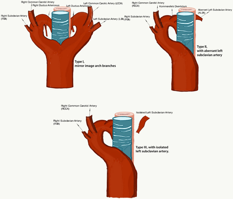
Right-sided aortic arch was first documented by Fioratti and Aglietti in 1763 [35]. In the adult population a right-sided aortic arch is often asymptomatic unless an eurysmal disease develops. The mortality associated with rupture, the morbidity caused by compression of mediastinal structures, and the complexity of surgery makes this condition clinically relevant [36]. A right aortic arch crosses the right mainstem bronchus and descends along the right side of the spine. The right-sided aortic arch is classified into three types [37,38,39]. Type I is categorized by aright-sided aortic arch with mirror image branching meaning the major arteries branching out from the arch are the left in nominate artery, followed by the right common carotid and right subclavian arteries. This type is usually associated with cyanotic congenital heart disease like Tetralogy of Fallot and truncus arteriosus [2,37,38]. Type IIis defined by a right-sided aortic arch with aberrant left subclavian artery as discussed in this case. It is associated with Kommerell’s diverticulum [39] and is rarely associated with other congenital heart diseases. Type IIIis classified by a right sided aortic arch with isolation of the left subclavian artery. In type III the left subclavian artery is connected to the pulmonary artery through the ductus arteriosus and it may be associated with subclavian steal syndrome and vertebrobasilar inefficiency. Type IIaccounts for approximately 40 % of all right-sided arches [39]. The most common is Type I [37,39].
Symptomatic formstypically present in childhood and are repaired then. In adults the symptoms may arise due to early atherosclerotic changes [40]. Dilatation of Kommerell’s diverticulum, resulting in compression of the surrounding structures, may cause shortness of breath, choking spells and dysphagia, as seen in our patient [41,42]. Other fearsome complications include aortic dissection that may present with chest pain [40]. The aberrant subclavian artery may be located posterior to the esophagus (most commonly), between the esophagus and trachea, or anterior to the trachea and it may cause compressive symptoms [1,3].
Pre-surgical evaluation of aortic arch anatomy is important for planning thoracic surgery and endovascular interventions. The presence of arch variants may influence the surgical approach taken. These arch anomalies can create complications in trans-radial coronary procedures, thoracic surgeries, and thyroid surgeries due to an abnormal recurrent laryngeal nerve course [44,45].
In pregnancy, the hemodynamic changes include increased cardiac output and expanded blood volume. Supine positioning reduces stroke volume and cardiac output and increases the heart rate due to compression of the aorta and vena cava from the enlarging uterus [46,47]. This enlargement of the uterus and compression of the vascular structures may have led to worsening of the symptoms in our patient.
5. Conclusion
We presented a rare case of Right-sided Aortic Arch with aberrant left subclavian artery in a pregnant female with review of the literature indicating only 31 cases reported between the years of 2011-2019in adults presenting with symptoms of a vascular ring. None of those cases involved a pregnant female. This case report and literature review highlights the importance of awareness of this diagnostic possibility among physicians, in order to broaden their differential diagnoses to include congenital anomalies, especially how subtle the findings on imaging may be. Diagnosed patients may benefit from surveillance or interventional therapy, from the prevention of misguided acute or long-term management, and avoidance of intraoperative surgical complications.
Figure 2.
Type I: right-sided aortic arch with mirror image aortic branches; Type II: right-sided aortic arch with aberrant left subclavian artery; Type III: right-sided aortic arch with isolated left subclavian artery.
Acknowledgements
This work is supported, in part, by the efforts of Dr. Moro O. Salifu M.D., M.P.H., M.B.A., M.A.C.P., Professor and Chairman of Medicine through NIH Grant number S21MD012474.
References
- [1].Cina CS, Althani H, Pasenau J, Abouzahr L. Kommerell's diverticulum and right-sided aortic arch: a cohort study and review of the literature. J Vasc Surg. 2004; 39(1): 131–139. [DOI] [PubMed] [Google Scholar]
- [2].Hara M, Kitase M, Satake M, et al. A case of right-sided aortic arch with isolation of the left subclavian artery: CT findings. Radiat Med. 2001; 19(1): 33–36. [PubMed] [Google Scholar]
- [3].Mubarak MY, Kamarul AT, Noordini MD. Right-sided Aortic Arch with Aberrant Left Subclavian Artery from Kommerell's Diverticulum. Iran J Radiol. 2011; 8(2): 103–106. [PMC free article] [PubMed] [Google Scholar]
- [4].Yang MH, Weng ZC, Weng YG, Chang HH. A right-sided aortic arch with Kommerell's diverticulum of the aberrant left subclavian artery presenting with syncope. J Chin Med Assoc. 2009; 72(5): 275–277. [DOI] [PubMed] [Google Scholar]
- [5].Arakoni R, Merrill R, Simon EL. Foreign body sensation: A rare case of dysphagia lusoria in a healthy female. Am J Emerg Med. 2018; 36(11): 2134 e2131–2134 e2132. [DOI] [PubMed] [Google Scholar]
- [6].Morosetti D, Di Stefano C, Mondillo M, et al. Right-sided aortic arch with mirror image branching and situs solitus: a case of a 79 years old woman. Radiol Case Rep. 2019; 14(10): 1246–1251. [DOI] [PMC free article] [PubMed] [Google Scholar]
- [7].Masuoka H, Miyauchi A, Higashiyama T, Yabuta T, Kihara M, Miya A. Right-sided aortic arch and aberrant left subclavian artery with or without a left nonrecurrent inferior laryngeal nerve. Head Neck. 2016; 38(10): E2508–2511. [DOI] [PMC free article] [PubMed] [Google Scholar]
- [8].Raymond SL, Gray SE, Peters KR, Fatima J. Right-sided aortic arch with aberrant left subclavian artery and Kommerell diverticulum. J Vasc Surg Cases Innov Tech. 2019; 5(3): 259–260. [DOI] [PMC free article] [PubMed] [Google Scholar]
- [9].Morishita A, Tomioka H, Katahira S, Hoshino T, Hanzawa K. Surgical Treatment for Kommerell's Diverticulum Associated with a Right-Sided Aortic Arch and an Aberrant Left Subclavian Artery: Endovascular or Hybrid. Ann Vasc Dis. 2019; 12(2): 228–232. [DOI] [PMC free article] [PubMed] [Google Scholar]
- [10].Zhao C, Sun Y, Wang Y, et al. Bicuspid aortic valve with right-sided aortic arch aneurysm and Kommerell's diverticulum. J Clin Ultrasound. 2019. [DOI] [PubMed] [Google Scholar]
- [11].Tanaka Y, Kitamura T, Irisawa Y, Miyaji K. Total arch replacement using the Gelweave Lupiae graft for an aneurysm arising from Kommerell's diverticulum with an aberrant left subclavian artery and right-sided aortic arch. Interact Cardiovasc Thorac Surg. 2018; 27(2): 307–309. [DOI] [PubMed] [Google Scholar]
- [12].Tempaku A, Kuroiwa T, Nishio A. Aberrant left subclavian artery occlusion in right-sided aortic artery associated with left cerebral infarction: A case report. Interv Neuroradiol. 2018; 24(3): 322–326. [DOI] [PMC free article] [PubMed] [Google Scholar]
- [13].Hamady M, Sharma PM, Patel R, Godfrey AD, Bicknell CD. Hybrid endovascular repair of aneurysmal right-sided aortic arch and Kommerell's diverticulum using a two-vessel branched stent graft: Case report and review of literature. SAGE Open Med Case Rep. 2017; 5: 2050313X17749082. [DOI] [PMC free article] [PubMed] [Google Scholar]
- [14].Wilinski J, Skwarek A, Kasprzyk A, Wilinski B, Kameczura T, Glowacki MD. Right-sided aortic arch with aberrant left subclavian artery and hearing loss in patient with abnormal cardiomediastinal contours in routine chest X-ray. Przegl Lek. 2017; 74(3): 125–128. [PubMed] [Google Scholar]
- [15].Bhatt TC, Muralidharan CG, Singh G, Jain NK. Kommerell's diverticulum: A rare aortic arch anomaly. Med J Armed Forces India. 2016; 72(Suppl 1): S80–S83. [DOI] [PMC free article] [PubMed] [Google Scholar]
- [16].Powell BL. Vascular ring presenting as dysphagia in an adult woman: a case report. Ann R Coll Surg Engl. 2017; 99(1): e3–e5. [DOI] [PMC free article] [PubMed] [Google Scholar]
- [17].Ahmed MM, Yoon DY, Desai SS, Pearce WH. Right-sided aortic arch with isolated left subclavian artery leading to subclavian steal syndrome. J Vasc Surg. 2016; 64(1): 233–234. [DOI] [PubMed] [Google Scholar]
- [18].Lococo F, Tusini N, Brandi L, et al. Successful Conservative Treatment of a Kommerell Aneurysm Associated With Right-Sided Aortic Arch. Vasc Endovascular Surg. 2016; 50(6): 458–459. [DOI] [PubMed] [Google Scholar]
- [19].Parikh P, Nasa M, Patil S, Patel J, Bhate P, Sawant P. A Rare Cause of Dysphagia--Kommerell's Diverticulum. J Assoc Physicians India. 2015; 63(1): 59–62. [PubMed] [Google Scholar]
- [20].Ohtani T, Yamazaki T, Ohtaki H, et al. Carotid Artery Stenting in Right-sided Aortic Arch: A Case Report. NMC Case Rep J. 2016; 3(1): 9–12. [DOI] [PMC free article] [PubMed] [Google Scholar]
- [21].Stefanczyk L, Szymczyk K, Stefanczyk K, Polguj M. The Presence of a Right Aortic Arch Associated with Severe Stenosis of the Right Common Carotid Artery and Steal Phenomenon. Ann Vasc Surg. 2015; 29(8): 1656 e1613–1657. [DOI] [PubMed] [Google Scholar]
- [22].Sierra-Galan LM, Shveid-Gerson D, Gomez-Garza G, Rey-Rodriguez A. Double incomplete aortic arch and Kommerell's Diverticulum as a cause of chronic cough. Arch Cardiol Mex. 2015; 85(2): 158–160. [DOI] [PubMed] [Google Scholar]
- [23].Inui T, Hashimoto H, Fujimoto K, et al. [Percutaneous transluminal angioplasty using a stent for an aberrant left subclavian artery stenosis with a right-sided aortic arch: a case report]. No Shinkei Geka. 2014; 42(10): 937–942. [DOI] [PubMed] [Google Scholar]
- [24].Lee CH, Son JW, Park JS. Comprehensive three-dimensional analysis of right-sided aortic arch with multiple vascular anomalies. BMC Cardiovasc Disord. 2014; 14:104. [DOI] [PMC free article] [PubMed] [Google Scholar]
- [25].Zhang M, Yuan Y, Hu Y, et al. Endovascular repair with the chimney technique for stanford type B aortic dissection involving right-sided arch with aberrant left subclavian artery. Ann Vasc Surg. 2014;28(7): 1798 e1797–1798 e1710. [DOI] [PubMed] [Google Scholar]
- [26].Batheeb NA, Habbab LM, Qattan NM. Symptomatic stenosis of left subclavian artery from Kommerell's diverticulum. Asian Cardiovasc Thorac Ann. 2015; 23(9): 1068–1071. [DOI] [PubMed] [Google Scholar]
- [27].Khalid S, Narayansamy S, Siddiqui S, Mohd K. Middle-aged woman with dysphagia to solid. Diagnosis: Right-sided aortic arch with an aberrant left subclavian artery. Saudi J Gastroenterol. 2014; 20(1): 76–77. [DOI] [PMC free article] [PubMed] [Google Scholar]
- [28].Samas J, Manetta F, Meyer DB. Repair of vascular ring with resection of kommerell diverticulum and transposition of aberrant left subclavian artery. Int J Angiol. 2013; 22(4): 243–244. [DOI] [PMC free article] [PubMed] [Google Scholar]
- [29].Ebner L, Huber A, Christe A. Right aortic arch and Kommerell's diverticulum associated with acute aortic dissection and pericardial tamponade. Acta Radiol Short Rep. 2013; 2(1): 2047981613476283. [DOI] [PMC free article] [PubMed] [Google Scholar]
- [30].Motoki M, Hattori K, Kato Y, et al. Endovascular repair of ruptured aberrant left subclavian artery with right aortic arch. Ann Thorac Surg. 2013; 95(2): 699–701. [DOI] [PubMed] [Google Scholar]
- [31].Yamashiro S, Nagano T, Kuniyoshi Y, Kise Y, Maeda T, Arakaki R. Endovascular repair of intrathoracic ruptured Kommerell's diverticulum. Asian Cardiovasc Thorac Ann. 2012; 20(5): 587–590. [DOI] [PubMed] [Google Scholar]
- [32].Suarez AE, Slivka R. Acute superior vena cava syndrome after rupture of Kommerell's diverticulum: a case report. Ann Emerg Med. 2013; 61(3): 348–350. [DOI] [PubMed] [Google Scholar]
- [33].Margolis J, Bilfinger T, Labropoulos N. A right-sided aortic arch and aberrant left subclavian artery with proximal segment hypoplasia. Interact Cardiovasc Thorac Surg. 2012; 14(3): 370–371. [DOI] [PMC free article] [PubMed] [Google Scholar]
- [34].Panduranga P, Al-Delamie T, Ratnam L, Al-Mukhaini M, Zachariah S. Repair of Kommerell's diverticulum with aberrant left subclavian artery in an elderly patient with right aortic arch and dysphagia lusoria. J Card Surg. 2011; 26(6): 637–640. [DOI] [PubMed] [Google Scholar]
- [35].Hastreiter AR, D'Cruz IA, Cantez T, Namin EP, Licata R. Right-sided aorta. I. Occurrence of right aortic arch in various types of congenital heart disease. II. Right aortic arch, right descending aorta, and associated anomalies. Br Heart J. 1966; 28(6): 722–739. [DOI] [PMC free article] [PubMed] [Google Scholar]
- [36].Karthekeyan BR, Sundar S, Rao S, Vakamudi M. Management of A Patient with Kommerrell's Aneurysm Causing Tracheal and Esophageal Compression. Indian J Anaesth. 2009; 53(3): 358–361. [PMC free article] [PubMed] [Google Scholar]
- [37].Turkvatan A, Buyukbayraktar FG, Olcer T, Cumhur T. Congenital anomalies of the aortic arch: evaluation with the use of multidetector computed tomography. Korean J Radiol. 2009; 10(2): 176–184. [DOI] [PMC free article] [PubMed] [Google Scholar]
- [38].Craatz S, Kunzel E, Spanel-Borowski K. Right-sided aortic arch and tetralogy of Fallot in humans--a morphological study of 10 cases. Cardiovasc Pathol. 2003; 12(4): 226–232. [DOI] [PubMed] [Google Scholar]
- [39].Chai OH, Han EH, Kim HT, Song CH. Right-sided aortic arch with the retroesophageal left subclavian artery as the fourth branch. Anat Cell Biol 2013; 46(2): 167–170. [DOI] [PMC free article] [PubMed] [Google Scholar]
- [40].Ota T, Okada K, Takanashi S, Yamamoto S, Okita Y. Surgical treatment for Kommerell's diverticulum. J Thorac Cardiovasc Surg. 2006; 131(3): 574–578. [DOI] [PubMed] [Google Scholar]
- [41].Polguj M, Chrzanowski L, Kasprzak JD, Stefanczyk L, Topol M, Majos A. The aberrant right subclavian artery (arteria lusoria): the morphological and clinical aspects of one of the most important variations--a systematic study of 141 reports. Scientific World Journal 2014; 2014: 292734. [DOI] [PMC free article] [PubMed] [Google Scholar]
- [42].Patiniotis TC, Mohajeri M, Hill DG. Right aortic arch with aberrant left subclavian artery: aneurysmal dilatation causing symptomatic compression of the right main bronchus in an adult. Aust N Z J Surg. 1995; 65(9): 690–692. [DOI] [PubMed] [Google Scholar]
- [43].Arazinska A, Polguj M, Szymczyk K, Kaczmarska M, Trebinski L, Stefanczyk L. Right aortic arch analysis - Anatomical variant or serious vascular defect? BMC Cardiovasc Disord. 2017; 17(1): 102. [DOI] [PMC free article] [PubMed] [Google Scholar]
- [44].Jakanani GC, Adair W. Frequency of variations in aortic arch anatomy depicted on multidetector CT. Clin Radiol. 2010; 65(6): 481–487. [DOI] [PubMed] [Google Scholar]
- [45].Faggioli GL, Ferri M, Freyrie A, et al. Aortic arch anomalies are associated with increased risk of neurological events in carotid stent procedures. Eur J Vasc Endovasc Surg. 2007; 33(4): 436–441. [DOI] [PubMed] [Google Scholar]
- [46].Meah VL, Cockcroft JR, Backx K, Shave R, Stohr EJ. Cardiac output and related haemodynamics during pregnancy: a series of meta-analyses. Heart. 2016; 102(7): 518–526. [DOI] [PubMed] [Google Scholar]
- [47].Metcalfe J, Ueland K. Maternal cardiovascular adjustments to pregnancy. Prog Cardiovasc Dis. 1974; 16(4): 363–374. [DOI] [PubMed] [Google Scholar]


