This nonrandomized controlled trial analyzes safety, biological, and functional outcomes associated with the infusion of rAAVrh74.MHCK7.micro-dystrophin gene transfer in a small group of patients with Duchenne muscular dystrophy.
Key Points
Question
Is rAAVrh74.MHCK7.micro-dystrophin gene transfer safe and well tolerated in patients with Duchenne muscular dystrophy?
Findings
In this nonrandomized controlled trial of 4 young patients with Duchenne muscular dystrophy, rAAVrh74.MHCK7.micro-dystrophin gene transfer was well tolerated, with minimal adverse events, and was associated with robust micro-dystrophin expression, reduced serum creatine kinase levels, and functional improvement as measured by the North Star Ambulatory Assessment.
Meaning
These results indicated the safe systemic delivery of micro-dystrophin transgene and targeted expression of functional micro-dystrophin protein product, suggesting the potential for rAAVrh74.MHCK7.micro-dystrophin to provide clinically meaningful functional improvement that is greater than the standard of care.
Abstract
Importance
Micro-dystrophin gene transfer shows promise for treating patients with Duchenne muscular dystrophy (DMD) using recombinant adeno-associated virus serotype rh74 (rAAVrh74) and codon-optimized human micro-dystrophin driven by a skeletal and cardiac muscle-specific promoter with enhanced cardiac expression (MHCK7).
Objective
To identify the 1-year safety and tolerability of intravenous rAAVrh74.MHCK7.micro-dystrophin in patients with DMD.
Design, Setting, and Participants
This open-label, phase 1/2a nonrandomized controlled trial was conducted at the Nationwide Children’s Hospital in Columbus, Ohio. It began on November 2, 2017, with a planned duration of follow-up of 3 years, ending in March 2021. The first 4 patients who met eligibility criteria were enrolled, consisting of ambulatory male children with DMD without preexisting AAVrh74 antibodies and a stable corticosteroid dose (≥12 weeks).
Interventions
A single dose of 2.0 × 10¹⁴ vg/kg rAAVrh74.MHCK7.micro-dystrophin was infused through a peripheral limb vein. Daily prednisolone, 1 mg/kg, started 1 day before gene delivery (30-day taper after infusion).
Main Outcomes and Measures
Safety was the primary outcome. Secondary outcomes included micro-dystrophin expression by Western blot and immunohistochemistry. Functional outcomes measured by North Star Ambulatory Assessment (NSAA) and serum creatine kinase were exploratory outcomes.
Results
Four patients were included (mean [SD] age at enrollment, 4.8 [1.0] years). All adverse events (n = 53) were considered mild (33 [62%]) or moderate (20 [38%]), and no serious adverse events occurred. Eighteen adverse events were considered treatment related, the most common of which was vomiting (9 of 18 events [50%]). Three patients had transiently elevated γ-glutamyltransferase, which resolved with corticosteroids. At 12 weeks, immunohistochemistry of gastrocnemius muscle biopsy specimens revealed robust transgene expression in all patients, with a mean of 81.2% of muscle fibers expressing micro-dystrophin with a mean intensity of 96% at the sarcolemma. Western blot showed a mean expression of 74.3% without fat or fibrosis adjustment and 95.8% with adjustment. All patients had confirmed vector transduction and showed functional improvement of NSAA scores and reduced creatine kinase levels (posttreatment vs baseline) that were maintained for 1 year.
Conclusions and Relevance
This trial showed rAAVrh74.MHCK7.micro-dystrophin to be well tolerated and have minimal adverse events; the safe delivery of micro-dystrophin transgene; the robust expression and correct localization of micro-dystrophin protein; and improvements in creatine kinase levels and NSAA scores. These findings suggest that rAAVrh74.MHCK7.micro-dystrophin can provide functional improvement that is greater than that observed under standard of care.
Trial Registration
ClinicalTrials.gov Identifier: NCT03375164
Introduction
Duchenne muscular dystrophy (DMD) is a rare, X-linked, fatal, degenerative neuromuscular disease caused by dystrophin gene (DMD) mutations. Estimated incidence worldwide is 1 in 5000 live male births.1,2 The DMD gene (OMIM 300377) encodes for dystrophin, a 427-kDa cytoskeletal protein required for sarcolemmal stability. Protein loss leads to susceptibility to repeated cycles of necrosis and regeneration as well as diminished regenerative muscle capacity, resulting in fat and connective tissue replacement (fibrosis).1 DMD is progressive, beginning with loss of ambulation between age 9 and 14 years, followed by respiratory complications and cardiac function decline, and ending in death.3,4,5
As standard of care options have changed, disease progression has improved.6 Corticosteroids have been reported to reduce inflammation7 and to delay the loss of ambulation (by approximately 3 years) and the decline of respiratory function.8 However, long-term corticosteroid use is associated with serious adverse effects, including bone fracture, infection, and gastrointestinal bleeding.7,8
Disease-modifying therapies, such as exon skipping, have been shown to produce functional dystrophin protein and slow the decline in ambulatory and pulmonary function after long-term use.9,10,11,12 However, dystrophin production is limited to patients amenable to select exon skipping. Novel therapies with a broader reach are still needed.
Gene transfer therapy has emerged as the most promising molecular approach for neuromuscular diseases. For spinal muscular atrophy (SMA), delivery of the SMN gene (SMN1 AC005031) to infants with SMA type 1 using adeno-associated virus (AAV) serotype 9 (onasemnogene abeparvovec; Zolgensma) has been shown to dramatically prolong life and improve function.13,14 In the original trial, 12 infants with disease onset before age 6 months who were treated with high-dose onasemnogene abeparvovec (2.0 × 10¹⁴ vg/kg) survived beyond 2 years, far exceeding the 8% predicted survival.13 The infants continue to gain strength, achieve long-term survival, and develop new motor milestones, demonstrating the medication’s safety beyond the trial.14,15
A translational DMD gene transfer trial is designed to achieve clinical efficacy without compromising safety. However, the large size of the DMD gene (2.4 MB) limits its ability to be packaged into AAV vector systems (with a capacity of approximately 4.7 kB). Seminal studies in the 1990s demonstrated the ability to deliver truncated forms of dystrophin to improve muscular function in murine models.16,17 The most notable evidence of shortened dystrophin protein retaining function was the case of a 61-year-old ambulatory patient with Becker muscular dystrophy who harbored a large (approximately 46%) deletion mutation of exons 17 to 48; this case report highlighted the critical domains essential for muscle function.18
We designed a micro-dystrophin transgene (AAVrh74.MHCK7.micro-dystrophin) that would enhance functional efficacy on delivery. This micro-dystrophin transgene contains the N-terminus for binding to f-actin; spectrin repeats 1 to 3 and 24; hinges 1, 2, and 4; and the cysteine-rich domain. Spectrin repeats 1 to 3 bind directly to the sarcolemma,19 providing enhanced resistance to eccentric contraction-induced injury.20,21 Spectrin repeat 24 is critical for microtubule binding, and the cysteine-rich domain is essential for binding to β-dystroglycan, leading to the restoration of dystrophin-associated protein complex.22
For clinical translation, we placed micro-dystrophin under the control of an MHCK7 promoter. The MHCK7 promoter is associated with high levels of expression in skeletal muscles, including the diaphragm, and includes an enhancer to especially drive expression in the heart, whereas expression in off-target tissues is minimal.23
Careful selection of the AAV capsid is also important to ensure transduction efficiency and tissue targeting while minimizing immune response. Selection of AAV serotypes that demonstrate muscle (skeletal and cardiac) tissue tropism is critical. The AAVrh74 (AAV serotype rh74) used in preclinical mouse and nonhuman primate experiments was isolated from rhesus monkeys on the basis of high tropism for skeletal and cardiac muscle as well as no adverse events.24 Being of nonhuman primate origin, it is hoped that AAVrh74 would decrease the likelihood of patients having preexisting immunity to the vector. To date, AAVrh74 as a delivery vehicle has been found to be safe and tolerable in human studies, with minimal immune response.25
Recently, we completed proof-of-principle, preclinical dosing experiments in the mdx mouse. In preclinical studies, systemic delivery of AAVrh74.MHCK7.micro-dystrophin at doses (2.0 × 10¹⁴ vg/kg) comparable to those shown to be safe and effective for SMA type 113 demonstrated widespread dystrophin expression to the diaphragm, skeletal, and cardiac muscles.26 These preclinical study findings were the impetus for bringing AAVrh74.MHCK7.micro-dystrophin forward in this pilot nonrandomized controlled trial of a young cohort of boys with DMD. Our intent was to identify the potential for muscle fiber rescue before fiber loss and high degree of endomysial connective tissue replacement. We hypothesized that robust gene expression and correct localization to the sarcolemma could protect muscle fibers from progressive decline typical of DMD.
Methods
Study Design and Participants
This phase 1/2a nonrandomized controlled trial tested the safety and biological efficacy of a single systemic infusion of rAAVrh74.MHCK7.micro-dystrophin in patients with DMD. The trial was approved by the institutional review board of Nationwide Children’s Hospital. All parents of minor participants signed informed consent in compliance with Code of Federal Regulations Title 21 Part 50 and the International Conference on Harmonization guidelines.27 Detailed inclusion and exclusion criteria are reported in the trial protocol (Supplement 1). We followed the Transparent Reporting of Evaluations With Nonrandomized Designs (TREND) reporting guideline.
This single-site trial was conducted at the Nationwide Children’s Hospital in Columbus, Ohio, and began on November 2, 2017. The duration of follow-up is 3 years, and the final date for follow-up is planned for March 2021.
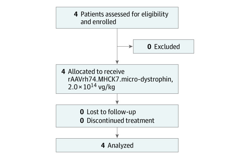
Patients were excluded if rAAVrh74 (recombinant adeno-associated virus serotype rh74) binding antibody was detected at a dilution greater than 1:400 at enrollment (lower limit of detection 1:25). Eligible patients were ambulatory boys (n = 4) aged 4 to 7 years with confirmed DMD gene mutations with frameshift (deletion or duplication) or premature stop codon mutation between exons 18 and 58 (exons not encoded by the micro-dystrophin transgene to limit immune responses to the transgene)28; creatine kinase (CK) level elevation of more than 1000 U/L (to convert unit per liter to microkat per liter, multiply by 0.0167); and below-average 100-m timed test29 (Figure 1). Eligible patients were required to be on a stable dose of corticosteroids for at least 12 weeks before entry. All patients were on a weekend dose30 of prednisone sodium (n = 1) or prednisolone sodium phosphate (n = 3). After gene transfer, they received daily prednisolone, 1 mg/kg, for 5 days in between the weekend dose for 30 days tapered over 2 to 4 weeks, depending on serum γ-glutamyltransferase (GGT) level after gene delivery (eTable 1 in Supplement 2).
Figure 1. Study Flow Diagram.
Other components of the chemistry panel serve as biomarkers for liver toxic effects, including the aminotransaminases, especially alanine aminotransferase, but distinguishing contributions from muscle vs liver can be difficult. In this trial, we also monitored bilirubin (direct and total), alkaline phosphatase, and albumin levels (eTable 2 in Supplement 2). Additional biomarkers might also be included in future trials, including glutamate dehydrogenase and ammonia, and depending on severity, ultrasonography and magnetic resonance imaging could be used to more clearly identify pathological changes in the liver.
Vector Production and Dosing Procedures
The cassette containing the MHCK7 promoter and micro-dystrophin transgene were packaged into the AAVrh74 capsid using the standard triple transfection protocol, as described elsewhere.24,31,32 Additional information on vector production and titering can be found in the eMethods in Supplement 2. Patients received a single dose of 2.0 × 10¹⁴ vg/kg rAAVrh74.MHCK7.micro-dystrophin in approximately 10 mL/kg. This dose was infused through peripheral limb vein over 1.25 to 1.5 hours in the pediatric intensive care unit at Nationwide Children’s Hospital.
Safety Assessments
Safety of intravenous delivery of rAAVrh74.MHCK7.micro-dystrophin was the primary outcome. Monitoring was conducted on days 1, 7, 14, 30, 60, 90, 180, 270 (9 months), and 365 (12 months). Safety assessments were performed at baseline and on the same days as monitoring and included physical examination, hematological analysis, serum chemistry panels (including liver function tests and CK level), urinalysis, and immunological assessment of response to rAAVrh74 and micro-dystrophin (serum antibodies and IFN-γ ELISpot [enzyme-linked immunospot] assays). Cardiac magnetic resonance imaging (3T Skyra; Siemens Medical Solutions USA, Inc) was assessed at baseline, 6 months, and 12 months. Serum CK level was an exploratory secondary outcome.
ELISpot Assay and Enzyme-Linked Immunosorbent Assay
Whole-blood samples were collected at study intervals and used for ELISpot to track immunological response to the viral capsid, AAVrh74, and the micro-dystrophin transgene product.
Enzyme-linked immunosorbent assays (ELISAs) were performed in 96-well Immulon 4HBX ELISA plates coated with 2 × 10⁹ vg AAVrh74 per well in carbonate buffer. Additional information can be found in the eMethods in Supplement 2.
Biological Assessments
Pretreatment and posttreatment (90-day) needle muscle biopsy specimens were obtained, with general anesthesia, from gastrocnemius muscles. An interventional radiologist (M.H.) guided by ultrasonography performed the procedures with a biopsy needle (Vacora; Bard Peripheral Vascular).33
Western Blot and Histological Study
Western blot was validated and performed under Good Clinical Laboratory Practice standards.34 Western blot was executed according to methods adapted from Charleston et al11 and Schnell et al.35 Dystrophin levels of treatment-blinded samples were calculated from a 5-point standard curve ranging from 5% to 80%. Information on analysis of picrosirius red staining, collagen quantification (percentage), and immunohistochemistry or immunofluorescence staining and quantification is provided in the eMethods in Supplement 2.
Functional Assessments
Muscle function was assessed using the North Star Ambulatory Assessment (NSAA), a 17-item measure of ambulatory functions with a score range of 0 to 34 (the highest score indicating perfect). Other functional outcomes included time to rise, 4-stair climb, 100-m timed test, and handheld dynamometry for knee extensors and flexors as well as elbow flexors and extensors (eTable 5 in Supplement 2).
Statistical Analysis
Baseline and demographic characteristics and safety and biopsy data were summarized using descriptive statistics. SAS, version 9.4 (SAS Institute Inc), was used for demographics and safety data, and Prism, version 5 (GraphPad Software), was used for biopsy data.
Results
Patient Demographics
Four male patients were screened for entry, with no screening failures. Patients with confirmed DMD, demonstrated by genotype and CK level elevations (Table 1), received a single intravenous infusion of 2.0 × 10¹⁴ vg/kg rAAVrh74.MHCK7.micro-dystrophin. Mean (SD) age at enrollment was 4.8 (1.0) years, mean (SD) weight was 18.1 (3.2) kg, and mean (SD) body mass index (BMI) (calculated as weight in kilograms divided by height in meters squared) was 16.3 (1.4). Demographics and baseline disease states, as indicated by CK levels and NSAA score, are shown in Table 1. Mean (SD) CK level at enrollment was 27 064.3 (6340.5) U/L, and the mean (SD) NSAA score was 20.5 (3.7) points.
Table 1. Baseline Demographics.
| Characteristic | Patient 1 | Patient 2 | Patient 3 | Patient 4 | Mean (SD) |
|---|---|---|---|---|---|
| Age, y | 5 | 4 | 6 | 4 | 4.8 (1.0) |
| Height, cm | 109.9 | 104.3 | 110 | 95.7 | 105.0 (6.7) |
| Weight, kg | 18.4 | 18.9 | 21.4 | 13.7 | 18.1 (3.2) |
| BMI | 15.2 | 17.4 | 17.7 | 15.0 | 16.3 (1.4) |
| DMD mutation | Deletion of exons 46-50 | Deletion of exons 46-49 | Premature stop codon exon 27 | Partial deletion of exon 44 | NA |
| Duration of steroids before treatment, mo | 5 | 15 | 23 | 11 | NA |
| Creatine kinase, U/L | 20 691 | 23 414 | 34 942 | 29 210 | 27 064.3 (6340.5) |
| NSAA score | 18 | 19 | 26 | 19 | 20.5 (3.7) |
| Time to rise, s | 3.7 | 3 | 3.9 | 4.1 | 3.68 (0.5) |
| 4-Stair climb, s | 3.4 | 3.8 | 1.9 | 4.8 | 3.48 (1.2) |
| 100 m, s | 49.3 | 49.9 | 59.3 | 67.2 | 56.43 (8.5) |
| 10 m, s | 5.1 | 4.3 | 4.7 | 5.4 | 4.88 (0.5) |
Abbreviations: BMI, body mass index (calculated as weight in kilograms divided by height in meters squared); DMD, Duchenne muscular dystrophy; NA, not applicable; NSAA, North Star Ambulatory Assessment.
SI conversion factor: To convert U/L to μkat/L, multiply by 0.0167.
Safety and Immunogenicity
All adverse events (n = 53) were considered mild (33 [62%]) or moderate (20 [38%]) (eTable 6 in Supplement 2); 35 adverse events were considered unrelated to treatment, and 18 were treatment related. The most common treatment-related adverse event was vomiting (9 of 18 events [50%]) after rAAVrh74 administration. No serious adverse events were detected from hematological and chemistry panels, which included liver function tests (eTable 2 in Supplement 2). Aminotransaminases were more difficult to assess in DMD because of skeletal muscle origin; nevertheless, aminotransaminase levels never reached more than 3 times the baseline levels for any patient. In 3 patients, moderate elevations of liver enzyme levels (GGT) increased to 4-fold greater than the upper normal limit (normal range: 8-78 U/L; baseline mean, 10 U/L), peaking at day 60 without clinical manifestations (peak value, 257-280 U/L) and then returning to normal levels with corticosteroids.
No adverse immune responses occurred, and no remarkable T-cell responses were observed toward 3 distinct peptide pools of micro-dystrophin corresponding to the N-terminus, middle, and C-terminus (eFigure 1 in Supplement 2). T-cell response toward AAV peptide pools 1 to 3 demonstrated transient increases as early as 14 days after gene delivery (eFigure 1 in Supplement 2), ranging from 50 to 352 spot-forming colonies per million peripheral blood mononuclear cells as measured through ELISpot. Elevations were not associated with liver enzymes or transgene expression. Expected increases in AAVrh74 antibodies were observed in all 4 patients, increasing by 14 days (range, 1:800-1:12 000) with post–gene therapy titers peaking around day 30 (range, 1:13-1:26 million) and remaining stable through 1 year (eFigure 1 in Supplement 2). None of the adverse events was associated with complement activation. Platelets remained within normal range (mean range, 232.2-398.5) (eTable 2 in Supplement 2).
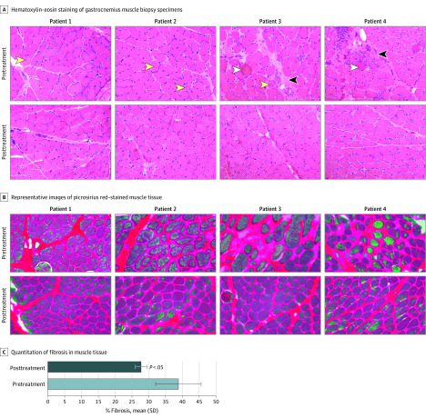
Evaluation by histopathological study showed that vector treatment was not associated with harmful alterations in the morphological features of muscle, demonstrated by the lack of remarkable alterations in central nucleation and the absence of ringed myofibers (Figure 2A). Vector treatment was not associated with increased muscle fibrosis but was associated with improvement in all 4 patients, demonstrated by a reduction in the percentage of collagen content in the muscle tissue after treatment compared with at baseline (mean [SD], 26.7% [8.4%]) (Figure 2B and C). Moreover, results from cardiac magnetic resonance imaging and echocardiography demonstrated no adverse findings in all 4 boys at the time points assessed and were within normal ranges for children with or without DMD36,37 (eTable 3 in Supplement 2). These results suggest a lack of toxic effects from vector exposure.
Figure 2. Lack of Vector-Associated Toxic Effects by Histopathological Assessments in 4 Patients.
A, Hematoxylin-eosin staining (original magnification ×20) shows unremarkable alterations in central nucleation (yellow arrowheads) of muscle fibers before and after treatment. Baseline (pretreatment) biopsy results show levels of necrosis (white arrowheads), stages of degeneration or regeneration, and increased connective tissue (black arrowheads). B, Picrosirius red staining shows a reduction in fibrosis after treatment. C, Fibrosis in tissue samples was measured by the percentage of collagen accumulation in tissue section. The error bars refer to the SD.
Micro-dystrophin Expression, Correct Localization, and Restoration of Dystrophin-Associated Protein Complex
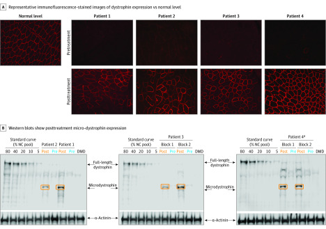
Successful gene transfer of micro-dystrophin was assessed by quantitative polymerase chain reaction for vector-specific genome molecular analysis, immunofluorescence (Figure 3A), and Western blot (Figure 3B). Transduction was confirmed in all 4 patients, showing a mean of 3.3 vector genome copies per nucleus detected by quantitative polymerase chain reaction (eTable 4 in Supplement 2), indicating the successful delivery of rAAVrh74.MHCK7.micro-dystrophin in skeletal muscle. At 12 weeks, immunohistochemistry of muscle biopsy specimens showed a mean of 81.2% micro-dystrophin-positive fibers in the gastrocnemius muscle, with a mean intensity of 96.0% (Figure 3A; eTable 4 in Supplement 2). Findings at 12 weeks were confirmed by Western blot (Figure 3B), in which mean micro-dystrophin expression in the sample was 74.3% of normal dystrophin expression level without adjustment for fat or fibrosis. When adjusted for fat or fibrosis, mean micro-dystrophin expression was 95.8%.
Figure 3. Systemic Administration of rAAVrh74.MHCK7.micro-dystrophin .
A, Frozen biopsied tissue sections of the gastrocnemius muscle were processed and stained with DYS3 antibody to detect micro-dystrophin. B, Micro-dystrophin compared with full-length protein was seen in 4 patients with Duchenne muscular dystrophy (DMD) before and after treatment (day 90) with rAAVrh74.MHCK7.micro-dystrophin. α-Actinin was used as a loading control, and sample from a patient was included as a negative control. NC indicates normal control.
aPatient 4 had a positive immunoreactive band that was above the upper limit of quantification and was diluted 1 to 4 before loading in the representative gel.
In addition to expression of micro-dystrophin at the sarcolemmal membrane, immunofluorescence for β-sarcoglycan, a critical component of dystrophin-associated protein complex, showed a robust increase in sarcolemma expression compared with pretreatment in all 4 patients (eFigure 2 in Supplement 2). This increase suggests that the micro-dystrophin transgene can promote restoration and reconstitution of the dystrophin-associated protein complex.
Functional Outcomes
All 4 patients showed improvements in NSAA scores and reductions in CK levels from baseline throughout the study and up to 1 year after treatment (Table 2). The 1-year NSAA score improvement was 7 points in patient 1, 8 points in patient 2, 2 points in patient 3, and 5 points in patient 4 (mean, 5.5 points). Patient 3 (aged 6 years) would be expected to decline, and thus the true Δ of improvement was likely greater than 2 points. The mean (SD) CK level at baseline was 27 064 (6340.5) U/L and after treatment was 8035 (3312.5) U/L. Patient 2 had CK levels that rose to greater than 40 000 U/L, and neither this participant nor any other participant had myoglobinuria.
Table 2. Micro-dystrophin Gene Transfer Association With Protection Against Muscle Damage.
| Patient | Parameter | Baseline | Day | Change from baseline at day 365 | |||||
|---|---|---|---|---|---|---|---|---|---|
| 30 | 60 | 90 | 180 | 270 | 365 | ||||
| 1 | NSAA | 18 | 22 | 24 | 23 | 25 | 26 | 25 | 7 |
| CK level, U/L | 20 691 | NA | 2984 | 2444 | 18 476 | 6317 | 11 073 | −46.48% | |
| 2 | NSAA | 19 | 21 | 22 | 25 | 27 | 27 | 27 | 8 |
| CK level, U/L | 23 414 | 10 427 | 4283 | 41 920 | 6209 | 17 614 | 10 494 | −55.18% | |
| 3 | NSAA | 26 | 28 | 28 | 30 | 30 | 28 | 28 | 2 |
| CK level, U/L | 34 942 | 10 430 | 2966 | 2546 | 9650 | 18 855 | 6410 | −81.66% | |
| 4 | NSAA | 19 | 20 | 20 | 25 | 24 | 27 | 24 | 5 |
| CK level, U/L | 29 210 | 7215 | 908 | 1382 | 2580 | 4262 | 4162 | −85.75% | |
Abbreviations: CK, creatine kinase; NA, not applicable; NSAA, North Star Ambulatory Assessment.
SI conversion factor: To convert U/L to μkat/L, multiply by 0.0167.
NSAA score improvement and the reduction in CK level appeared to be the most sensitive measures, but only a larger sample size and a clinical trial will validate improved motor function. There were different magnitudes of improvement across various other functional outcomes measured, including time to rise, 4-stair climb, 100-m timed test, and handheld dynamometry (eTable 5 in Supplement 2). Variability in clinical outcomes was associated with multiple factors, including age and disease severity. Activity can also suddenly increase serum CK level, leading to levels far above the mean. To avoid this, families should modify activity for at least 1 day before blood draw. An ongoing placebo-controlled randomized clinical trial includes a much greater sample size and will provide further insight for assessment of outcome measures.38
Discussion
Results of this pilot nonrandomized controlled trial provided biological and clinical evidence of systemic gene therapy for potential use in treating patients with DMD. These findings follow the success reported for exon-skipping therapy for patients with DMD and gene therapy for patients with SMA.12,13 Specifically, findings to date have shown the advantages of exon-skipping therapy for promoting the expression of even small amounts of truncated dystrophin in muscle that can positively alter the DMD course.9,12 Moreover, the natural history of a patient with Becker muscular dystrophy who harbored a dramatically shortened dystrophin suggests that expression of micro-dystrophin protein can lead to relatively mild dystrophinopathy.18 Together these findings suggest the possibility that targeted muscular expression of micro-dystrophin may lessen DMD severity.
Adverse event findings were minimal with rAAVrh74.MHCK7.micro-dystrophin. In this trial, the only vector-related adverse event was elevated transaminase and GGT levels, which was previously reported in other trials.39,40 Nausea and vomiting were the most common clinical adverse effects of AAV administration seen shortly (1-4 weeks) after gene delivery. No clinical manifestations associated with elevated liver enzymes were observed, and the elevated enzyme levels gradually resolved over 4 to 10 weeks. In this trial and a previous study,13 glucocorticoids were used beginning 1 day before gene delivery and continuing until GGT reached less than 150 U/L. It is speculative to suggest that glucocorticoids may be associated with suppression or resolution of the inflammatory infiltrate provoked by degrading capsids, but we have seen recurrent elevated liver enzymes as glucocorticoids were tapered, followed by GGT decrease after reinitiating the drug.
In this trial, gene transfer was done with AAVrh74. Preclinical studies have reported that micro-dystrophin delivery in an mdx mouse model under control of the MHCK7 promoter was associated with robust cardiac and skeletal muscle expression without toxic effects, providing compelling reasons to assess this association in a clinical trial. In this study, enrollment criteria included patients with DMD with out-of-frame deletions between exons 18 and 58 (exons not encoded by micro-dystrophin) to minimize the theoretical concern for immune response to the transgene.28 However, no identified immune responses to micro-dystrophin were seen, supporting the conduct of registration trials applicable to all DMD gene mutations.
We hypothesized that robust gene expression and correct localization to the sarcolemma could protect muscle fibers from progressive decline typical of DMD, but the expression of the micro-dystrophin and degree of protection provided require further time to evaluate. In addition, cells that were not transduced still have the potential for CK leak. It was encouraging that at the 90-day period after gene delivery, muscle biopsy specimens showed substantial dystrophin expression. The mean percentage of dystrophin-positive muscle fibers reached 81.2% (compared with normal biopsies in which dystrophin was detected in 100% of fibers), with a mean intensity at the sarcolemma of 96% of normal mean intensity.
Assessing long-term results from a small cohort of patients with DMD is challenging, but we believe that the value of this preliminary report is justified in several ways. First, the safety of systemic delivery using the rAAVrh74.MHCK7.micro-dystrophin cassette at a high dose was demonstrated. Early indications of efficacy favored moving forward with the current dose of virus and this relatively simple, noninvasive delivery method. We avoided dose escalation, circumventing poor results and unnecessary AAV exposure as previously encountered.13 Second, the potential advantage of micro-dystrophin for gene transfer was shown, given that CK levels were markedly lower compared with baseline. CK levels are typically high in patients with DMD because of the leakage from muscles, and the decrease in CK levels after micro-dystrophin gene therapy suggests stabilization of the sarcolemma. However, the trial also showed a potential caveat for using CK level as an outcome measure if assessed at 1 time point, as we did in this study. We attributed the high CK level as a response to intense motor activity.41 An important concern for gene transfer therapy is the potential for declining outcome over time. Until the entire portfolio of gene therapies expands through multiple applications, we can only speculate on long-term efficacy on the basis of single-gene administration. Data from a long-term follow-up in a clinical trial of 12 patients with SMA who were treated with a dose range of virus (Zolgensma15) similar to the dose delivered in this micro-dystrophin trial showed sustained efficacy of the therapy for 5 years.
It is important to review the available published natural history data in patients aged 4 to 7 years to help contextualize the findings from this trial. Patients with DMD generally showed improved physical function up to age 7 years,42,43,44,45,46,47 a finding that complicates the interpretation of the treatment effect of a therapeutic agent compared with that of the natural disease course.46 Recent data from the Collaborative Trajectory Analysis Project demonstrated that approximately 2 points constituted a clinically meaningful change of the NSAA total score over a 48-week period.47
Early studies reported that approximately 40% of patients up to age 7 years showed some improvement of their NSAA scores over 12 months, but a sizeable proportion did not improve.44 Furthermore, the literature reported that the expected higher NSAA scores in patients up to age 7 years ranged from approximately 0.7 to 3 points per year.42,45,46 In a 12-month longitudinal cohort study, Mazzone et al44 found a 0.7-point increase in NSAA score from baseline in patients up to 7 years of age who were receiving continuous corticosteroid treatment. Ricotti et al42 reported a functional gain of approximately 1.5 points per year on NSAA scores in patients aged 4 to 7 years, with an additional improvement of 1.3 points per year in boys who had started glucocorticoids at an age younger than 5 years compared with boys who started glucocorticoids at an age older than 5 years.
In an extensive systematic natural history study, Muntoni et al45 reported findings of a cohort of approximately 395 patients aged 2 to 17 years from the UK North Star registry. Mean trajectory of NSAA total score peaked at age 6.3 years, and the population mean NSAA total score initially increased at a rate of approximately 3 units per year, which, following the peak, approached a rate of decline of approximately 3 units per year. The North Star registry cohort included patients with DMD mutations that were generally associated with slower disease progression than typically seen in DMD (eg, 44 skip-amenable patients).
In the present study, the 1-year NSAA score improvement was 7 points in patient 1, 8 points in patient 2, 2 points in patient 3, and 5 points in patient 4 (mean, 5.5 points). Patient 3 (aged 6 years) would be expected to decline, and thus the true Δ of improvement was likely greater than 2 points. Patient 3 also showed the least improvement; however, the NSAA score of this patient constituted a clinically meaningful change according to data from the Collaborative Trajectory Analysis Project.47 All 4 patients had a clinically meaningful NSAA score improvement and had such improvements as soon as day 90. This finding suggests an overall improvement vs natural history and acquisition of activities that were greater than that expected in patients with DMD who were receiving standard-of-care treatment.
These results, along with biological and clinical markers of efficacy, provide proof-of-concept support for the continuation of clinical trials to assess rAAVrh74.MHCK7.micro-dystrophin using single-dose gene transfer in patients with DMD. A randomized clinical trial with a much larger sample size of boys with DMD is under way.38
Limitations
This nonrandomized trial yielded encouraging results for AAVrh74.MHCK7.micro-dystrophin. However, the safety and efficacy of this therapy will need to be confirmed in a randomized clinical trial.
Conclusions
This trial demonstrated that a single systemic infusion of rAAVrh74.MHCK7.micro-dystrophin was well tolerated and had a favorable safety profile. In addition, it described the delivery of transgene to the nuclei and the robust expression and proper localization of micro-dystrophin, which coincided with marked reductions in CK levels and improvement in functional measurements such as NSAA score. These preliminary results, albeit in a small number of patients, suggest the potential for rAAVrh74.MHCK7.micro-dystrophin to provide clinically meaningful functional improvement that is greater than that observed with standard-of-care treatment, including corticosteroids.
Trial Protocol
eMethods
eReferences
eFigure 1. Immune Responses to the AAVrh74 Capsid Protein and Micro-dystrophin
eFigure 2. Micro-dystrophin Gene Transfer Increases β-Sarcoglycan Expression
eTable 1. Weekend Prednisone Dose Maintained During Clinical Trial
eTable 2. Serum Chemistry Profiles
eTable 3. Cardiac Magnetic Resonance Imaging at Baseline and Post Treatment
eTable 4. Quantitative Summary of Protein Expression by Intensity and Percentage of
Dystrophin-Positive Fibers and Transduction by qPCR of Vector Genome
eTable 5. Additional Function Outcome Measures Assessed Throughout the Study in Each Patient
eTable 6. List of Adverse Events by System Organ Class
References
- 1.Mendell JR, Lloyd-Puryear M. Report of MDA muscle disease symposium on newborn screening for Duchenne muscular dystrophy. Muscle Nerve. 2013;48(1):21-26. doi: 10.1002/mus.23810 [DOI] [PubMed] [Google Scholar]
- 2.Mendell JR, Shilling C, Leslie ND, et al. Evidence-based path to newborn screening for Duchenne muscular dystrophy. Ann Neurol. 2012;71(3):304-313. doi: 10.1002/ana.23528 [DOI] [PubMed] [Google Scholar]
- 3.Bello L, Morgenroth LP, Gordish-Dressman H, Hoffman EP, McDonald CM, Cirak S; CINRG investigators . DMD genotypes and loss of ambulation in the CINRG Duchenne Natural History Study. Neurology. 2016;87(4):401-409. doi: 10.1212/WNL.0000000000002891 [DOI] [PMC free article] [PubMed] [Google Scholar]
- 4.Birnkrant DJ, Bushby K, Bann CM, et al. ; DMD Care Considerations Working Group . Diagnosis and management of Duchenne muscular dystrophy, part 2: respiratory, cardiac, bone health, and orthopaedic management. Lancet Neurol. 2018;17(4):347-361. doi: 10.1016/S1474-4422(18)30025-5 [DOI] [PMC free article] [PubMed] [Google Scholar]
- 5.Birnkrant DJ, Bushby K, Bann CM, et al. ; DMD Care Considerations Working Group . Diagnosis and management of Duchenne muscular dystrophy, part 3: primary care, emergency management, psychosocial care, and transitions of care across the lifespan. Lancet Neurol. 2018;17(5):445-455. doi: 10.1016/S1474-4422(18)30026-7 [DOI] [PMC free article] [PubMed] [Google Scholar]
- 6.Henricson EK, Abresch RT, Cnaan A, et al. ; CINRG Investigators . The cooperative international neuromuscular research group Duchenne natural history study: glucocorticoid treatment preserves clinically meaningful functional milestones and reduces rate of disease progression as measured by manual muscle testing and other commonly used clinical trial outcome measures. Muscle Nerve. 2013;48(1):55-67. doi: 10.1002/mus.23808 [DOI] [PMC free article] [PubMed] [Google Scholar]
- 7.Liu D, Ahmet A, Ward L, et al. A practical guide to the monitoring and management of the complications of systemic corticosteroid therapy. Allergy Asthma Clin Immunol. 2013;9(1):30. doi: 10.1186/1710-1492-9-30 [DOI] [PMC free article] [PubMed] [Google Scholar]
- 8.Rice JB, White AG, Scarpati LM, Wan G, Nelson WW. Long-term systemic corticosteroid exposure: a systematic literature review. Clin Ther. 2017;39(11):2216-2229. doi: 10.1016/j.clinthera.2017.09.011 [DOI] [PubMed] [Google Scholar]
- 9.Khan N, Eliopoulos H, Han L, et al. ; Eteplirsen Investigators and the CINRG DNHS Investigators . Eteplirsen treatment attenuates respiratory decline in ambulatory and non-ambulatory patients with Duchenne muscular dystrophy. J Neuromuscul Dis. 2019;6(2):213-225. doi: 10.3233/JND-180351 [DOI] [PMC free article] [PubMed] [Google Scholar]
- 10.Aartsma-Rus A, Verschuuren JJGM, Campion GV, van Ommen GB, van Deutekom JCT. Overview on DMD exon skipping. Methods Mol Biol. 2012;867:97-116. doi: 10.1007/978-1-61779-767-5_7 [DOI] [PubMed] [Google Scholar]
- 11.Charleston JS, Schnell FJ, Dworzak J, et al. Eteplirsen treatment for Duchenne muscular dystrophy: exon skipping and dystrophin production. Neurology. 2018;90(24):e2146-e2154. doi: 10.1212/WNL.0000000000005680 [DOI] [PubMed] [Google Scholar]
- 12.Cirak S, Arechavala-Gomeza V, Guglieri M, et al. Exon skipping and dystrophin restoration in patients with Duchenne muscular dystrophy after systemic phosphorodiamidate morpholino oligomer treatment: an open-label, phase 2, dose-escalation study. Lancet. 2011;378(9791):595-605. doi: 10.1016/S0140-6736(11)60756-3 [DOI] [PMC free article] [PubMed] [Google Scholar]
- 13.Mendell JR, Al-Zaidy S, Shell R, et al. Single-dose gene-replacement therapy for spinal muscular atrophy. N Engl J Med. 2017;377(18):1713-1722. doi: 10.1056/NEJMoa1706198 [DOI] [PubMed] [Google Scholar]
- 14.Lowes LP, Alfano LN, Arnold WD, et al. Impact of age and motor function in a phase 1/2A study of infants with SMA type 1 receiving single-dose gene replacement therapy. Pediatr Neurol. 2019;98:39-45. doi: 10.1016/j.pediatrneurol.2019.05.005 [DOI] [PubMed] [Google Scholar]
- 15.Al-Zaidy SA, Kolb SJ, Lowes L, et al. AVXS-101 (onasemnogene abeparvovec) for SMA1: comparative study with a prospective natural history cohort. J Neuromuscul Dis. 2019;6(3):307-317. doi: 10.3233/JND-190403 [DOI] [PubMed] [Google Scholar]
- 16.Duan D. Systemic AAV micro-dystrophin gene therapy for Duchenne muscular dystrophy. Mol Ther. 2018;26(10):2337-2356. doi: 10.1016/j.ymthe.2018.07.011 [DOI] [PMC free article] [PubMed] [Google Scholar]
- 17.Wells DJ, Wells KE, Asante EA, et al. Expression of human full-length and minidystrophin in transgenic mdx mice: implications for gene therapy of Duchenne muscular dystrophy. Hum Mol Genet. 1995;4(8):1245-1250. doi: 10.1093/hmg/4.8.1245 [DOI] [PubMed] [Google Scholar]
- 18.England SB, Nicholson LV, Johnson MA, et al. Very mild muscular dystrophy associated with the deletion of 46% of dystrophin. Nature. 1990;343(6254):180-182. doi: 10.1038/343180a0 [DOI] [PubMed] [Google Scholar]
- 19.Zhao J, Kodippili K, Yue Y, et al. Dystrophin contains multiple independent membrane-binding domains. Hum Mol Genet. 2016;25(17):3647-3653. doi: 10.1093/hmg/ddw210 [DOI] [PMC free article] [PubMed] [Google Scholar]
- 20.Nelson DM, Lindsay A, Judge LM, et al. Variable rescue of microtubule and physiological phenotypes in mdx muscle expressing different miniaturized dystrophins. Hum Mol Genet. 2018;27(12):2090-2100. doi: 10.1093/hmg/ddy113 [DOI] [PMC free article] [PubMed] [Google Scholar]
- 21.Harper SQ, Hauser MA, DelloRusso C, et al. Modular flexibility of dystrophin: implications for gene therapy of Duchenne muscular dystrophy. Nat Med. 2002;8(3):253-261. doi: 10.1038/nm0302-253 [DOI] [PubMed] [Google Scholar]
- 22.Suzuki A, Yoshida M, Yamamoto H, Ozawa E. Glycoprotein-binding site of dystrophin is confined to the cysteine-rich domain and the first half of the carboxy-terminal domain. FEBS Lett. 1992;308(2):154-160. doi: 10.1016/0014-5793(92)81265-N [DOI] [PubMed] [Google Scholar]
- 23.Salva MZ, Himeda CL, Tai PW, et al. Design of tissue-specific regulatory cassettes for high-level rAAV-mediated expression in skeletal and cardiac muscle. Mol Ther. 2007;15(2):320-329. doi: 10.1038/sj.mt.6300027 [DOI] [PubMed] [Google Scholar]
- 24.Rodino-Klapac LR, Janssen PM, Montgomery CL, et al. A translational approach for limb vascular delivery of the micro-dystrophin gene without high volume or high pressure for treatment of Duchenne muscular dystrophy. J Transl Med. 2007;5:45. doi: 10.1186/1479-5876-5-45 [DOI] [PMC free article] [PubMed] [Google Scholar]
- 25.Mendell JR, Rodino-Klapac LR, Rosales-Quintero X, et al. Limb-girdle muscular dystrophy type 2D gene therapy restores alpha-sarcoglycan and associated proteins. Ann Neurol. 2009;66(3):290-297. doi: 10.1002/ana.21732 [DOI] [PMC free article] [PubMed] [Google Scholar]
- 26. Potter RA, Griffin DA, Heller KN, et al. Dose escalation study of systemically delivered AAVrh74.MHCK7.micro-dystrophin in the mdx mouse model of DMD. Data presented at: 2018 American Society of Gene & Cell Therapy Annual Meeting; May 16-18, 2018; Chicago, Illinois. [Google Scholar]
- 27.Dixon JR. The International Conference on Harmonization Good Clinical Practice guideline. Qual Assur. 1998;6(2):65-74. doi: 10.1080/105294199277860 [DOI] [PubMed] [Google Scholar]
- 28.Mendell JR, Campbell K, Rodino-Klapac L, et al. Dystrophin immunity in Duchenne’s muscular dystrophy. N Engl J Med. 2010;363(15):1429-1437. doi: 10.1056/NEJMoa1000228 [DOI] [PMC free article] [PubMed] [Google Scholar]
- 29.Alfano LN, Miller NF, Berry KM, et al. The 100-meter timed test: normative data in healthy males and comparative pilot outcome data for use in Duchenne muscular dystrophy clinical trials. Neuromuscul Disord. 2017;27(5):452-457. doi: 10.1016/j.nmd.2017.02.007 [DOI] [PubMed] [Google Scholar]
- 30.Escolar DM, Hache LP, Clemens PR, et al. Randomized, blinded trial of weekend vs daily prednisone in Duchenne muscular dystrophy. Neurology. 2011;77(5):444-452. doi: 10.1212/WNL.0b013e318227b164 [DOI] [PMC free article] [PubMed] [Google Scholar]
- 31.Sondergaard PC, Griffin DA, Pozsgai ER, et al. AAV.dysferlin overlap vectors restore function in dysferlinopathy animal models. Ann Clin Transl Neurol. 2015;2(3):256-270. doi: 10.1002/acn3.172 [DOI] [PMC free article] [PubMed] [Google Scholar]
- 32.Schnepp BC, Jensen RL, Chen CL, Johnson PR, Clark KR. Characterization of adeno-associated virus genomes isolated from human tissues. J Virol. 2005;79(23):14793-14803. doi: 10.1128/JVI.79.23.14793-14803.2005 [DOI] [PMC free article] [PubMed] [Google Scholar]
- 33.Tarnopolsky MA, Pearce E, Smith K, Lach B. Suction-modified Bergström muscle biopsy technique: experience with 13,500 procedures. Muscle Nerve. 2011;43(5):717-725. doi: 10.1002/mus.21945 [DOI] [PubMed] [Google Scholar]
- 34.Stevens W. Good clinical laboratory practice (GCLP): the need for a hybrid of good laboratory practice and good clinical practice guidelines/standards for medical testing laboratories conducting clinical trials in developing countries. Qual Assur. 2003;10(2):83-89. doi: 10.1080/10529410390262727 [DOI] [PubMed] [Google Scholar]
- 35.Schnell FJ, Frank D, Fletcher S, Johnsen RD, Wilton SD. Challenges of interpreting dystrophin content by western blot. US Neurol. 2019;15(1):40-46. doi: 10.17925/USN.2019.15.1.40 [DOI] [Google Scholar]
- 36.Silva MC, Meira ZM, Gurgel Giannetti J, et al. Myocardial delayed enhancement by magnetic resonance imaging in patients with muscular dystrophy. J Am Coll Cardiol. 2007;49(18):1874-1879. doi: 10.1016/j.jacc.2006.10.078 [DOI] [PubMed] [Google Scholar]
- 37.Kawel-Boehm N, Maceira A, Valsangiacomo-Buechel ER, et al. Normal values for cardiovascular magnetic resonance in adults and children. J Cardiovasc Magn Reson. 2015;17:29. doi: 10.1186/s12968-015-0111-7 [DOI] [PMC free article] [PubMed] [Google Scholar]
- 38.A randomized, double-blind, placebo-controlled study of SRP-9001 for Duchenne muscular dystrophy (DMD). ClinicalTrials.gov Identifier: NCT03769116. Updated March 30, 2020. Accessed February 12, 2020. https://clinicaltrials.gov/ct2/show/NCT03769116
- 39.Manno CS, Pierce GF, Arruda VR, et al. Successful transduction of liver in hemophilia by AAV-Factor IX and limitations imposed by the host immune response. Nat Med. 2006;12(3):342-347. Published correction appears in Nat Med. 2006;12(5):592. [DOI] [PubMed] [Google Scholar]
- 40.Nathwani AC, Tuddenham EG, Rangarajan S, et al. Adenovirus-associated virus vector-mediated gene transfer in hemophilia B. N Engl J Med. 2011;365(25):2357-2365. doi: 10.1056/NEJMoa1108046 [DOI] [PMC free article] [PubMed] [Google Scholar]
- 41.Doriguzzi C, Palmucci L, Mongini T, Chiadò-Piat L, Restagno G, Ferrone M. Exercise intolerance and recurrent myoglobinuria as the only expression of Xp21 Becker type muscular dystrophy. J Neurol. 1993;240(5):269-271. doi: 10.1007/BF00838159 [DOI] [PubMed] [Google Scholar]
- 42.Ricotti V, Ridout DA, Pane M, et al. ; UK NorthStar Clinical Network . The NorthStar Ambulatory Assessment in Duchenne muscular dystrophy: considerations for the design of clinical trials. J Neurol Neurosurg Psychiatry. 2016;87(2):149-155. doi: 10.1136/jnnp-2014-309405 [DOI] [PMC free article] [PubMed] [Google Scholar]
- 43.McDonald CM, Henricson EK, Abresch RT, et al. ; PTC124-GD-007-DMD Study Group . The 6-minute walk test and other clinical endpoints in Duchenne muscular dystrophy: reliability, concurrent validity, and minimal clinically important differences from a multicenter study. Muscle Nerve. 2013;48(3):357-368. doi: 10.1002/mus.23905 [DOI] [PMC free article] [PubMed] [Google Scholar]
- 44.Mazzone E, Vasco G, Sormani MP, et al. Functional changes in Duchenne muscular dystrophy: a 12-month longitudinal cohort study. Neurology. 2011;77(3):250-256. doi: 10.1212/WNL.0b013e318225ab2e [DOI] [PubMed] [Google Scholar]
- 45.Muntoni F, Domingos J, Manzur AY, et al. ; UK NorthStar Network . Categorising trajectories and individual item changes of the North Star Ambulatory Assessment in patients with Duchenne muscular dystrophy. PLoS One. 2019;14(9):e0221097. doi: 10.1371/journal.pone.0221097 [DOI] [PMC free article] [PubMed] [Google Scholar]
- 46.Ricotti V, Ridout DA, Scott E, et al. ; NorthStar Clinical Network . Long-term benefits and adverse effects of intermittent versus daily glucocorticoids in boys with Duchenne muscular dystrophy. J Neurol Neurosurg Psychiatry. 2013;84(6):698-705. doi: 10.1136/jnnp-2012-303902 [DOI] [PubMed] [Google Scholar]
- 47.Wong B, Signorovitch J, Staunton H, et al. Estimating clinically meaningful change thresholds in the North Star Ambulatory Assessment (NSAA) and four-stair climb (4SC) in Duchenne muscular dystrophy (DMD). Neuromuscul Disord. 2019;29:S106. doi: 10.1016/j.nmd.2019.06.251 [DOI] [Google Scholar]
Associated Data
This section collects any data citations, data availability statements, or supplementary materials included in this article.
Supplementary Materials
Trial Protocol
eMethods
eReferences
eFigure 1. Immune Responses to the AAVrh74 Capsid Protein and Micro-dystrophin
eFigure 2. Micro-dystrophin Gene Transfer Increases β-Sarcoglycan Expression
eTable 1. Weekend Prednisone Dose Maintained During Clinical Trial
eTable 2. Serum Chemistry Profiles
eTable 3. Cardiac Magnetic Resonance Imaging at Baseline and Post Treatment
eTable 4. Quantitative Summary of Protein Expression by Intensity and Percentage of
Dystrophin-Positive Fibers and Transduction by qPCR of Vector Genome
eTable 5. Additional Function Outcome Measures Assessed Throughout the Study in Each Patient
eTable 6. List of Adverse Events by System Organ Class


