PLOS ONE logoLink to PLOS ONE
. 2020 Jun 16;15(6):e0234700. doi: 10.1371/journal.pone.0234700

Comparison of the change in QuantiFERON-TB Gold Plus and QuantiFERON-TB Gold In-Tube results after preventive therapy for latent tuberculosis infection

Ock-Hwa Kim 1,#, Kyung-Wook Jo 1,#, Shinhee Park 2, Yong-Ha Jo 3, Mi-Na Kim 4, Heungsup Sung 4, Tae Sun Shim 1,*
Editor: Katalin Andrea Wilkinson5
PMCID: PMC7297367  PMID: 32544206

Abstract

Background

We investigated changes in the interferon-γ levels before and after treatment of latent tuberculosis infection (LTBI) using QuantiFERON-TB Gold Plus (QFT-Plus) and QuantiFERON-TB Gold In-Tube (QFT-GIT) assays. The objective was to assess whether QFT-Plus could serve as a biomarker of LTBI treatment response.

Methods

We prospectively enrolled 44 individuals whose baseline QFT-GIT and QFT-Plus showed positive results at a tertiary referral center in South Korea between March 2017 and March 2018. The results of the QFT-Plus assay were defined as positive if either or both of the antigen tubes (TB1 and/or TB2) were positive. After LTBI treatment, both tests were repeated.

Results

The mean age of the participants was 47.6 years. The QFT-GIT and QFT-Plus assays revealed positive results in 42/44 (95.5%) and 41/44 (93.2%) participants after LTBI treatment, showing overall agreement of 93.2%, with a Cohen’s kappa value of 0.37 (fair agreement). The differences between pre- and post-LTBI treatment interferon-γ levels were measured using the QFT-GIT and QFT-Plus assays. No significant differences were noted among the 3 values: the median difference in interferon-γ value with QFT-GIT, QFT-Plus TB1, and QFT-Plus TB2 was 0.211 IU/mL (IQR, −0.337–3.347), 0.025 IU/mL (IQR, −0.338–1.368), and 0.180 IU/mL (IQR, −0.490–2.278), respectively (P = 0.401).

Conclusion

The change in interferon-γ levels before and after LTBI treatment measured using the QFT-Plus assay showed a similar trend to that of the QFT-GIT assay. Considering that the QFT-GIT assay is not a useful biomarker of LTBI treatment response, QFT-Plus also appears not to be useful for this purpose.

Introduction

Latent tuberculosis infection (LTBI) is defined as a state wherein humans are infected with Mycobacterium tuberculosis without any clinical symptoms, radiological abnormality, or microbiological evidence of infection [1,2]. According to the World Health Organization, approximately 2–3 billion people in the world are affected by LTBI [3]. Given that approximately 5%–10% of immunocompetent persons with LTBI will develop overt tuberculosis (TB) in their lifetime [4], treatment of LTBI has direct implications for global prevention and control efforts against TB.

The World Health Organization guidelines recommend the use of a tuberculin skin test or interferon-γ release assay (IGRA) for detection of LTBI in high-income and upper middle-income countries that have an estimated TB incidence of <100 per 100,000 population [5]. Until recently, two IGRAs have been commercialized: the T-SPOT.TB assay (Oxford Immunotec, Abingdon, United Kingdom) and the QuantiFERON-TB Gold In-Tube (QFT-GIT) assay (Cellestis/Qiagen, Carnegie, Australia) [6,7]. The QFT-GIT assay measures interferon-γ (IFN-γ) concentration to detect M. tuberculosis infection using a single mixture of synthetic peptides (ESAT-6, CFP-10, and TB7.7) to stimulate T cells [6]. Although the ESAT-6 and CFP-10 antigens stimulate both CD4+ and CD8+ T cells to release IFN-γ, these two epitopes mainly stimulate CD4+ T cells [8]. However, when exposed to M. tuberculosis antigens, both CD4+ and CD8+ T cells secrete IFN-γ [9]. Therefore, the newest generation of the QFT assay, QuantiFERON-TB Gold Plus (QFT-Plus) was recently developed and launched in 2015. In contrast to QFT-GIT assay, QFT-Plus has two TB antigen tubes: TB antigen tube 1 (TB1) and TB antigen tube 2 (TB2). TB1 contains long peptides from ESAT-6 and CFP-10, which are designed to stimulate an immune response from CD4+ T cells. In contrast, TB2 contains relatively short peptides that elicit immune responses from both CD4+ and CD8+ T cells. The TB7.7 present in the QFT-GIT has been removed in the QFT-Plus assay [8,10].

One of the most frustrating problems in the treatment of patients with LTBI is that the IGRA results are not a surrogate marker of response to LTBI therapy. Previous studies have shown no differences in the decline in QFT-GIT or reversion to negative results between patients who received LTBI treatment and those who received placebo or no treatment [11,12]. Given the reported positive correlation between the CD8+ T-cell response against TB antigens and higher CD8+ T-cell responses in the context of recent exposure to M. tuberculosis [13,14], we postulated that the CD8+ response would be more sensitive to LTBI treatment compared with the CD4+ response. Therefore, in this study, we examined the changes in IFN-γ levels as measured using QFT-Plus and QFT-GIT assays before and after LTBI treatment. The objective was to assess whether QFT-Plus can be employed as a useful biomarker of LTBI treatment response.

Materials and methods

Study population

This prospective trial was conducted between March 2017 and March 2018 at a tertiary referral center in South Korea, where the incidence of TB is intermediate (80–100/100,000 people per year) [15]. Adults aged 18–80 years, who were candidates for an LTBI screening test for various reasons (such as healthcare personnel undergoing screening, contact investigation, and candidate recipients of tumor necrosis factor inhibitor therapy) were prospectively recruited. All enrolled participants were examined using the following strategy to rule out the possibility of active TB. All participants were interviewed by the attending physician regarding the presence of any respiratory symptoms that were suggestive of active TB, such as cough, sputum, or hemoptysis, in addition to undergoing a simple chest radiography (CXR). For those with radiographic anomalies suggestive of TB, further evaluation was performed using sputum acid-fast bacilli smear and culture to rule out active TB or lung disease due to nontuberculous mycobacteria. Moreover, present and former CXR findings were compared to identify the presence of newly developed lesions.

At baseline, both QFT-GIT and QFT-Plus were performed for all participants. Then, among the participants with a positive QFT-GIT assay, those whose QFT-Plus assay (TB antigen tube 1 [TB1] or TB antigen tube 2 [TB2]) results were also positive were selected for the present study. After the completion of LTBI treatment, both tests were performed again for all participants to assess the post-treatment changes in IFN-γ level. The exclusion criteria were as follows: (i) those whose baseline QFT-GIT and/or QFT-Plus tests (both TB1 and TB2) revealed a negative result; (ii) those who did not receive QFT tests after LTBI treatment; (iii) participants who did not complete the treatment; and (iv) participants with an indeterminate QFT result. The trial protocol was approved by the institutional review board (IRB) at the Asan Medical Center (IRB No.: 2017–0004). Written informed consent was obtained from all participants before enrollment.

The QFT test assay

The QFT-Plus kits were donated by Qiagen and were used according to the manufacturer’s instructions at our center [16]. QFT-GIT was also performed at our center as per the manufacturer’s instructions [17]. We defined QFT-GIT results as positive (1) if the nil value was ≤8.0 IU/mL, (2) the TB antigen minus the nil IFN-γ value was ≥0.35 IU/mL, and (3) at least 25% of the nil value, irrespective of the mitogen minus nil value [18]. The same criteria applied to the QFT-Plus assay. However, the results of the QFT-Plus assay were defined as positive if either or both of the TB antigen tubes (TB1 and/or TB2) were positive [18]. If antigen-nil was <0.35 IU/mL or <25% of the nil value, when the mitogen was ≥0.5 IU/mL, the result was considered negative. If (1) nil was >8 IU/mL or (2) antigen-nil ≥0.35 IU/mL and <25% of the nil value when the nil was ≤8.0 IU/mL and the mitogen was <0.5 IU/mL, the results were considered indeterminate. The IFN-γ levels in the antigen tubes >10 IU/mL were classified as 10 IU/mL.

LTBI treatment regimen

During the study period, daily isoniazid and rifampin for 3 months were selected as the primary regimen for LTBI treatment. Based on the discretion of the attending physician, a regimen of rifampin for 4 months could also be prescribed. Treatment completion was defined as intake of >80% of all prescribed medication within 4 months for isoniazid and rifampin and within 6 months for rifampin.

Statistical analysis

All analyses were performed using SPSS software (version 20.0; SPSS, Chicago, IL, USA). The data are presented as mean (± standard deviation) or median (interquartile range [IQR]) for continuous variables and as percent for discrete variables. The Friedman test was used to compare the IFN-γ levels measured in different antigen-containing tubes before LTBI treatment and changes in the paired IFN-γ levels before and after LTBI treatment. In cases where there was a significant difference among the three values, a post hoc analysis using the Wilcoxon signed rank test for multiple comparisons was performed with the application of Bonferroni correction.

To qualitatively compare the results of the QFT-GIT and QFT-Plus assays after LTBI treatment, positive, negative, and overall percent agreements were calculated using the QFT-GIT results as the reference standard. Concordance between the QFT-GIT and QFT-Plus assays was assessed using kappa coefficients; kappa values of ≤0, 0.01–0.20, 0.21–0.40, 0.41–0.60, 0.61–0.80, and ≥0.81 were interpreted to indicate no, slight, fair, moderate, substantial, and almost perfect agreement, respectively [19].

Results

Study participants

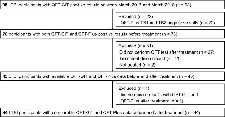
We enrolled 44 participants whose baseline QFT-GIT and QFT-Plus assay showed positive results and who received repeat tests of the QFT-GIT and QFT-Plus assay after LTBI treatment (Fig 1). The mean age was 47.6 years. The most common indication for LTBI testing was baseline or periodic screening tests for healthcare workers, followed by LTBI screening for the day care center workers. Almost all (42/44, 95.5%) of the participants completed treatment with isoniazid and rifampin for 3 months, without any change in the regimen. The baseline characteristics of the 44 participants are shown in Table 1. Among the 44 participants, most (41/44, 93.2%) were positive with both QFT-Plus TB1 and TB2, with the exception of 3 participants, 1 of who was positive with QFT-Plus TB1 only, whereas the other 2 were positive with QFT-Plus TB2 only (Table 2).

Fig 1. Study flow chart.

Fig 1

LTBI, latent tuberculosis infection; QFT-GIT, QuantiFERON-TB Gold In-Tube; QFT-Plus, QuantiFERON-TB Gold Plus.

Table 1. Clinical characteristics of the study participants.

Characteristics Total (n = 44)
Age (years) 47.6 ± 11.4
Female sex 29 (65.9%)
Body mass index at initiation of treatment (kg/m2) 23.6 ± 3.1
Current or past smoker 9 (20.5%)
Previous history of TB 0 (0.0%)
Indication for LTBI treatment
    Health care workers 19 (43.2%)
    Day care center workers 10 (22.7%)
    Treatment with TNF-α inhibitor 7 (15.9%)
    Close contact with persons with TB 4 (9.1%)
    CXR findings suggestive of healed TB 3 (6.8%)
    Recent TB infection (<2 years) 1 (2.3%)
Treatment regimen
    3 months of isoniazid and rifampin 42 (95.5%)
    4 months of rifampin 2 (4.5%)

Abbreviations: TB, tuberculosis; LTBI, latent tuberculosis infection; TNF, tumor necrosis factor; CXR, chest X-ray.

Data are reported as mean (± standard deviation) or as frequencies (%).

Table 2. The results of QFT assays at baseline and after the completion of preventive therapy.

At baseline After LTBI treatment
Positive N (%) Positive N (%)
QFT-GIT 44 (100) 42 (95.5)
QFT-Plus TB1 or TB2 44 (100) 41 (93.2)
QFT-Plus TB1 and TB2 41 (93.2) 41 (93.2)
QFT-Plus only TB1 1 (2.3) 0 (0)
QFT-Plus only TB2 2 (4.5) 0 (0)

Abbreviations: QFT, QuantiFERON; LTBI, latent tuberculosis infection; QFT-GIT, QuantiFERON-TB Gold In-Tube; QFT-Plus, QuantiFERON-TB Gold Plus.

Baseline QFT-GIT and QFT-Plus value

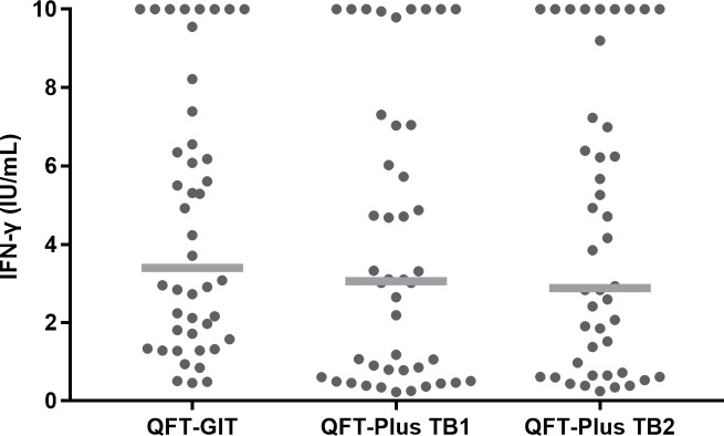
Fig 2 shows the IFN-γ levels of 44 participants with LTBI measured using the QFT-GIT and QFT-Plus (TB1 or TB2) assay before preventive treatment. Overall, statistical significance was noted among the three baseline IFN-γ values. That is, the median IFN-γ level value of the QFT-GIT assay was slightly higher than that of the QFT-Plus assay; the median IFN-γ level of QFT-GIT was 3.395 IU/mL (IQR, 1.615–7.180), the median TB1 QFT-Plus antigen IFN-γ level was 3.060 IU/mL (IQR, 0.655–7.045), and the median TB2 QFT-Plus antigen IFN-γ level was 2.880 IU/mL (IQR, 0.668–7.170) (P = 0.040). However, the difference was not statistically significant between the QFT-GIT and QFT-Plus TB1 (Z = −1.743, P = 0.081) or between the QFT-GIT and QFT-Plus TB2 (Z = −1.367, P = 0.172) assays.

Fig 2. IFN-γ levels measured using different antigen-containing tubes before preventive therapy.

Fig 2

IFN-γ concentrations from each participant are shown (●). Median levels are indicated by the gray lines. IFN-γ, interferon-γ; QFT-GIT, QuantiFERON-TB Gold In-Tube; QFT-Plus, QuantiFERON-TB Gold Plus.

The results of QFT assays after completion of LTBI treatment

After LTBI treatment, the QFT-GIT and QFT-Plus assays revealed positive results in 42/44 (95.5%) and 41/44 (93.2%) participants. Among the 44 participants with positive QFT-Plus assay results at baseline, 41/44 (93.2%) responded to both TB1 and TB2 at baseline, and the proportion did not change after treatment completion. In the remaining 3 participants, the QFT-Plus assay reverted to negative after LTBI treatment. The detailed results of the QFT-Plus according to the participants who responded to TB1 and/or TB2 peptides are shown in Table 2.

Comparison of the QFT-GIT and QFT-Plus assays after completion of LTBI treatment

Then, we qualitatively compared the results of the QFT-GIT and QFT-Plus assays after completion of LTBI treatment. The QFT-Plus assay showed agreement with the QFT-GIT assay in 40/42 (95.2%) positive tests and 1/2 (50.0%) negative tests and had an overall agreement of 41/44 (93.2%) among all participants, with a Cohen’s kappa value of 0.37 (fair agreement). Among the 44 participants enrolled in our study, the QFT-GIT and QFT-Plus assays after completion of LTBI treatment were discordant in 3 (6.8%) participants (Table 3).

Table 3. Qualitative comparison of QFT-GIT and QFT-Plus assays after preventive therapy.

QFT-Plus result QFT-GIT result % agreement (95% CI) Kappa value
Positive Negative Positive Negative Overall
Positive 40 1 95.2 (83.4–99.5) 50.0 (9.5–90.6) 93.2 (81.1–98.3) 0.37 (−0.19–0.92)
Negative 2 1

Abbreviations: QFT-GIT, QuantiFERON-TB Gold In-Tube; QFT-Plus, QuantiFERON-TB Gold Plus.

Changes in IFN-γ levels of two QFT assays after completion of LTBI treatment

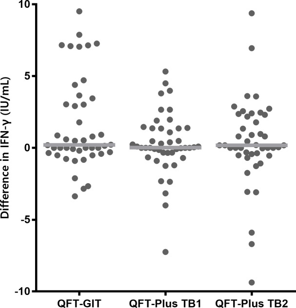
Next, we calculated the differences between the pre- and post-LTBI treatment IFN-γ levels (post-treatment IFN-γ level minus pre-treatment IFN-γ level), measured using the QFT-GIT and QFT-Plus assays. The median duration from baseline QFT tests to initiation of LTBI treatment was 42 days (IQR, 26–65). The follow-up QFT tests were performed at a median interval of 2 days (IQR, 0–8.8) after the completion of LTBI treatment. As shown in Fig 3, no significant differences were noted among the three values. The median difference in the IFN-γ value of QFT-GIT was 0.211 IU/mL (IQR, −0.337–3.347), the median difference in TB1 QFT-Plus antigen IFN-γ level was 0.025 IU/mL (IQR, −0.338–1.368), and the median difference in TB2 QFT-Plus antigen IFN-γ level was 0.180 IU/mL (IQR, −0.490–2.278) (P = 0.401). We also evaluated the overall median IFN-γ levels before and after treatment, the results of which are shown in the supporting information (S1 Fig and S1 Table). In addition, when we compared the IFN-γ response to TB1 and TB2 peptides at the same time point, we observed similar levels of IFN-γ and a positive correlation between IFN-γ values in response to TB1 and TB2 stimulation both at baseline and after LTBI treatment (S2 Fig).

Fig 3. Differences in IFN-γ levels before and after preventive therapy.

Fig 3

The differences in IFN-γ concentrations before and after preventive therapy for each participant are shown (●). Median levels are indicated by gray lines. IFN-γ, interferon-γ; QFT-GIT, QuantiFERON-TB Gold In-Tube; QFT-Plus, QuantiFERON-TB Gold Plus.

Discussion

Since the QFT-Plus has been developed, several studies have compared the performance of QFT-GIT and QFT-Plus for the diagnosis of active TB [20], contact screening [10], LTBI [21], and screening of healthcare personnel [18]. However, no study has yet analyzed the QFT-Plus responses before and after LTBI treatment compared with the QFT-GIT assay result. To the best of our knowledge, this is the first study that evaluates this issue. The most important finding of the present study was that the trend of changes in the IFN-γ levels before and after LTBI treatment measured using the QFT-Plus assay (either TB1 or TB2) was similar to that observed with the QFT-GIT assay. Our findings suggest that QFT-Plus is unlikely to be a useful biomarker of LTBI treatment response, based on earlier reports [11,22] that showed that the QFT-GIT cannot be used as the biomarker of LTBI treatment response.

Previous studies have reported that IGRA is probably not a useful biomarker of treatment efficacy in the context of LTBI, because LTBI treatment appears to have no effect on the results of serial IGRA testing. In their prospective trial involving 82 patients with LTBI, Johnson et al. had found no differences with respect to the decline in QFT-GIT or reversion to negative results after isoniazid preventive therapy or with observation alone [11]. Another placebo-controlled randomized trial revealed no differences in IFN-γ enzyme-linked immunosorbent spot test (ELISPOT) reversion over time between the treatment and placebo arms [12]. Bastos et al. also reported frequent within-participant variability in the QFT-GIT responses, which was not associated with LTBI treatment in high-risk tuberculin skin test-positive participants [22]. The present study compared the difference in the IFN-γ value measured using the QFT-Plus assay with the QFT-GIT result, without using a placebo (or observation) arm. However, given QFT-GIT is known to have no usefulness as a marker for LTBI treatment [11,22], and the differences between pre- and post-treatment IFN-γ values showed a similar trend between QFT-GIT and QFT-Plus (TB1 or TB2) assays in the present study, we can conclude that QFT-Plus does not appear to be a useful biomarker either.

In the case of active TB, previous studies have shown that the M. tuberculosis specific CD8+ T-cell response declines with anti-TB treatment and could be a surrogate marker of anti-TB therapy response [23,24]. In a prospective cohort study of 50 patients with culture-confirmed TB, a significant difference was observed in the M. tuberculosis specific CD8+ T-cell response after anti-TB treatment, but not in the CD4+ T-cell response [23]. This decline in response was noted over 24 weeks of treatment, with the earliest change observed at 8 weeks of therapy. In addition, Kamada et al. had suggested that QFT-Plus could be used as a potential tool for monitoring the efficacy of anti-TB therapy by showing a decline in the surrogate CD8+ T-cell response (difference “TB2 minus TB1”) at the end of treatment [24]. A recently published study reported a reduction in IFN-γ production over time in response to both TB1- and TB2-peptide stimulation in 28 patients with active TB [25]. Conversely, in the present study, we did not note any significant differences between the baseline and post-treatment TB1 and TB2 response in participants with LTBI (Fig 3). We believe that the difference in response between active TB and LTBI can be attributed to the differences with respect to the mycobacterial load [26] or the persistence of immunoreactive cells due to the persistence of antigen or mycobacteria after LTBI treatment [27].

Our findings are in contrast with those of a recent study by Petruccioli et al. in which LTBI therapy significantly reduced IFN-γ levels and the number of responders to TB1- and TB2- peptide stimulation [25]. The most plausible explanation for this disparity could be differences in the patient population. In the study by Petruccioli et al., 80.3% (37/46) of patients with LTBI had been recent contacts of patients with pulmonary TB, whereas the majority of participants in our study were healthcare and day care center workers or candidates for TNF inhibitor therapy. That is, only 11.4% (5/44, 4 in close contact with people with TB and 1 recently converted, Table 1) were likely to have been infected recently. Given that another previous study had also shown that the likelihood of tuberculin skin test reversion to negative following treatment with isoniazid in a group of US Navy personnel was strongly associated with the recency of infection [28], different results might have been observed if we mainly enrolled participants who were likely to have been infected recently. Although the findings were generally the same when we stratified our participants according to the possibility of recent infection (S3 Fig), it was likely that the number of recently infected participants was too small to reveal statistical significance.

Although we generally did not observe a statistically significant trend in terms of changes in IFN-γ level before and after LTBI treatment, some participants had a reduction in the TB1 and/or TB2 response on follow-up tests. This decline could be attributed to the natural course of IFN-γ response rather than to the effects of LTBI treatment, considering the findings of a previous study showing that approximately half of the contacts of patients with TB with initial positive ELISPOT assay had shown a negative result on repeat ELISPOT assay at 3 months, in the absence of treatment [29].

IFN-γ response failed to distinguish between active infection and cleared infection because of the persistence of antigen-specific memory T cells induced by prior exposure to M. tuberculosis [30]. In the present study, as depicted in Fig 2, we frequently observed increases in IFN-γ levels. This increase could have been due to continued exposure to M. tuberculosis in a high-incidence setting or persistent infection with latent bacilli. We also noted a post-treatment decline in IFN-γ levels in some of our study participants, which could have been caused by a natural decline in T-cell frequencies [12,31]. These factors collectively explain the reason why IGRA is of no use as a biomarker of infection clearance efficacy following LTBI treatment in previous studies as well as the present one. The well-documented inherent variability of the IGRA test is another important factor to be considered [32]. Considering this inherent IGRA test variability, we performed our analysis again after stratifying our participants according to the baseline IFN-γ value of QFT-Plus (either TB1 or TB2) as previously described [25]: 0.2–0.34 IU/mL; 0.35–0.7 IU/mL; and >0.7 IU/mL. The overall results are shown in the supporting information (S4 Fig). Reversions tended to be more in the uncertainty zone, which had been defined as baseline IFN-γ value 0.2–0.7 IU/mL in previous studies [25,33]. This result suggests that some of the reversions could be attributable to inherent variability.

As shown in Fig 2, the stimulated IFN-γ values obtained with QFT-Plus were statistically significantly lower than those obtained with the QFT-GIT assay. This finding was consistent with those of previous studies [8,20] and is probably because the QFT-Plus assay released less IFN-γ than the QFT-GIT assay because the QFT-Plus assay does not contain TB7.7. A previous study had shown that the epitope TB7.7 is highly specific for TB [34].

The present study has several limitations. Most significantly, it was conducted at a single referral center and included a relatively small number of cases. In particular, it is possible that the number of our study participants was insufficient to detect the difference between 2 tests, considering the test variability of the QFT-GIT [32,35] and QFT-Plus assays [36]. In addition, we compared only the differences in the IFN-γ values of the QFT-GIT and QFT-Plus assay before and after LTBI treatment, without using a placebo or observation arm. Moreover, the follow-up QFT tests were performed almost immediately after the end of LTBI treatment. It remained unclear whether we would have different results if the follow-up tests were checked at a later period.

In conclusion, we found no difference with respect to the change in IFN-γ levels before and after preventive treatment measured using the QFT-Plus assay and QFT-GIT assay in 44 participants with LTBI. Considering that the QFT-GIT assay is not a useful biomarker of response to LTBI treatment, the QFT-Plus also does not appear to be useful as a biomarker of LTBI treatment response.

Supporting information

S1 Fig. Changes in IFN-γ levels before and after preventive therapy.

IFN-γ concentrations from each participant are shown (•). Median levels are indicated by heavy lines. IFN-γ, interferon-γ; QFT-GIT, QuantiFERON-TB Gold In-Tube; QFT-Plus, QuantiFERON-TB Gold Plus.

(DOCX)

S2 Fig. Comparison of IFN-γ response to QFT-Plus antigen TB1 and TB2 at baseline and after LTBI treatment.

(A) IFN-γ response to QFT-Plus antigen TB1 and TB2 is similar both at baseline and after LTBI treatment. (B) Positive correlation between IFN-γ values in response to TB1 and TB2 stimulation both at baseline and after LTBI treatment (Baseline: r = 0.912, P < 0.001, after LTBI treatment: r = 0.892, P <0.001). IFN-γ, interferon-γ; QFT-Plus, QuantiFERON-TB Gold Plus; LTBI, latent tuberculosis infection.

(DOCX)

S3 Fig. Changes in IFN-γ levels before and after preventive therapy according to the possibility of recent infection.

(A) The LTBI participants who were likely to have been infected recently; (B) the other participants. Difference in IFN-γ concentrations before and after preventive therapy for each participant are shown (●). Median levels are indicated by gray lines. IFN-γ, interferon-γ; QFT-GIT, QuantiFERON-TB Gold In-Tube; QFT-Plus, QuantiFERON-TB Gold Plus; LTBI, latent tuberculosis infection.

(DOCX)

S4 Fig. Distribution of IFN-γ values before and after preventive therapy according to the baseline IFN-γ results.

IFN-γ concentrations from each participant are shown (•). IFN-γ, interferon-γ; QFT-Plus, QuantiFERON-TB Gold Plus.

(DOCX)

S1 Table. Median IFN-γ levels before and after preventive therapy.

(DOCX)

Acknowledgments

We thank Qiagen for providing the QuantiFERON-TB Plus kits free of charge.

Data Availability

All relevant data are within the manuscript and its Supporting Information files.

Funding Statement

The authors received no specific funding for this work.

References

  • 1.Lee SH. Diagnosis and treatment of latent tuberculosis infection. Tuberc Respir Dis (Seoul). 2015; 78: 56–63. 10.4046/trd.2015.78.2.56 [DOI] [PMC free article] [PubMed] [Google Scholar]
  • 2.Kim HW, Kim JS. Treatment of latent tuberculosis infection and its clinical efficacy. Tuberc Respir Dis (Seoul). 2018; 81: 6–12. 10.4046/trd.2017.0052 [DOI] [PMC free article] [PubMed] [Google Scholar]
  • 3.World Health Organization. Global tuberculosis report 2018. Geneva: World Health Organization; 2018. [Google Scholar]
  • 4.Horsburgh CR Jr., Rubin EJ. Clinical practice. Latent tuberculosis infection in the United States. N Engl J Med. 2011; 364: 1441–1448. 10.1056/NEJMcp1005750 [DOI] [PubMed] [Google Scholar]
  • 5.Getahun H, Matteelli A, Abubakar I, Aziz MA, Baddeley A, Barreira D, et al. Management of latent Mycobacterium tuberculosis infection: WHO guidelines for low tuberculosis burden countries. Eur Respir J. 2015; 46: 1563–1576. 10.1183/13993003.01245-2015 [DOI] [PMC free article] [PubMed] [Google Scholar]
  • 6.Pai M, Denkinger CM, Kik SV, Rangaka MX, Zwerling A, Oxlade O, et al. Gamma interferon release assays for detection of Mycobacterium tuberculosis infection. Clin Microbiol Rev. 2014; 27: 3–20. 10.1128/CMR.00034-13 [DOI] [PMC free article] [PubMed] [Google Scholar]
  • 7.UNITAID, World Health Organization. Tuberculosis diagnostics technology and market landscape 2014. 3th ed Geneva: World Health Organization; 2014. [Google Scholar]
  • 8.Yi L, Sasaki Y, Nagai H, Ishikawa S, Takamori M, Sakashita K, et al. Evaluation of QuantiFERON-TB Gold Plus for detection of Mycobacterium tuberculosis infection in Japan. Sci Rep. 2016; 6: 30617 10.1038/srep30617 [DOI] [PMC free article] [PubMed] [Google Scholar]
  • 9.Jasenosky LD, Scriba TJ, Hanekom WA, Goldfeld AE. T cells and adaptive immunity to Mycobacterium tuberculosis in humans. Immunol Rev. 2015; 264: 74–87. 10.1111/imr.12274 [DOI] [PubMed] [Google Scholar]
  • 10.Barcellini L, Borroni E, Brown J, Brunetti E, Campisi D, Castellotti PF, et al. First evaluation of QuantiFERON-TB Gold Plus performance in contact screening. Eur Respir J. 2016; 48: 1411–1419. 10.1183/13993003.00510-2016 [DOI] [PubMed] [Google Scholar]
  • 11.Johnson JL, Geldenhuys H, Thiel BA, Toefy A, Suliman S, Pienaar B, et al. Effect of isoniazid therapy for latent TB infection on QuantiFERON-TB gold in-tube responses in adults with positive tuberculin skin test results in a high TB incidence area: a controlled study. Chest. 2014; 145: 612–617. 10.1378/chest.13-1232 [DOI] [PMC free article] [PubMed] [Google Scholar]
  • 12.Adetifa IM, Ota MO, Jeffries DJ, Lugos MD, Hammond AS, Battersby NJ, et al. Interferon-gamma ELISPOT as a biomarker of treatment efficacy in latent tuberculosis infection: a clinical trial. Am J Respir Crit Care Med. 2013; 187: 439–445. 10.1164/rccm.201208-1352OC [DOI] [PubMed] [Google Scholar]
  • 13.Rozot V, Vigano S, Mazza-Stalder J, Idrizi E, Day CL, Perreau M, et al. Mycobacterium tuberculosis-specific CD8+ T cells are functionally and phenotypically different between latent infection and active disease. Eur J Immunol. 2013; 43: 1568–1577. 10.1002/eji.201243262 [DOI] [PMC free article] [PubMed] [Google Scholar]
  • 14.Nikolova M, Markova R, Drenska R, Muhtarova M, Todorova Y, Dimitrov V, et al. Antigen-specific CD4- and CD8-positive signatures in different phases of Mycobacterium tuberculosis infection. Diagn Microbiol Infect Dis. 2013; 75: 277–281. 10.1016/j.diagmicrobio.2012.11.023 [DOI] [PubMed] [Google Scholar]
  • 15.Kim JH, Yim JJ. Achievements in and challenges of tuberculosis control in South Korea. Emerg Infect Dis. 2015; 21: 1913–1920. 10.3201/eid2111.141894 [DOI] [PMC free article] [PubMed] [Google Scholar]
  • 16.Qiagen. QuantiFERON-TB Gold Plus (QFT-Plus) ELISA package insert. Qiagen: Hilden, Germany; 2015. [Google Scholar]
  • 17.Cellestis Limited. QuantiFERON-TB Gold In-Tube package insert. Cellestis Limited: Carnegie, VIC, Australia; 2006. [Google Scholar]
  • 18.Theel ES, Hilgart H, Breen-Lyles M, McCoy K, Flury R, Breeher LE, et al. Comparison of the QuantiFERON-TB Gold Plus and QuantiFERON-TB Gold In-Tube interferon gamma release assays in patients at risk for tuberculosis and in health care workers. J Clin Microbiol. 2018; 56 10.1128/JCM.00614-18 [DOI] [PMC free article] [PubMed] [Google Scholar]
  • 19.McHugh ML. Interrater reliability: the kappa statistic. Biochem Med (Zagreb). 2012; 22: 276–282. [PMC free article] [PubMed] [Google Scholar]
  • 20.Horne DJ, Jones BE, Kamada A, Fukushima K, Winthrop KL, Siegel SAR, et al. Multicenter study of QuantiFERON-TB Gold Plus in patients with active tuberculosis. Int J Tuberc Lung Dis. 2018; 22: 617–621. 10.5588/ijtld.17.0721 [DOI] [PubMed] [Google Scholar]
  • 21.Chien JY, Chiang HT, Lu MC, Ko WC, Yu CJ, Chen YH, et al. QuantiFERON-TB Gold Plus is a more sensitive screening tool than QuantiFERON-TB Gold in-tube for latent tuberculosis infection among older adults in long-term care facilities. J Clin Microbiol. 2018; 56 10.1128/JCM.00427-18 [DOI] [PMC free article] [PubMed] [Google Scholar]
  • 22.Bastos ML, Menzies D, Belo MT, Teixeira EG, de Abreu ST, Antas PR, et al. Changes in QuantiFERON-TB Gold In-Tube results during treatment for tuberculous infection. Int J Tuberc Lung Dis. 2013; 17: 909–916. 10.5588/ijtld.12.0927 [DOI] [PubMed] [Google Scholar]
  • 23.Nyendak MR, Park B, Null MD, Baseke J, Swarbrick G, Mayanja-Kizza H, et al. Mycobacterium tuberculosis specific CD8(+) T cells rapidly decline with antituberculosis treatment. PLoS One. 2013; 8: e81564 10.1371/journal.pone.0081564 [DOI] [PMC free article] [PubMed] [Google Scholar]
  • 24.Kamada A, Amishima M. QuantiFERON-TB Gold Plus as a potential tuberculosis treatment monitoring tool. Eur Respir J. 2017; 49 10.1183/13993003.01976-2016 [DOI] [PubMed] [Google Scholar]
  • 25.Petruccioli E, Chiacchio T, Vanini V, Cuzzi G, Codecasa LR, Ferrarese M, et al. Effect of therapy on Quantiferon-Plus response in patients with active and latent tuberculosis infection. Sci Rep. 2018; 8: 15626 10.1038/s41598-018-33825-w [DOI] [PMC free article] [PubMed] [Google Scholar]
  • 26.Canadian Lung Association, Canadian Thoracic Society, Public Health Agency of Canada, Centre for Communicable Diseases and Infection Control (Canada). Canadian Tuberculosis Standards. 7th ed Ottawa: Public Health Agency of Canada; 2014. [Google Scholar]
  • 27.Tapaninen P, Korhonen A, Pusa L, Seppala I, Tuuminen T. Effector memory T-cells dominate immune responses in tuberculosis treatment: antigen or bacteria persistence? Int J Tuberc Lung Dis. 2010; 14: 347–355. [PubMed] [Google Scholar]
  • 28.Houk VN, Kent DC, Sorensen K, Baker JH. The eradication of tuberculosis infection by isoniazid chemoprophylaxis. Arch Environ Health. 1968; 16: 46–50. 10.1080/00039896.1968.10665013 [DOI] [PubMed] [Google Scholar]
  • 29.Hill PC, Brookes RH, Fox A, Jackson-Sillah D, Jeffries DJ, Lugos MD, et al. Longitudinal assessment of an ELISPOT test for Mycobacterium tuberculosis infection. PLoS Med. 2007; 4: e192 10.1371/journal.pmed.0040192 [DOI] [PMC free article] [PubMed] [Google Scholar]
  • 30.Borgstrom E, Andersen P, Andersson L, Julander I, Kallenius G, Maeurer M, et al. Detection of proliferative responses to ESAT-6 and CFP-10 by FASCIA assay for diagnosis of Mycobacterium tuberculosis infection. J Immunol Methods. 2011; 370: 55–64. 10.1016/j.jim.2011.05.008 [DOI] [PubMed] [Google Scholar]
  • 31.Mori T, Harada N, Higuchi K, Sekiya Y, Uchimura K, Shimao T. Waning of the specific interferon-gamma response after years of tuberculosis infection. Int J Tuberc Lung Dis. 2007; 11: 1021–1025. [PubMed] [Google Scholar]
  • 32.Metcalfe JZ, Cattamanchi A, McCulloch CE, Lew JD, Ha NP, Graviss EA. Test variability of the QuantiFERON-TB gold in-tube assay in clinical practice. Am J Respir Crit Care Med. 2013; 187: 206–211. 10.1164/rccm.201203-0430OC [DOI] [PMC free article] [PubMed] [Google Scholar]
  • 33.Nemes E, Rozot V, Geldenhuys H, Bilek N, Mabwe S, Abrahams D, et al. Optimization and interpretation of serial QuantiFERON testing to measure acquisition of Mycobacterium tuberculosis infection. Am J Respir Crit Care Med. 2017; 196: 638–648. 10.1164/rccm.201704-0817OC [DOI] [PMC free article] [PubMed] [Google Scholar]
  • 34.Aagaard C, Brock I, Olsen A, Ottenhoff TH, Weldingh K, Andersen P. Mapping immune reactivity toward Rv2653 and Rv2654: two novel low-molecular-mass antigens found specifically in the Mycobacterium tuberculosis complex. J Infect Dis. 2004; 189: 812–819. 10.1086/381679 [DOI] [PubMed] [Google Scholar]
  • 35.Tagmouti S, Slater M, Benedetti A, Kik SV, Banaei N, Cattamanchi A, et al. Reproducibility of interferon gamma (IFN-gamma) release assays. a systematic review. Ann Am Thorac Soc. 2014; 11: 1267–1276. 10.1513/AnnalsATS.201405-188OC [DOI] [PMC free article] [PubMed] [Google Scholar]
  • 36.Knierer J, Gallegos Morales EN, Schablon A, Nienhaus A, Kersten JF. QFT-Plus: a plus in variability?—Evaluation of new generation IGRA in serial testing of students with a migration background in Germany. J Occup Med Toxicol. 2017; 12: 1 10.1186/s12995-016-0148-z [DOI] [PMC free article] [PubMed] [Google Scholar]

Decision Letter 0

Joan A Caylà

17 Sep 2019

PONE-D-19-16329

Comparison of the change in QuantiFERON-TB Gold Plus and QuantiFERON-TB Gold In-Tube results after preventive therapy for latent tuberculosis infection

PLOS ONE

Dear Prof. Shim,

Thank you for submitting your manuscript to PLOS ONE. After careful consideration, we feel that it has merit but does not fully meet PLOS ONE’s publication criteria as it currently stands. Therefore, we invite you to submit a revised version of the manuscript that addresses the points raised during the review process.

Please, improve the paper according to the referee's comments (see below).

We would appreciate receiving your revised manuscript by Nov 01 2019 11:59PM. When you are ready to submit your revision, log on to https://www.editorialmanager.com/pone/ and select the 'Submissions Needing Revision' folder to locate your manuscript file.

If you would like to make changes to your financial disclosure, please include your updated statement in your cover letter.

To enhance the reproducibility of your results, we recommend that if applicable you deposit your laboratory protocols in protocols.io, where a protocol can be assigned its own identifier (DOI) such that it can be cited independently in the future. For instructions see: http://journals.plos.org/plosone/s/submission-guidelines#loc-laboratory-protocols

Please include the following items when submitting your revised manuscript:

  • A rebuttal letter that responds to each point raised by the academic editor and reviewer(s). This letter should be uploaded as separate file and labeled 'Response to Reviewers'.

  • A marked-up copy of your manuscript that highlights changes made to the original version. This file should be uploaded as separate file and labeled 'Revised Manuscript with Track Changes'.

  • An unmarked version of your revised paper without tracked changes. This file should be uploaded as separate file and labeled 'Manuscript'.

Please note while forming your response, if your article is accepted, you may have the opportunity to make the peer review history publicly available. The record will include editor decision letters (with reviews) and your responses to reviewer comments. If eligible, we will contact you to opt in or out.

We look forward to receiving your revised manuscript.

Kind regards,

Joan A Caylà, PhD, MD

Academic Editor

PLOS ONE

Journal Requirements:

1. When submitting your revision, we need you to address these additional requirements.

Please ensure that your manuscript meets PLOS ONE's style requirements, including those for file naming. The PLOS ONE style templates can be found at

http://www.journals.plos.org/plosone/s/file?id=wjVg/PLOSOne_formatting_sample_main_body.pdf and http://www.journals.plos.org/plosone/s/file?id=ba62/PLOSOne_formatting_sample_title_authors_affiliations.pdf

[Note: HTML markup is below. Please do not edit.]

Reviewers' comments:

Reviewer's Responses to Questions

Comments to the Author

1. Is the manuscript technically sound, and do the data support the conclusions?

The manuscript must describe a technically sound piece of scientific research with data that supports the conclusions. Experiments must have been conducted rigorously, with appropriate controls, replication, and sample sizes. The conclusions must be drawn appropriately based on the data presented.

Reviewer #1: Yes

Reviewer #2: Yes

**********

2. Has the statistical analysis been performed appropriately and rigorously?

Reviewer #1: I Don't Know

Reviewer #2: Yes

**********

3. Have the authors made all data underlying the findings in their manuscript fully available?

The PLOS Data policy requires authors to make all data underlying the findings described in their manuscript fully available without restriction, with rare exception (please refer to the Data Availability Statement in the manuscript PDF file). The data should be provided as part of the manuscript or its supporting information, or deposited to a public repository. For example, in addition to summary statistics, the data points behind means, medians and variance measures should be available. If there are restrictions on publicly sharing data—e.g. participant privacy or use of data from a third party—those must be specified.

Reviewer #1: No

Reviewer #2: Yes

**********

4. Is the manuscript presented in an intelligible fashion and written in standard English?

PLOS ONE does not copyedit accepted manuscripts, so the language in submitted articles must be clear, correct, and unambiguous. Any typographical or grammatical errors should be corrected at revision, so please note any specific errors here.

Reviewer #1: Yes

Reviewer #2: Yes

**********

5. Review Comments to the Author

Please use the space provided to explain your answers to the questions above. You may also include additional comments for the author, including concerns about dual publication, research ethics, or publication ethics. (Please upload your review as an attachment if it exceeds 20,000 characters)

Reviewer #1: The authors seek to examine whether the QFT plus before and after LTBI treatment was significantly different and if this could be used to as a biomarker for LTBI treatment response. A cohort of LTBI patients who had both the QFT-IT and the QFT-plus before and after LTBI treatment were examined in this analysis. Treatment of LTBI was managed as either 3 months of INH and rifampin or 4 months of rifampin. This area is novel as there have not been publications to address the use of QFT-Plus although the data for QFT-IT have not been promising. The data suggest that there is no overt evidence that the QFT-Plus can be used as a marker for LTBI treatment success. However, the manuscript lacks quite a bit of clinically relevant information that can be gleaned from the data set for the readership to appreciate. There are also important methodologic details that are missing that are outlined below.

Major issues

1. Was active TB excluded from those recruited into the LTBI study and how?

2. How long after treatment was the post treatment blood QFT draw performed?

3. It is worthwhile to have more descriptive components of the data. Was there ever discordance between the QFT-IT and the QFT-plus?

4. The statistical analysis in figure 2 (line 153) states that the p-value= 0.06 which is a trend. Was this the pre-hoc testing? Where does the statement, “ median IFN-g level value of the QFT-GIT assay was slightly higher than those of the QFT-PLUS assay” come from?

5. Figure 3 is unnecessary. Showing the log transformed data does not help clinicians as these numbers bear no significance and the data is already shown in Figure 2.

6. One of the main questions presented in the introduction was “to assess whether QFT-plus can be employed as a useful biomarker of LTBI treatment response”. While data in figure 4 shows the mean difference in IFN level before and after LTBI treatment, it would also be helpful to know if the overall mean between those before and after treatment were significantly different.

7. It is notable in figure 4 that there are some individuals that increase in IFN-g after treatment in the QFT-plus groups but not the QFT-IT. In general, were the trends between pre and post treatment similar between assays (i.e., when a reduction in IFN-g was observed by QFT-IT, was it there also an observed reduction in the QFT-Plus?) Were there any notable clinical issues between those who increased in IFN after treatment (e.g., recent exposure, treatment group as INH has been noted to increase during the course of treatment, time from ending treatment)?

Minor issues:

1. The introduction discusses the difference in TB1 and TB2 within the QFT plus assay and it is presumed that the QFTplus has both TB1 and TB 2 stimulation. Line 88 sates “those whose QFT-PLIS assay (TB1 or TB2) results…”. I presume that this should be written as (TB1 and TB2) but perhaps it is one or the other. Is one set of stimulators (TB 1 vs TB2) more likely to be positive then than the other based on the dataset given? And is one more likely to decrease than the other after treatment with LTBI?

2. In each figure, do the lines represent means or medians? If the data are not normally distributed, then it should be medians.

Reviewer #2: This is a straight-forward comparison of the response of 2 different versions of the Quantiferon test to treatment for latent tuberculosis infection. The response of the older version, QFT gold IT, has been previously tested and shown not to change significantly with LTBI treatment. The response of the newer version, QFT Plus, has been examined I a limited number of subjects. There are reasons to think that the QFT Plus may reflect an earlier CD 8 cell response and thus might be more influenced by LTBI treatment, thus, providing the rationale for the study. However the investigators found that the response did not change after treatment.

I think the study is sound, although the numbers are small, and the information is useful. However, my main observation is that the in the population studied there were only 5 subjects who might have been infected recently. The authors note that in an earlier study showed a significant decline in IFNG response following LTBI treatment and speculate that reason for the different results may be related to the fact that most of the subjects in the earlier study were likely to have been infected recently. Stronger evidence to this point is found in a study of tuberculin skin test responses following treatment with isoniazid in a group of US Navy personnel. The likelihood of skin test reversion to negative was strongly associated with the recency of infection.

(V. N. Houk MC, USN , D. C. Kent MC, USN , K. Sorensen MC, USNR

& J. H. Baker MC, USN (1968) The Eradication of Tuberculosis Infection by Isoniazid

Chemoprophylaxis, Archives of Environmental Health: An International Journal, 16:1, 46-50, DOI:

10.1080/00039896.1968.10665013)

I would suggest that the authors incorporate the findings from this study into their discussion in support of their speculation that recency of infection .influenced their findings

**********

6. PLOS authors have the option to publish the peer review history of their article (what does this mean?). If published, this will include your full peer review and any attached files.

If you choose “no”, your identity will remain anonymous but your review may still be made public.

Do you want your identity to be public for this peer review? For information about this choice, including consent withdrawal, please see our Privacy Policy.

Reviewer #1: No

Reviewer #2: No

[NOTE: If reviewer comments were submitted as an attachment file, they will be attached to this email and accessible via the submission site. Please log into your account, locate the manuscript record, and check for the action link "View Attachments". If this link does not appear, there are no attachment files to be viewed.]

While revising your submission, please upload your figure files to the Preflight Analysis and Conversion Engine (PACE) digital diagnostic tool, https://pacev2.apexcovantage.com/. PACE helps ensure that figures meet PLOS requirements. To use PACE, you must first register as a user. Registration is free. Then, login and navigate to the UPLOAD tab, where you will find detailed instructions on how to use the tool. If you encounter any issues or have any questions when using PACE, please email us at figures@plos.org. Please note that Supporting Information files do not need this step.

PLoS One. 2020 Jun 16;15(6):e0234700. doi: 10.1371/journal.pone.0234700.r002

Author response to Decision Letter 0


5 Nov 2019

To the Editor of PLOS ONE

Dear Joan A Caylà:

We would like to thank you and the reviewers for the helpful comments pertaining to our manuscript entitled “Comparison of the change in QuantiFERON-TB Gold Plus and QuantiFERON-TB Gold In-Tube results after preventive therapy for latent tuberculosis infection” (manuscript ID: PONE-D-19-16329). We believe that the manuscript has been significantly improved after incorporation of the comments provided.

The manuscript has been revised in accordance with your suggestions. Our point-by-point responses to the reviewers’ comments are provided below.

Please note that we have changed the order of the authors (Ock-Hwa Kim and Kyung-Wook Jo) in the revised manuscript.

We hope that the manuscript is now suitable for publication in PLOS ONE.

Sincerely,

Tae Sun Shim, MD, PhD

Address: Division of Pulmonary and Critical Care Medicine,

University of Ulsan College of Medicine, Asan Medical Center,

88 Olympic-ro 43-gil, Songpa-gu, Seoul 05505, South Korea

Phone: 82-2-3010-3892; Fax: 82-2-3010-6968

E-mail: shimts@amc.seoul.kr

Responses to Reviewer 1:

The authors seek to examine whether the QFT plus before and after LTBI treatment was significantly different and if this could be used to as a biomarker for LTBI treatment response. A cohort of LTBI patients who had both the QFT-IT and the QFT-plus before and after LTBI treatment were examined in this analysis. Treatment of LTBI was managed as either 3 months of INH and rifampin or 4 months of rifampin. This area is novel as there have not been publications to address the use of QFT-Plus although the data for QFT-IT have not been promising. The data suggest that there is no overt evidence that the QFT-Plus can be used as a marker for LTBI treatment success. However, the manuscript lacks quite a bit of clinically relevant information that can be gleaned from the data set for the readership to appreciate. There are also important methodologic details that are missing that are outlined below.

Major issues

1. Was active TB excluded from those recruited into the LTBI study and how?

� Thank you for your comments. We have added the following sentences in the Materials and methods section of the revised manuscript.

“All enrolled subjects were examined using the following strategy to rule out the possibility of active tuberculosis. All subjects were interviewed by the attending physician regarding the presence of any respiratory symptoms that were suggestive of active tuberculosis, such as cough, sputum, or hemoptysis. In addition, all patients underwent simple chest radiography. For those with radiographic anomalies suggestive of tuberculosis, further evaluation was performed using sputum acid-fast bacilli smear and culture to rule out active tuberculosis or lung disease due to nontuberculous mycobacteria. Moreover, present and former CXR findings were compared to identify the presence of newly developed lesions.”

(Page 4, lines 86–93 in the Methods section of the revised manuscript)

2. How long after treatment was the post treatment blood QFT draw performed?

� Thank you for your comment. As mentioned in lines 182–183 of the original manuscript, follow-up QFT tests were performed at a median interval of 2 days (IQR 0–8.8) after the completion of LTBI treatment.

3. It is worthwhile to have more descriptive components of the data. Was there ever discordance between the QFT-IT and the QFT-plus?

� Thank you for your insightful comment. As per your comment, we have analyzed if there was a discordance between the QFT-GIT and the QFT-Plus assays after LTBI treatment.

1) We have added following sentences in the Materials and methods section of the revised manuscript.

“To qualitatively compare the results of the QFT-GIT and QFT-Plus assays after LTBI treatment, positive, negative, and overall percent agreements were calculated using the results of QFT-GIT as the reference standard. Concordance between the QFT-GIT and QFT-Plus assays was assessed using kappa coefficients; kappa values of ≤0, 0.01–0.20, 0.21–0.40, 0.41–0.60, 0.61–0.80, and ≥0.81 were interpreted to indicate no, slight, fair, moderate, substantial, and almost perfect agreement, respectively [19].”

(Page 6, lines 134–139 in the Materials and methods section of the revised manuscript)

2) Furthermore, we have added the following sentences and Table 2 in the Results section of the revised manuscript.

“We qualitatively compared the results of the QFT-GIT and QFT-Plus assays after LTBI treatment. The QFT-Plus assay showed agreement with the QFT-GIT assay in 40/42 (95.2%) positive tests and 1/2 (50.0%) negative tests and had an overall agreement of 41/44 (93.2%) among all subjects, with a Cohen’s kappa value of 0.37 (fair agreement). Among the 44 subjects enrolled in our study, the QFT-GIT and QFT-Plus assays after LTBI treatment were discordant in 3 (6.8%) subjects (Table 2).” (Table 2 was in the attached file, "Response to reviewers".)

(Page 9, lines 195–page 10, line 203 in the Results section of the revised manuscript)

4. The statistical analysis in figure 2 (line 153) states that the p-value= 0.06 which is a trend. Was this the pre-hoc testing? Where does the statement, “median IFN-g level value of the QFT-GIT assay was slightly higher than those of the QFT-PLUS assay” come from?

� Thank you for your valuable comment.

1) Statistical analysis demonstrated in Figure 2 was a pre-hoc analysis using the Friedman test. We performed the Friedman test again and found that the P value was actually 0.040 but was miswritten as 0.060. We apologize for this error. There was a statistically significant difference in the IFN-γ levels measured in different antigen-containing tubes before LTBI treatment. The P value has been revised from 0.060 to 0.040 in the Abstract and Results sections of the revised manuscript as follows:

“The baseline IFN-γ levels were different among the three values; the median IFN-γ level with QFT-GIT, QFT-Plus TB1, and QFT-Plus TB2 was 3.395 IU/mL (interquartile range [IQR] 1.615–7.180), 3.060 IU/mL (IQR 0.655–7.045), and 2.880 IU/mL (IQR 0.668–7.170) (P = 0.040), respectively.”

(Page 2, lines 30–33 in the Abstract of the revised manuscript)

“Overall, statistical significance was noted among the three baseline IFN-γ values; that is, the median IFN-γ level of QFT-GIT TB antigen-nil levels was 3.395 IU/mL (IQR 1.615–7.180), the median TB1 QFT-Plus antigen IFN-γ level (TB1-Nil) was 3.060 IU/mL (IQR 0.655–7.045), and the median TB2 QFT-Plus antigen IFN-γ level (TB2-Nil) was 2.880 IU/mL (IQR 0.668–7.170) (P = 0.040).”

(Page 8, lines 162–166 in the Results section of the revised manuscript)

2) Post hoc analysis using Wilcoxon signed rank tests was conducted with the application of Bonferroni correction, resulting in a significance level of P < 0.017. There were no significant differences in the baseline IFN-γ values between QFT-GIT and QFT-Plus TB1 (Z = -1.743, P = 0.081) or between the QFT-GIT and QFT-Plus TB2 assays (Z = -1.367, P = 0.172).

We have added the following sentence concerning post hoc analysis to the Materials and methods section of the revised manuscript.

“In cases where there was a significant difference among the three values, post hoc analysis using Wilcoxon signed rank test for multiple comparisons was performed with the application of Bonferroni correction.”

(Page 6, lines 130–133 in the Materials and methods section of the revised manuscript)

We have revised following sentence by adding the results of the post hoc analysis in the Results section of the revised manuscript.

“The median IFN-γ level value of the QFT-GIT assay was slightly higher than that of the QFT-Plus assay; however, the difference was not statistically significant between the QFT-GIT and QFT-Plus TB1 (Z = -1.743, P = 0.081) or between QFT-GIT and QFT-Plus TB2 (Z = -1.367, P = 0.172) assays.”

(Page 8, line 166–169 in the Results of the revised manuscript)

5. Figure 3 is unnecessary. Showing the log transformed data does not help clinicians as these numbers bear no significance and the data is already shown in Figure 2.

� Thank you for your comments. We completely agree with your opinion and we have deleted Figure 3 and the related sentences in the revised manuscript.

6. One of the main questions presented in the introduction was “to assess whether QFT-plus can be employed as a useful biomarker of LTBI treatment response”. While data in figure 4 shows the mean difference in IFN level before and after LTBI treatment, it would also be helpful to know if the overall mean between those before and after treatment were significantly different.

� Thank you for your insightful comment. In addition to the median difference in the IFN-γ levels before and after LTBI treatment, we also re-analyzed whether the overall median IFN-γ levels before and after treatment were significantly different. We have represented the IFN-γ levels as median values because the data are not normally distributed.

The median IFN-γ levels before and after preventive therapy

QFT-GIT QFT-Plus TB1 QFT-Plus TB2

Before treatment 3.395 3.060 2.880

After treatment 6.804 2.905 3.880

As shown in the Table and Figure above (Table and Figure was in the attached file, "Response to reviewers".), the median IFN-γ levels measured using the QFT-GIT and QFT-Plus TB2 assays increased between baseline and the end of LTBI treatment (Wilcoxon signed rank test P = 0.008, 0.167, respectively). Although they were not statistically significant, the median IFN-γ levels of the QFT-Plus TB1 assay decreased after LTBI treatment (Wilcoxon signed rank test P = 0.247).

7. It is notable in figure 4 that there are some individuals that increase in IFN-g after treatment in the QFT-plus groups but not the QFT-IT. In general, were the trends between pre and post treatment similar between assays (i.e., when a reduction in IFN-g was observed by QFT-IT, was it there also an observed reduction in the QFT-Plus?) Were there any notable clinical issues between those who increased in IFN after treatment (e.g., recent exposure, treatment group as INH has been noted to increase during the course of treatment, time from ending treatment)?

� Thank you for your comment.

1) Among the 44 subjects enrolled in our study, both the QFT-GIT and QFT-Plus (TB1 or TB2) assays showed similar trends (increase or decrease in IFN-γ) between pre- and post-treatment in 21 subjects. When an increase in IFN-γ levels was observed in 14 subjects by QFT-GIT, an increase in IFN-γ levels was also observed in the QFT-Plus assay.

2) The 14 subjects who showed an increase in IFN-γ levels after LTBI treatment in both QFT-GIT and QFT-Plus (TB1 or TB2) contained one subject with recently converted; however, none of the subjects in this group were in close contact with people with tuberculosis. All the subjects in this group were treated with isoniazid and rifampin for 3 months. The median duration from the completion of LTBI treatment to the follow-up QFT tests was 5.5 days (IQR 0.8–18.5). We could not find any notable clinical issues in the subjects with increased IFN-γ levels after LTBI treatment.

Minor issues:

1. The introduction discusses the difference in TB1 and TB2 within the QFT plus assay and it is presumed that the QFTplus has both TB1 and TB 2 stimulation. Line 88 sates “those whose QFT-PLIS assay (TB1 or TB2) results…”. I presume that this should be written as (TB1 and TB2) but perhaps it is one or the other. Is one set of stimulators (TB 1 vs TB2) more likely to be positive then than the other based on the dataset given? And is one more likely to decrease than the other after treatment with LTBI?

1) In the present study, the results of the QFT-Plus assay were defined as positive if either one (TB1 or TB2) or both of the TB antigen tubes were positive as outlined previously (J Clin Microbiol. 2018; 56(7): e00614-18). We had already described this in the original manuscript.

(Page 5, lines 104–105 in the Methods of the original manuscript)

2)

Comparison of QFT-Plus TB1 and QFT-Plus TB2

TB1 result

TB2 result Positive Negative

Positive 41 2

Negative 1 0

As shown in the Table above (Table was in the attached file, "Response to reviewers".), among the 44 subjects enrolled in our study, almost all patients (41/44, 93.2%) were positive with both QFT-Plus TB1 and TB2, with the exception of three subjects, one of whom was positive with QFT-Plus TB1 only, whereas two of whom were positive with QFT-Plus TB2 only.

The median differences in the IFN-γ levels using different antigen-containing tubes before and after LTBI treatment are shown in Figure 3 of the revised manuscript (Figure 4 in the original manuscript). There was no significant difference noted among the three values.

According to your comment #6, we also analyzed whether the overall median IFN-γ levels were significantly different before and after treatment. As shown in our response to comment #6, the median IFN-γ levels measured using the QFT-Plus TB2 assay tended to increase between baseline and the end of LTBI treatment (Wilcoxon signed rank test P = 0.167). However, the median IFN-γ levels of the QFT-Plus TB1 assay tended to decrease after LTBI treatment (Wilcoxon signed rank test P = 0.247).

2. In each figure, do the lines represent means or medians? If the data are not normally distributed, then it should be medians.

� Thank you for your comment. The gray lines in each figure represent median values. We have added this information to each of the figure legends.

Responses to Reviewer 2:

This is a straight-forward comparison of the response of 2 different versions of the Quantiferon test to treatment for latent tuberculosis infection. The response of the older version, QFT gold IT, has been previously tested and shown not to change significantly with LTBI treatment. The response of the newer version, QFT Plus, has been examined I a limited number of subjects. There are reasons to think that the QFT Plus may reflect an earlier CD 8 cell response and thus might be more influenced by LTBI treatment, thus, providing the rationale for the study. However the investigators found that the response did not change after treatment.

I think the study is sound, although the numbers are small, and the information is useful. However, my main observation is that the in the population studied there were only 5 subjects who might have been infected recently. The authors note that in an earlier study showed a significant decline in IFNG response following LTBI treatment and speculate that reason for the different results may be related to the fact that most of the subjects in the earlier study were likely to have been infected recently. Stronger evidence to this point is found in a study of tuberculin skin test responses following treatment with isoniazid in a group of US Navy personnel. The likelihood of skin test reversion to negative was strongly associated with the recency of infection.

(V. N. Houk MC, USN , D. C. Kent MC, USN , K. Sorensen MC, USNR

& J. H. Baker MC, USN (1968) The Eradication of Tuberculosis Infection by Isoniazid

Chemoprophylaxis, Archives of Environmental Health: An International Journal, 16:1, 46-50, DOI:

10.1080/00039896.1968.10665013)

I would suggest that the authors incorporate the findings from this study into their discussion in support of their speculation that recency of infection influenced their findings

� Thank you for your valuable comments. As you have correctly pointed out, the majority of the enrolled subjects in the present study were not patients who were likely to have been infected recently. Therefore, different results may have been observed if we studied recently infected patients, such as those in close contact with people with tuberculosis.

As the reviewer has noted, we have already described our opinion concerning these points in the Discussion section of the original manuscript as follows:

“Our findings also contrast with those of a recent study in Japan in which LTBI therapy significantly decreased IFN-γ levels and the number of responders to TB1- and TB2- peptide stimulation [24]. The most plausible explanation of this difference may be the difference of patient population. In that study, 80.3% (37/46) of patients with LTBI were recent contacts of patients with pulmonary tuberculosis, whereas the majority of our subjects were healthcare and day-care center workers or candidates for TNF inhibitor therapy (Table 1). In a previous study, approximately half of all contacts of tuberculosis patients with initial positive ELISPOT assay showed negative result on repeat ELISPOT assay at 3 months, in the absence of any treatment [27]. Therefore, the decline of TB1 and TB2 response on follow-up tests in tuberculosis contacts with positive QFT assays in previous study may be attributable to the natural course of IFN-γ response in a substantial proportion of patients, rather than to the effects of LTBI treatment. Further studies are needed to better elucidate this issue in a different study population.”

In the revised manuscript, we have added following sentences to this paragraph according to the reviewer’s suggestion.

“Our findings are in contrast with those of a recent study in Japan in which LTBI therapy significantly decreased IFN-γ levels and the number of responders to TB1- and TB2- peptide stimulation [25]. The most plausible explanation for this difference may be differences in the patient population. In the previous study, 80.3% (37/46) of patients with LTBI were recent contacts of patients with pulmonary tuberculosis, whereas the majority of subjects in our study were healthcare and day-care center workers or candidates for TNF inhibitor therapy. That is, only 11.4% (5/44, four subjects in close contact with people with tuberculosis and one subject with recently converted, Table 1) were likely to have been infected recently. Given that another previous study has also shown that the likelihood of tuberculin skin test reversion to negative following treatment with isoniazid in a group of US Navy personnel was strongly associated with the recency of infection [28], different results may have been observed if we enrolled patients who were likely to have been infected recently. In this context, the decline of the TB1 and TB2 response on follow-up tests in the subjects with positive QFT assays in the present study may be attributable to the natural course of IFN-γ response in a substantial proportion of patients, rather than to the effects of LTBI treatment. This speculation was supported by a previous study showing that approximately half of the contacts of tuberculosis patients with initial positive ELISPOT assay showed a negative result on repeat ELISPOT assay at 3 months, in the absence of any treatment [29]. Therefore, further studies are needed to better elucidate this issue in a different study population.”

(Page 11, lines 248–page 12, line 266 in the Discussion section of the revised manuscript)

Attachment

Submitted filename: Response to reviewers.docx

Decision Letter 1

Katalin Andrea Wilkinson

21 Apr 2020

PONE-D-19-16329R1

Comparison of the change in QuantiFERON-TB Gold Plus and QuantiFERON-TB Gold In-Tube results after preventive therapy for latent tuberculosis infection

PLOS ONE

Dear Prof. Shim,

Thank you for submitting your manuscript to PLOS ONE. After careful consideration, we feel that it has merit but does not fully meet PLOS ONE’s publication criteria as it currently stands. Therefore, we invite you to submit a revised version of the manuscript that addresses the points raised during the review process.

We would appreciate receiving your revised manuscript by Jun 05 2020 11:59PM. When you are ready to submit your revision, log on to https://www.editorialmanager.com/pone/ and select the 'Submissions Needing Revision' folder to locate your manuscript file.

If you would like to make changes to your financial disclosure, please include your updated statement in your cover letter.

To enhance the reproducibility of your results, we recommend that if applicable you deposit your laboratory protocols in protocols.io, where a protocol can be assigned its own identifier (DOI) such that it can be cited independently in the future. For instructions see: http://journals.plos.org/plosone/s/submission-guidelines#loc-laboratory-protocols

Please include the following items when submitting your revised manuscript:

  • A rebuttal letter that responds to each point raised by the academic editor and reviewer(s). This letter should be uploaded as separate file and labeled 'Response to Reviewers'.

  • A marked-up copy of your manuscript that highlights changes made to the original version. This file should be uploaded as separate file and labeled 'Revised Manuscript with Track Changes'.

  • An unmarked version of your revised paper without tracked changes. This file should be uploaded as separate file and labeled 'Manuscript'.

Please note while forming your response, if your article is accepted, you may have the opportunity to make the peer review history publicly available. The record will include editor decision letters (with reviews) and your responses to reviewer comments. If eligible, we will contact you to opt in or out.

We look forward to receiving your revised manuscript.

Kind regards,

Katalin Andrea Wilkinson, PhD

Academic Editor

PLOS ONE

[Note: HTML markup is below. Please do not edit.]

Reviewers' comments:

Reviewer's Responses to Questions

Comments to the Author

1. If the authors have adequately addressed your comments raised in a previous round of review and you feel that this manuscript is now acceptable for publication, you may indicate that here to bypass the “Comments to the Author” section, enter your conflict of interest statement in the “Confidential to Editor” section, and submit your "Accept" recommendation.

Reviewer #1: All comments have been addressed

Reviewer #3: (No Response)

**********

2. Is the manuscript technically sound, and do the data support the conclusions?

The manuscript must describe a technically sound piece of scientific research with data that supports the conclusions. Experiments must have been conducted rigorously, with appropriate controls, replication, and sample sizes. The conclusions must be drawn appropriately based on the data presented.

Reviewer #1: Yes

Reviewer #3: (No Response)

**********

3. Has the statistical analysis been performed appropriately and rigorously?

Reviewer #1: Yes

Reviewer #3: (No Response)

**********

4. Have the authors made all data underlying the findings in their manuscript fully available?

The PLOS Data policy requires authors to make all data underlying the findings described in their manuscript fully available without restriction, with rare exception (please refer to the Data Availability Statement in the manuscript PDF file). The data should be provided as part of the manuscript or its supporting information, or deposited to a public repository. For example, in addition to summary statistics, the data points behind means, medians and variance measures should be available. If there are restrictions on publicly sharing data—e.g. participant privacy or use of data from a third party—those must be specified.

Reviewer #1: Yes

Reviewer #3: (No Response)

**********

5. Is the manuscript presented in an intelligible fashion and written in standard English?

PLOS ONE does not copyedit accepted manuscripts, so the language in submitted articles must be clear, correct, and unambiguous. Any typographical or grammatical errors should be corrected at revision, so please note any specific errors here.

Reviewer #1: Yes

Reviewer #3: (No Response)

**********

6. Review Comments to the Author

Please use the space provided to explain your answers to the questions above. You may also include additional comments for the author, including concerns about dual publication, research ethics, or publication ethics. (Please upload your review as an attachment if it exceeds 20,000 characters)

Reviewer #1: There remain just a few minor revisions but otherwise the manuscript is acceptable for publication:

1. Statistics for figure 2- While the authors redid their statistics to find that there were differences in the results of baseline IFN-measurement between QFT-TB, TB1-QFT-plus and TB QFT plus. This would not be unusual given that the antigens used in each assay were different. While this fact may be fine for the results section of the manuscript, it may not be that critical for the abstract. Because the “objective was to assess whether QFT-Plus can serve as a biomarker of LTBI treatment response”, at a minimum the abstract should state either the number (and percentage) of positive results of QFT-PLUS and QFT-TB in the pre LTBI and post LTBI treatment groups to address the primary objective.

2. In the revised manuscript line 259-261, the authors state “the decline of the TB1 and TB2 responses on follow tests in the subjects with positive QFT assays in the present study” is not true. These were not statically significant and therefore this statement should be modified or removed from the revised manuscript.

Reviewer #3: Dear Katalin Andrea Wilkinson,

Thank you very much for sending this manuscript to me. This paper is worth publishing; however, some major revision is recommended.

Special attention to response to TB1 or TB2 peptides should consider.

Recent review articles in this issue should consider.

Can Interferon-γ Release Assays Be Useful for Monitoring the Response to Anti-tuberculosis Treatment?: A Systematic Review and Meta-analysis

Archivum Immunologiae et Therapiae Experimentalis volume 68, Article number: 4 (2020)

Comparison of the QuantiFERON-TB Gold Plus and QuantiFERON-TB Gold In-Tube interferon-γ release assays: A systematic review and meta-analysis

Advances in Medical Sciences. Volume 64, Issue 2, September 2019, Pages 437-443

How many had positive response to either TB1 or TB2 peptides?

The authors should stratify the QFT Plus results according to the ability of the subjects to respond to both TB1 and TB2 peptides (“TB1 and TB2”), only to TB1 (“only TB1”) or only to TB2 (“only TB2”).

The proportion of “only TB1” or “only TB2” responders change overtime or not?

Comparing the IFN-γ response to TB1 and TB2 peptides at the same time point is very important.

How was the median of TB1 and TB2 response at baseline and at the end of preventive therapy?

Did the IFN-γ production we stratified in LTBI subjects according to the time of exposure to Mtb?

In Petruccioli study, IFN-γ production decreased significantly in recent LTBI at the end of TB preventive therapy, in response to TB1 peptides. (see Effect of therapy on Quantiferon-Plus response in patients with active and latent tuberculosis infection Scientific reports | (2018) 8:15626)

it has been suggested the use of an uncertain zone for a better interpretation of the test resultsTo evaluate the distribution of IFN-γ values in the uncertain zone, authors should report their results stratifying the base-line results as follow: <0.2 IU/mL; ranging in 0.2–0.34 IU/mL; ranging in 0.35–0–7 IU/mL; >0.7 IU/Ml.

Effect of therapy on Quantiferon-Plus response in patients with active and latent tuberculosis infection Scientific reports | (2018) 8:15626

**********

7. PLOS authors have the option to publish the peer review history of their article (what does this mean?). If published, this will include your full peer review and any attached files.

If you choose “no”, your identity will remain anonymous but your review may still be made public.

Do you want your identity to be public for this peer review? For information about this choice, including consent withdrawal, please see our Privacy Policy.

Reviewer #1: No

Reviewer #3: Yes: Shima Mahmoudi

[NOTE: If reviewer comments were submitted as an attachment file, they will be attached to this email and accessible via the submission site. Please log into your account, locate the manuscript record, and check for the action link "View Attachments". If this link does not appear, there are no attachment files to be viewed.]

While revising your submission, please upload your figure files to the Preflight Analysis and Conversion Engine (PACE) digital diagnostic tool, https://pacev2.apexcovantage.com/. PACE helps ensure that figures meet PLOS requirements. To use PACE, you must first register as a user. Registration is free. Then, login and navigate to the UPLOAD tab, where you will find detailed instructions on how to use the tool. If you encounter any issues or have any questions when using PACE, please email us at figures@plos.org. Please note that Supporting Information files do not need this step.

Attachment

Submitted filename: letter.docx

PLoS One. 2020 Jun 16;15(6):e0234700. doi: 10.1371/journal.pone.0234700.r004

Author response to Decision Letter 1


28 May 2020

To the Editor of PLOS ONE

Dear Katalin Andrea Wilkinson:

We would like to thank you and the reviewers for the helpful comments pertaining to our manuscript entitled “Comparison of the change in QuantiFERON-TB Gold Plus and QuantiFERON-TB Gold In-Tube results after preventive therapy for latent tuberculosis infection” (manuscript ID: PONE-D-19-16329R1). We believe that the manuscript has been significantly improved after incorporation of the comments provided.

The manuscript has been revised in accordance with your suggestions. Our point-by-point responses to the reviewers’ comments are provided below and attached as a file named "Response to reviewers".

We hope that the manuscript is now suitable for publication in PLOS ONE.

Sincerely,

Tae Sun Shim, MD, PhD

Address: Division of Pulmonary and Critical Care Medicine,

University of Ulsan College of Medicine, Asan Medical Center,

88 Olympic-ro 43-gil, Songpa-gu, Seoul 05505, South Korea

Phone: 82-2-3010-3892; Fax: 82-2-3010-6968

E-mail: shimts@amc.seoul.kr

Responses to Reviewer 1:

There remain just a few minor revisions but otherwise the manuscript is acceptable for publication:

1. Statistics for figure 2- While the authors redid their statistics to find that there were differences in the results of baseline IFN-measurement between QFT-TB, TB1-QFT-plus and TB QFT plus. This would not be unusual given that the antigens used in each assay were different. While this fact may be fine for the results section of the manuscript, it may not be that critical for the abstract. Because the “objective was to assess whether QFT-Plus can serve as a biomarker of LTBI treatment response”, at a minimum the abstract should state either the number (and percentage) of positive results of QFT-PLUS and QFT-TB in the pre LTBI and post LTBI treatment groups to address the primary objective.

� Thank you for your valuable comments. We have deleted the sentence about the baseline IFN-γ levels and have added the following text, as well as Table 2, concerning the number (and percentage) of positive results of the QFT-Plus and QFT-GIT assays after LTBI treatment in the Abstract and the Results sections of the revised manuscript.

“Results: The mean age of the participants was 47.6 years. The QFT-GIT and QFT-Plus assays revealed positive results in 42/44 (95.5%) and 41/44 (93.2%) participants after LTBI treatment, showing overall agreement of 93.2%, with a Cohen’s kappa value of 0.37 (fair agreement). The differences between pre- and post-LTBI treatment interferon-γ levels were measured using the QFT-GIT and QFT-Plus assays. No significant differences were noted among the 3 values: the median difference in interferon-γ value with QFT-GIT, QFT-Plus TB1, and QFT-Plus TB2 was 0.211 IU/mL (IQR, −0.337–3.347), 0.025 IU/mL (IQR, −0.338–1.368), and 0.180 IU/mL (IQR, −0.490–2.278), respectively (P = 0.401).”

(Page 2, lines 33–35 in the Abstract of the revised manuscript)

“Table 2. The results of QFT assays at baseline and after the completion of preventive therapy.”

(Page 8–9, lines 168–172 in the Results section of the revised manuscript)

“After LTBI treatment, the QFT-GIT and QFT-Plus assays revealed positive results in 42/44 (95.5%) and 41/44 (93.2%) participants. Among the 44 participants with positive QFT-Plus assay results at baseline, 41/44 (93.2%) responded to both TB1 and TB2 at baseline, and the proportion did not change after treatment completion. In the remaining 3 participants, the QFT-Plus assay reverted to negative after LTBI treatment. The detailed results of the QFT-Plus according to the participants who responded to TB1 and/or TB2 peptides are shown in Table 2.”

(Page 9–10, lines 191–196 in the Results section of the revised manuscript)

2. In the revised manuscript line 259-261, the authors state “the decline of the TB1 and TB2 responses on follow tests in the subjects with positive QFT assays in the present study” is not true. These were not statically significant and therefore this statement should be modified or removed from the revised manuscript.

� We have modified the text in the Discussion section of the revised manuscript as follows:

“Although we generally did not observe a statistically significant trend in terms of changes in IFN-γ level before and after LTBI treatment, some participants had a reduction in the TB1 and/or TB2 response on follow-up tests. This decline could be attributed to the natural course of IFN-γ response rather than to the effects of LTBI treatment, considering the findings of a previous study showing that approximately half of the contacts of patients with TB with initial positive ELISPOT assay had shown a negative result on repeat ELISPOT assay at 3 months, in the absence of treatment [29].”

(Page 14, lines 292–298 in the Discussion section of the revised manuscript)

Responses to Reviewer 3:

This paper is worth publishing; however, some major revision is recommended.

Special attention to response to TB1 or TB2 peptides should consider.

Recent review articles in this issue should consider.

1. How many had positive response to either TB1 or TB2 peptides?

� Thank you for your valuable comments.

1) First of all, before we revise our present study, we want to highlight some of the differences between the present study and the study, “Effect of therapy on Quantiferon-Plus response in patients with active and latent tuberculosis infection,” on which you commented (Petruccioli E, Chiacchio T, Vanini V, et al. Effect of therapy on Quantiferon-Plus response in patients with active and latent tuberculosis infection. Sci Rep. 2018; 8: 15626.).

In the Petruccioli study, they have analyzed whether the overall median IFN-γ levels before and after treatment measured using only QFT-Plus were significantly different, using the Wilcoxon signed rank test.

However, the present study measured the median value of the differences between pre- and post-treatment IFN-γ to evaluate the change in each individual. Moreover, we compared the difference in the IFN-γ value measured using the QFT-Plus assay (TB1 or TB2) with the QFT-GIT result using the Friedman test.

2) Among the 44 participants enrolled in our study, most (41/44, 93.2%) were positive with both QFT-Plus TB1 and TB2, with the exception of 3 participants, 1 of who was positive with QFT-Plus TB1 only, whereas the other 2 were positive with QFT-Plus TB2 only.

We have thus added the following text to the Results section of the revised manuscript:

“Among the 44 participants, most (41/44, 93.2%) were positive with both QFT-Plus TB1 and TB2, with the exception of 3 participants, 1 of who was positive with QFT-Plus TB1 only, whereas the other 2 were positive with QFT-Plus TB2 only (Table 2).”

(Page 7, lines 154–157 in the Results section of the revised manuscript)

2. The authors should stratify the QFT Plus results according to the ability of the subjects to respond to both TB1 and TB2 peptides (“TB1 and TB2”), only to TB1 (“only TB1”) or only to TB2 (“only TB2”). The proportion of “only TB1” or “only TB2” responders change overtime or not?

� We have stratified the study participants according to the results of the QFT-Plus assay with 2 different TB antigen tubes (TB1 and TB2).

Among the 44 participants with positive QFT-Plus assay results, 41/44 (93.2%) responded to both TB1 and TB2 at baseline, and the proportion did not change at the end of therapy. The proportion of only-TB1 and only-TB2 responders changed to 0% over time. In other words, 3 participants had QFT-Plus reversions after LTBI treatment. The participant (1/41, 2.4%) who had responded to both TB1 and TB2 and the other 2 (2/2, 100%) participants who had responded to only TB2 at baseline showed QFT-Plus reversion after LTBI treatment. The participant who had responded to only TB1 at baseline responded to both TB1 and TB2 at the end of therapy, and reversion was not observed.

We have added Table 2 and the following text to the Results section of the revised manuscript.

“Table 2. The results of QFT assays at baseline and after the completion of preventive therapy."

(Page 8–9, lines 168–172 in the Results section of the revised manuscript)

“Among the 44 participants with positive QFT-Plus assay results at baseline, 41/44 (93.2%) responded to both TB1 and TB2 at baseline, and the proportion did not change after treatment completion. In the remaining 3 participants, the QFT-Plus assay reverted to negative after LTBI treatment. The detailed results of the QFT-Plus according to the participants who responded to TB1 and/or TB2 peptides are shown in Table 2.”

(Page 9–10, lines 192–196 in the Results section of the revised manuscript)

3. Comparing the IFN-γ response to TB1 and TB2 peptides at the same time point is very important.

� We have analyzed IFN-γ response to TB1 and TB2 stimulation at baseline and after LTBI treatment. Comparing the IFN-γ response to TB1 and TB2 peptides at the same time point, we observed similar levels of IFN-γ. In addition, we found a positive correlation between the IFN-γ values in response to TB1 and TB2 stimulation both at baseline and after LTBI treatment (Baseline: r = 0.912, P < 0.001; after LTBI treatment: r = 0.892, P <0.001), as described in the Petruccioli study.

We have added the following text and the Figure above to the Results section of the revised manuscript:

“In addition, when we compared the IFN-γ response to TB1 and TB2 peptides at the same time point, we observed similar levels of IFN-γ and a positive correlation between IFN-γ values in response to TB1 and TB2 stimulation both at baseline and after LTBI treatment. (S2 Fig).”

(Pages 11, lines 225–228 in the Results section of the revised manuscript)

4. How was the median of TB1 and TB2 response a baseline and at the end of preventive therapy?

� In addition to the median difference in the IFN-γ levels before and after LTBI treatment, we also re-analyzed whether the overall median IFN-γ levels before and after treatment were significantly different. We have represented the IFN-γ levels as median values because the data were not normally distributed.

Median IFN-γ levels measured using the QFT-GIT and QFT-Plus TB2 assays increased between baseline and the end of LTBI treatment (Wilcoxon signed rank test P = 0.008, 0.167, respectively). Although they were not statistically significant, the median IFN-γ levels of the QFT-Plus TB1 assay decreased after LTBI treatment (Wilcoxon signed rank test P = 0.247).

We have added the following sentence, as well as the Figure and Table above, to the Results section of the revised manuscript.

“We also evaluated the overall median IFN-γ levels before and after treatment, the results of which are shown in the supporting information (S1 Fig and S1 Table).”

(Page 11, lines 223–225 in the Results section of the revised manuscript)

5. Did the IFN-γ production we stratified in LTBI subjects according to the time of exposure to Mtb? In Petruccioli study, IFN-γ production decreased significantly in recent LTBI at the end of TB preventive therapy, in response to TB1 peptides. (see Effect of therapy on Quantiferon-Plus response in patients with active and latent tuberculosis infection Scientific reports | (2018) 8:15626)

� In the Petruccioli study, the subjects with LTBI were divided into those with remote infection (at least 3 years) and those with recent infection (no more than 3 months). However, as described at line 281–284 in the Discussion section of the revised manuscript, the majority of the participants in our study were healthcare and day care center workers or candidates for TNF inhibitor therapy, and we could not evaluate the time of exposure to M. tuberculosis.

Therefore, we divided the study participants into the those who were likely to have been infected recently (4 participants in close contact with people with tuberculosis and 1 recently converted) and the other participants, then observed IFN-γ production.

1) Among the 5 participants who were likely to have been infected recently, 4/5 (80.0%) showed a positive response in QFT-GIT and QFT-Plus (TB1 or TB2) after the completion of therapy, whereas in the other group, 35/36 (97.2%) and 34/36 (94.4%) responded after the treatment in QFT-GIT, and QFT-Plus (TB1 or TB2), respectively.

2) Analyzing the quantitative results, the median IFN-γ levels measured using the QFT-GIT assay, QFT-Plus TB1, and QFT-Plus TB2 increased between baseline and the end of LTBI treatment in the LTBI participants with probable recent infection (Wilcoxon signed rank test P = 0.686, 0.893, 0.686, respectively).

In the other group, the median IFN-γ levels measured using the QFT-GIT assay, QFT-Plus TB1, and QFT-Plus TB2 also increased between baseline and the end of LTBI treatment (Wilcoxon signed rank test P = 0.004, 0.248, 0.209, respectively) (See the Figures and Tables above).

3) In the LTBI participants who were likely to have been infected recently, the median difference of the IFN-γ value of QFT-GIT was −0.187 IU/mL (IQR, −2.131–0.895), the median difference of TB1 QFT-Plus antigen IFN-γ level was −0.080 IU/mL (IQR, −0.795–1.950), and the median difference of TB2 QFT-Plus antigen IFN-γ level was 0.050 IU/mL (IQR, −0.425–1.320) (P = 0.819).

In the other group, the median difference of the IFN-γ value of QFT-GIT was 0.228 IU/mL (IQR, −0.296–3.650), the median difference of TB1 QFT-Plus antigen IFN-γ level was 0.050 IU/mL (IQR, −0.330–1.370), and the median difference of TB2 QFT-Plus antigen IFN-γ level was 0.180 IU/mL (IQR, −0.510–2.370) (P = 0.454).

We have added the following sentence and the Figures above to the Discussion section of the revised manuscript:

“Although the findings were generally the same when we stratified our participants according to the possibility of recent infection (S3 Fig), it was likely that the number of recently infected participants was too small to reveal statistical significance.”

(Page 14, line 288–291 in the Discussion section of the revised manuscript)

6. It has been suggested the use of an uncertain zone for a better interpretation of the test results. To evaluate the distribution of IFN-γ values in the uncertain zone, authors should report their results stratifying the base-line results as follow: <0.2 IU/mL; ranging in 0.2–0.34 IU/mL; ranging in 0.35–0–7 IU/mL; >0.7 IU/Ml. (see Effect of therapy on Quantiferon-Plus response in patients with active and latent tuberculosis infection Scientific reports | (2018) 8:15626)

� Considering the inherent variability of the IGRA test, we performed our analysis again after stratifying our participants according to the baseline IFN-γ value of QFT-Plus (either TB1 or TB2), as previously described in the Petruccioli study: 0.2–0.34 IU/mL; 0.35–0.7 IU/mL; and >0.7 IU/mL. We found that 75% (33/44) of the participants had a baseline IFN-γ value >0.7 IU/mL in the QFT-Plus TB1 and TB2 assay, respectively.

Among 3 participants who had QFT-Plus reversions after LTBI treatment, 2 were in the uncertainty zone, which had been defined as baseline IFN-γ value 0.2–0.7 IU/mL in a previous study (Nemes E, Rozot V, Geldenhuys H, et al. Optimization and interpretation of serial QuantiFERON testing to measure acquisition of Mycobacterium tuberculosis infection. Am J Respir Crit Care Med. 2017; 196: 638–648.). The majority of the LTBI participants with IFN-γ values at baseline >0.7 IU/mL also had a response >0.7 IU/mL after the completion of preventive therapy.

We have added the following text, as well as the Figure above, to the Discussion section of the revised manuscript:

“Considering this inherent IGRA test variability, we performed our analysis again after stratifying our participants according to the baseline IFN-γ value of QFT-Plus (either TB1 or TB2) as previously described [25]: 0.2–0.34 IU/mL; 0.35–0.7 IU/mL; and >0.7 IU/mL. The overall results are shown in the supporting information (S4 Fig). Reversions tended to be more in the uncertainty zone, which had been defined as baseline IFN-γ value 0.2–0.7 IU/mL in previous studies [25,33]. This result suggests that some of the reversions could be attributable to inherent variability.”

(Page 14–15, lines 308–314 in the Discussion section of the revised manuscript)

Attachment

Submitted filename: Response to reviewers.docx

Decision Letter 2

Katalin Andrea Wilkinson

2 Jun 2020

Comparison of the change in QuantiFERON-TB Gold Plus and QuantiFERON-TB Gold In-Tube results after preventive therapy for latent tuberculosis infection

PONE-D-19-16329R2

Dear Dr. Shim,

We are pleased to inform you that your manuscript has been judged scientifically suitable for publication and will be formally accepted for publication once it complies with all outstanding technical requirements.

Within one week, you will receive an e-mail containing information on the amendments required prior to publication. When all required modifications have been addressed, you will receive a formal acceptance letter and your manuscript will proceed to our production department and be scheduled for publication.

Shortly after the formal acceptance letter is sent, an invoice for payment will follow. To ensure an efficient production and billing process, please log into Editorial Manager at https://www.editorialmanager.com/pone/, click the "Update My Information" link at the top of the page, and update your user information. If you have any billing related questions, please contact our Author Billing department directly at authorbilling@plos.org.

If your institution or institutions have a press office, please notify them about your upcoming paper to enable them to help maximize its impact. If they will be preparing press materials for this manuscript, you must inform our press team as soon as possible and no later than 48 hours after receiving the formal acceptance. Your manuscript will remain under strict press embargo until 2 pm Eastern Time on the date of publication. For more information, please contact onepress@plos.org.

With kind regards,

Katalin Andrea Wilkinson, PhD

Academic Editor

PLOS ONE

Additional Editor Comments (optional):

Reviewers' comments:

Acceptance letter

Katalin Andrea Wilkinson

5 Jun 2020

PONE-D-19-16329R2

Comparison of the change in QuantiFERON-TB Gold Plus and QuantiFERON-TB Gold In-Tube results after preventive therapy for latent tuberculosis infection

Dear Dr. Shim:

I'm pleased to inform you that your manuscript has been deemed suitable for publication in PLOS ONE. Congratulations! Your manuscript is now with our production department.

If your institution or institutions have a press office, please let them know about your upcoming paper now to help maximize its impact. If they'll be preparing press materials, please inform our press team within the next 48 hours. Your manuscript will remain under strict press embargo until 2 pm Eastern Time on the date of publication. For more information please contact onepress@plos.org.

If we can help with anything else, please email us at plosone@plos.org.

Thank you for submitting your work to PLOS ONE and supporting open access.

Kind regards,

PLOS ONE Editorial Office Staff

on behalf of

Associate Professor Katalin Andrea Wilkinson

Academic Editor

PLOS ONE

Associated Data

    This section collects any data citations, data availability statements, or supplementary materials included in this article.

    Supplementary Materials

    S1 Fig. Changes in IFN-γ levels before and after preventive therapy.

    IFN-γ concentrations from each participant are shown (•). Median levels are indicated by heavy lines. IFN-γ, interferon-γ; QFT-GIT, QuantiFERON-TB Gold In-Tube; QFT-Plus, QuantiFERON-TB Gold Plus.

    (DOCX)

    S2 Fig. Comparison of IFN-γ response to QFT-Plus antigen TB1 and TB2 at baseline and after LTBI treatment.

    (A) IFN-γ response to QFT-Plus antigen TB1 and TB2 is similar both at baseline and after LTBI treatment. (B) Positive correlation between IFN-γ values in response to TB1 and TB2 stimulation both at baseline and after LTBI treatment (Baseline: r = 0.912, P < 0.001, after LTBI treatment: r = 0.892, P <0.001). IFN-γ, interferon-γ; QFT-Plus, QuantiFERON-TB Gold Plus; LTBI, latent tuberculosis infection.

    (DOCX)

    S3 Fig. Changes in IFN-γ levels before and after preventive therapy according to the possibility of recent infection.

    (A) The LTBI participants who were likely to have been infected recently; (B) the other participants. Difference in IFN-γ concentrations before and after preventive therapy for each participant are shown (●). Median levels are indicated by gray lines. IFN-γ, interferon-γ; QFT-GIT, QuantiFERON-TB Gold In-Tube; QFT-Plus, QuantiFERON-TB Gold Plus; LTBI, latent tuberculosis infection.

    (DOCX)

    S4 Fig. Distribution of IFN-γ values before and after preventive therapy according to the baseline IFN-γ results.

    IFN-γ concentrations from each participant are shown (•). IFN-γ, interferon-γ; QFT-Plus, QuantiFERON-TB Gold Plus.

    (DOCX)

    S1 Table. Median IFN-γ levels before and after preventive therapy.

    (DOCX)

    Attachment

    Submitted filename: Response to reviewers.docx

    Attachment

    Submitted filename: letter.docx

    Attachment

    Submitted filename: Response to reviewers.docx

    Data Availability Statement

    All relevant data are within the manuscript and its Supporting Information files.


    Articles from PLoS ONE are provided here courtesy of PLOS

    RESOURCES