eLife logoLink to eLife
. 2020 May 22;9:e56096. doi: 10.7554/eLife.56096

Oomycete small RNAs bind to the plant RNA-induced silencing complex for virulence

Florian Dunker 1, Adriana Trutzenberg 1, Jan S Rothenpieler 1, Sarah Kuhn 1, Reinhard Pröls 2, Tom Schreiber 3, Alain Tissier 3, Ariane Kemen 4, Eric Kemen 4, Ralph Hückelhoven 2, Arne Weiberg 1,
Editors: Axel A Brakhage5, Christian S Hardtke6
PMCID: PMC7297541  PMID: 32441255

Abstract

The exchange of small RNAs (sRNAs) between hosts and pathogens can lead to gene silencing in the recipient organism, a mechanism termed cross-kingdom RNAi (ck-RNAi). While fungal sRNAs promoting virulence are established, the significance of ck-RNAi in distinct plant pathogens is not clear. Here, we describe that sRNAs of the pathogen Hyaloperonospora arabidopsidis, which represents the kingdom of oomycetes and is phylogenetically distant from fungi, employ the host plant’s Argonaute (AGO)/RNA-induced silencing complex for virulence. To demonstrate H. arabidopsidis sRNA (HpasRNA) functionality in ck-RNAi, we designed a novel CRISPR endoribonuclease Csy4/GUS reporter that enabled in situ visualization of HpasRNA-induced target suppression in Arabidopsis. The significant role of HpasRNAs together with AtAGO1 in virulence was revealed in plant atago1 mutants and by transgenic Arabidopsis expressing a short-tandem-target-mimic to block HpasRNAs, that both exhibited enhanced resistance. HpasRNA-targeted plant genes contributed to host immunity, as Arabidopsis gene knockout mutants displayed quantitatively enhanced susceptibility.

Research organism: A. thaliana, Other

Introduction

Plant small RNAs (sRNAs) regulate gene expression via the Argonaute (AGO)/RNA-induced silencing complex (RISC), which is crucial for tissue development, stress physiology and activating immunity (Chen, 2009; Huang et al., 2016; Khraiwesh et al., 2012). The fungal plant pathogen Botrytis cinerea, secretes sRNAs that hijack the plant AGO/RISC in Arabidopsis, and B. cinerea sRNAs induce host gene silencing to support virulence (Weiberg et al., 2013), a mechanism known as cross-kingdom RNA interference (ck-RNAi) (Weiberg et al., 2015). In fungal-plant interactions, ck-RNAi is bidirectional, as plant-originated sRNAs are secreted into fungal pathogens and trigger gene silencing of virulence genes (Cai et al., 2018; Zhang et al., 2016). It is currently not known, how important ck-RNAi is for pathogen virulence in general and whether other kingdoms of microbial pathogens, such as oomycetes, transfer sRNAs into hosts to support virulence.

Oomycetes comprise some of the most notorious plant pathogens and belong to the eukaryotic phylum stramenopiles, which diverged from animals, plants and fungi over 1.5 billion years ago (Parfrey et al., 2011). Here, we demonstrate that sRNAs of the downy mildew causing oomycete Hyaloperonospora arabidopsidis are associated with the host plant's Arabidopsis thaliana AGO1/RISC and that these mobile oomycete sRNAs are crucial for virulence by silencing plant host defence genes.

Results

Oomycete sRNAs associate with the plant AGO1

We used the oomycete Hyaloperonospora arabidopsidis isolate Noco2 as an inoculum that is virulent on the host plant A. thaliana ecotype Col-0 (Knoth et al., 2007). We presumed that H. arabidopsidis can produce sRNAs, as sRNA biogenesis genes like RNA-dependent RNA polymerases (RDRs) and Dicer-like (DCL) were discovered in the genome (Bollmann et al., 2016). In order to identify oomycete sRNAs that were expressed during infection and might be transferred into plant cells, we performed two types of sRNA-seq experiments. First, we sequenced sRNAs isolated from total RNA extracts at 4 and 7 days post inoculation (dpi) together with mock-treated plants. Second, we sequenced sRNAs isolated from AtAGO1 immunopurification (AtAGO1-IP) samples to seek for translocated oomycete sRNAs. We chose AtAGO1-IP for sequencing, because AtAGO1 is constitutively expressed and forms the major RISC in Arabidopsis (Vaucheret, 2008), and sRNAs of fungal pathogens were previously found to be associated with AtAGO1 during infection (Wang et al., 2016; Weiberg et al., 2013). An overview of A. thaliana and H. arabidopsidis sRNA (HpasRNA) read numbers identified in all sRNA-seq experiments is given in Supplementary file 1. Size profiles of HpasRNA reads in total sRNA samples depicted two major peaks of 21 nucleotides (nt) and 25 nt (Figure 1a), suggesting that at least two categories of sRNAs occurred in this oomycete species. Similar sRNA size profiles were previously reported for plant pathogenic Phytophthora species (Fahlgren et al., 2013; Jia et al., 2017). The identified HpasRNAs mapped in different amounts to distinct regions of a H. arabidopsidis reference genome including ribosomal RNA (rRNA), transfer RNA (tRNA), small nuclear/nucleolar RNA (snRNA/snoRNA), protein-coding messenger RNA (mRNA, cDNA) and non-annotated regions (Figure 1—figure supplement 1a). After filtering out rRNA, tRNA and snRNA/snoRNA reads, HpasRNAs mapping to protein-coding genes and non-annotated regions still displayed 21 nt as well as 25 nt size enrichment (Figure 1—figure supplement 1b) with 5’ terminal uracil (U) enrichment (Figure 1b). We also identified HpasRNA reads in the AtAGO1-IP sRNA-seq data providing evidence that HpasRNAs associated with this host AGO-RISC. The AtAGO1-associated HpasRNAs revealed a strong enrichment for 21 nt reads with 5’ terminal U preference (Figure 1c). AtAGO1 is known to bind preferentially endogenous 21 nt sRNAs with 5’ terminal U (Mi et al., 2008), and we confirmed such AtAGO1-binding preference to endogenous Arabidopsis sRNAs in our dataset (Figure 1—figure supplement 1c). Therefore, we suspected that HpasRNAs bound to AtAGO1 during infection might have the potential to silence plant genes. To follow this line, we focussed on 133 unique HpasRNA reads that were present in the sRNA-seq data of total RNAs from infected samples with read counts > 5 reads per million and in at least one read in the AtAGO1-IP sRNA-seq dataset. Among those, 34 HpasRNAs were predicted to target as a minimum one A. thaliana mRNA with stringent cut-off criteria. Most of the AtAGO1-bound HpasRNAs with predicted Arabidopsis target genes mapped to non-annotated, intergenic regions in the H. arabidopsidis genome (Supplementary file 2). These HpasRNAs were found to be enriched in AtAGO1-IP data compared to AtAGO2-IP in an additional comparative AGO-IP sRNA-seq experiment (Supplementary file 2).

Figure 1. HpasRNAs translocated into the plant AtAGO1 and induced host target silencing in infected plant cells.

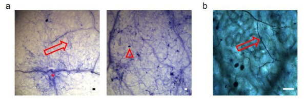
(a) Size profile of HpasRNAs revealed two size peaks at 21 nt and 25 nt at 4 and 7 dpi. (b) The frequency of the first nucleotide at 5’ terminal positions of HpasRNAs mapping to cDNAs or non-annotated regions revealed bias towards uracil. (c) Size distribution and first nucleotide analysis of AtAGO1-associated HpasRNAs showed size preference at 21 nt with 5’ terminal uracil. (d) A novel Csy4/GUS reporter construct was assembled to detect HpasRNA-directed gene silencing, reporting GUS activity if HpasRNAs were functional to suppress Csy4 expression sequence-specificly. (e) GUS staining of infected leaves at two magnifications revealed sequence-specific reporter silencing at 4 dpi. Csy4 with HpasRNA2 and HpasRNA90 target sequences (ts) is depicted on the top and with random scrambled ts on the bottom. Red arrows indicate H. arabidopsidis hyphae in the higher magnification images. Scale bars indicate 50 µm. Numbers in the micrographs indicate number of leaves showing GUS activity per total leaves inspected.

Figure 1.

Figure 1—figure supplement 1. Insights into the small RNAome of H. arabidopsidis and Arabidopsis.

Figure 1—figure supplement 1.

(a) HpasRNAs mapped to distinct coding and to non-coding genomic regions. (b) Relative read counts and size distribution of HpasRNAs mapped to different genomic regions at 4 and 7 dpi. (c) Size distribution and first nucleotide analysis of AtAGO1-associated sRNAs of A. thaliana.
Figure 1—figure supplement 2. Stem-loop RT-PCR revealed HpasRNA2, HpasRNA30 and HpasRNA90 expression at 4 and 7 dpi in three biological replicates.

Figure 1—figure supplement 2.

Total RNA served as loading control.
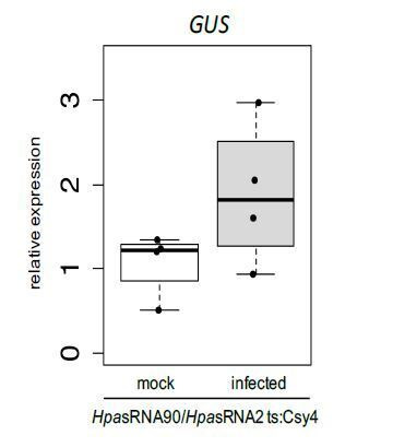
Figure 1—figure supplement 3. Relative expression of AtAED3 and AtWNK2 was measured in mock-treated or H. arabidopsidis inoculated plants.

Figure 1—figure supplement 3.

arabidopsidis-infected WT and atago1-27 seedlings before and at 4 and 7 dpi by qRT-PCR using AtActin as a reference in two independent infection experiments (a, b). The bars within the graphs represent the average of n ≥ three biological replicates, and letters indicate groups of statistically significant difference within one time point by ANOVA followed by TukeyHSD with p≤0.05. Numbers below the graphs give change-fold factors of H. arabidopsidis-infected versus mock-treated samples.
Figure 1—figure supplement 4. 5’ RACE PCR did not provide evidence for pathogen sRNA mediated target cleavage.

Figure 1—figure supplement 4.

(a) Agarose gel images show RACE-PCR bands amplified at the predicted cleavage size of 440 bp for AtAED3 and 530 bp for AtWNK2, marked with asterisks. (b) Mapping schemes of AtWNK2 and AtAED3 mRNA reads indicated the ends of RACE-PCR fragments corresponding to a as revealed by next generation sequencing. (c) Numbers of 5’ end position of AtWNK2 and AtAED3 mRNAs at the predicted HpasRNA2 or HpasRNA90 target sites.
Figure 1—figure supplement 5. The reporter was neither activated by an endogenous miRNA target site nor by a distinct pathogen.

Figure 1—figure supplement 5.

(a) Csy4 repressor reporter with HpasRNA2 and HpasRNA90 target sequence (ts) is depicted on the top and with AtmiRNA164 ts of the AtCUC2 target gene on the bottom. GUS staining of infected leaves at two magnifications revealed sequence-specific reporter silencing at 4 dpi in HpasRNA2/HpasRNA90 ts construct but not in AtmiRNA164 ts. (b) The Csy4 reporter was not activated by the oomycete pathogen Phytophthora capsici. At 2 dpi, P. capsici formed a dense hyphal network, but no pathogen-associated GUS activity was observed (upper panel). In two of the five inspected leaf discs GUS activity was detected in cell clusters, but these were independent of pathogen presence (lower panel). The numbers indicate leaves with GUS activity per total inspected leaves in this experiment. Red arrows indicate H. arabidopsidis or P. capsici hyphae. Scale bars indicate 50 µm.

Two predicted Arabidopsis mRNAs targets of HpasRNAs are down-regulated upon infection

In the following assays to investigate the function of HpasRNAs in ck-RNAi, we chose the AtAGO1-enriched sRNA candidates HpasRNA2 and HpasRNA90. These two HpasRNAs were predicted to target the Arabidopsis WITH NO LYSINE (K) KINASE (AtWNK)2 and the extracellular protease APOPLASTIC, ENHANCED DISEASE SUSCEPTIBILITY1-DEPENDENT (AtAED)3, respectively (Supplementary file 2). We focussed on these two HpasRNAs and target genes, because AtWNK2 and AtAED3 mRNA levels were lower in leaves infected with a virulent H. arabidopsidis strain compared to an avirulent in a previous RNA-seq study (Asai et al., 2014), suggesting a negative impact of H. arabidopsidis proliferation on target transcript accumulation. Further on, members of the WNK protein family as well as AtAED3 have been previously linked to plant stress response and immunity, respectively (Balakireva and Zamyatnin, 2018; Cao-Pham et al., 2018). We confirmed expression of HpasRNA2 and HpasRNA90 in infected plants at 4 and 7 dpi by stem-loop reverse transcriptase (RT)-PCR (Figure 1—figure supplement 2). We then performed quantitative (q)RT-PCR to measure AtWNK2 and AtAED3 mRNAs expressed in whole seedling leaves of wild type (WT) plants upon H. arabidopsidis infection or mock treatment. We used the atago1-27 mutant as a control line, because we anticipated that target suppression should fail in this mutant. Indeed, AtAED3 was significantly down-regulated upon H. arabidopsidis inoculation at 7 dpi, and AtWNK2 expression indicated moderate suppression at 4 dpi in WT plants, when compared to mock-treated plants (Figure 1—figure supplement 3a). Because the down-regulation effects were rather moderate, we repeated this experiment with a second independent H. arabidopsidis inoculation that validated the qRT-PCR results (Figure 1—figure supplement 3b). In support of AtAGO1-mediated target silencing through HpasRNAs, WT-like suppression of AtWNK2 and AtAED3 was not observed in the atago1-27 background (Figure 1—figure supplement 3). However, AtAED3 expression data also indicated down-regulation upon mock treatment during the course of the experiment that might have been caused by the almost 100% relative air humidity during the assay. Moreover, higher transcript levels were measured in atago1-27 before infection when compared to WT plants.

As Arabidopsis target transcripts displayed expressional down-regulation upon H. arabidopsidis infection in WT plants, we wanted to explore, if HpasRNAs guided mRNA slicing of AtWNK2 and AtAED3 through the host AtAGO1/RISC during infection. AtAGO1 possesses RNA cleavage activity on AtmiRNA-guided target mRNAs at the position 10/11 counted from the 5’ end of the miRNA (Mallory and Bouché, 2008). We performed 5’ rapid amplification of cDNA-ends (RACE)-PCR analysis to determine the 5’ ends of target transcripts in RNAs isolated from infected plants pooled from 4 and 7 dpi. We isolated PCR products at the predicted cleavage sizes (Figure 1—figure supplement 4a) for next generation sequencing analysis. In total, we obtained 58,954 and 88,697 reads mapping to AtWNK2 and AtAED3, respectively. However, only a small fraction of reads (639 for AtWNK2 and 17 for AtAED3) mapped at the predicted target sites, while most reads aligned to further 3’ downstream regions indicating rapid RNA degradation (Figure 1—figure supplement 4b). The 5’ ends that matched to the predicted target sites did not display any predominant peak at the expected cleavage position 10/11, but were rather scattered over the entire target sites (Figure 1—figure supplement 4c). Therefore, RACE-PCR did not support HpasRNA-guided cleavage of the Arabidopsis target mRNAs.

HpasRNAs translocate into Arabidopsis and induce host gene silencing in infected plant cells

To further examine if translocation of HpasRNAs into Arabidopsis was sufficient to induce plant gene silencing during infection, we designed a novel in situ silencing reporter. This reporter is based on the CRISPR endonuclease Csy4 that specifically binds to and cleaves a short RNA sequence motif (Haurwitz et al., 2010). We fused this cleavage motif to a β-glucuronidase (GUS) reporter gene to mark it for degradation by Csy4 (Figure 1d). Further on, we cloned the native AtWNK2 and AtAED3 target sequences of HpasRNA2 and HpasRNA90 as flanking tags to the Csy4 coding sequence that turned Csy4 into a target of these HpasRNAs. If HpasRNAs would be capable of silencing effectively the Csy4 transgene, we expected an activation of GUS. Moreover, we constructed control reporters with either a scrambled target sequence or with the binding sequence taken from the endogenous AtmiRNA164 target gene AtCUC2 (Nikovics et al., 2006) instead of the HpasRNA2/HpasRNA90 target sequences. With these control reporters, we intended to test if any HpasRNA2/HpasRNA90-independent suppression of Csy4 or any ck-RNAi-unrelated effect could result in GUS activation. Using the AtmiR164 target site, we anticipated to induce infection-independent local Csy4 silencing, because AtmiR164 expression in young, developing leaves was previously described to be locally restricted to defined regions at the leaf teeth and in the apical meristem (Nikovics et al., 2006). To simulate AtWNK2 target mRNA expression level of the Csy4 reporter transgene, we used a 2 kb-DNA fragment upstream of the AtWNK2 start codon as a promoter sequence for all reporter constructs.

We transformed the reporter variants into Arabidopsis to examine the silencing efficiency of HpasRNAs on predicted plant targets upon infection. In each experiment, we tested at least three individual T1 lines per construct, and all plants appeared to be fully compatible with H. arabidopsidis. Csy4 successfully blocked GUS activity in plant cells that were not close to H. arabidopsidis infection sites (Figure 1e), providing evidence for functional GUS repression by Csy4. Plants expressing Csy4 transcripts fused to HpasRNA2 and HpasRNA90 target sequences highlighted GUS activation along the H. arabidopsidis hyphal infection front (Figure 1e). This experiment provided visual insights into the effective plant gene silencing by pathogen sRNAs, and thus let us assume that efficient sRNA translocation from the pathogen into the host cell occurred. GUS activity emerged only around the pathogen hyphae indicating that ck-RNAi did not spread further into distal regions away from primary infection sites. In contrast, Csy4 linked to a randomly scrambled or AtmiRNA164 target sequence did not express GUS activation around the H. arabidopsidis hyphae (Figure 1e, Figure 1—figure supplement 5a). We concluded that GUS activity induced by H. arabidopsidis in plants expressing Csy4 fused to HpasRNA2/HpasRNA90 target sites was neither due to target sequence-unspecific regulation of Csy4 or GUS nor due to pathogen-triggered regulation of the AtWNK2 promoter. Moreover, reporter plants did also not display any local GUS activity at infection sites when inoculated with the unrelated oomycete pathogen Phytophthora capsici (Figure 1—figure supplement 5b). This result further supported that the GUS reporter was activated specifically by HpasRNAs and not by infection stress.

Arabidopsis atago1 exhibited enhanced disease resistance against downy mildew

Over one hundred HpasRNAs were detected to associate with the plant AGO1/RISC during infection, with 34 HpasRNAs being predicted to silence 49 plant targets including stress-related genes (Supplementary file 2). Such HpasRNAs can induce host target gene silencing at the infection site (Figure 1e). Based on these observations, we hypothesized that AtAGO1 was relevant for H. arabidopsidis to suppress plant defence genes for infection. To test this hypothesis, we compared the disease outcome of atago1-27 with WT plants. The atago1-27 line represents a hypomorphic mutant, and developmental alterations are relatively mild compared to other atago1 mutant alleles (Morel et al., 2002). Therefore, this atago1 mutant line was suitable to perform pathogen infection assays. We stained infected leaves with Trypan Blue that visualized H. arabidopsidis infection structures and indicated plant cell death using a bright-field light microscope. The atago1-27 plants exhibited a remarkable change of the disease phenotype by exhibiting dark Trypan Blue-stained host cells around hyphae instead of unstained plant cells colonized with H. arabidopsidis haustoria in WT plants (Figure 2a). We interpreted this disease phenotype in atago1-27 plants as trailing necrosis of plant cells, which has been described for sub-compatible A. thaliana/H. arabidopsidis interactions (Coates and Beynon, 2010). Indeed, the trailing necrosis co-occurred with enhanced disease resistance, because H. arabidopsidis DNA content was strongly reduced (Figure 2b) and the number of H. arabidopsidis conidiospores was significantly lower in atago1-27 (Figure 2c). Pathogen DNA content was also reduced in atago1-27 cotyledons (Figure 2—figure supplement 1a) without displaying the trailing necrosis (Figure 2—figure supplement 1b). This reduced disease phenotype was linked to atago1 mutations, as independent hypomorphic mutant alleles of atago1-45 and atago1-46 also displayed trailing necrosis after H. arabidopsidis inoculation, albeit to a smaller extent (Figure 2—figure supplement 1c). On the contrary, atago2-1 and atago4-2 did neither exhibit trailing necrosis nor reduced oomycete biomass (Figure 2—figure supplement 1d–e). We confirmed that HpasRNA2 and HpasRNA90 preferably bound to AtAGO1 compared to AtAGO2 by AtAGO-IP coupled to stem-loop RT-PCR (Figure 2—figure supplement 2). This result was consistent with the observed reduced disease level in the atago1 mutant lines in contrast to atago2-1.

Figure 2. Arabidopsis atago1 exhibited enhanced disease resistance against H. arabidopsidis.

(a) Trypan Blue-stained microscopy images showed trailing necrosis around hyphae in atago1-27, but no necrosis on WT seedling leaves at 7 dpi. Red arrow in WT marks H. arabidopsidis haustorium, red arrow in atago1-27 indicates trailing necrosis. (b) H. arabidopsidis genomic DNA was quantified in atago1-27 and WT plants by qPCR at 4 dpi relative to plant genomic DNA represented by n ≥ four biological replicates. (c) Numbers of conidiospores per gram leaf fresh weight (FW) in atago1-27 and WT plants at 7 dpi are represented by four biological replicates. (d) Trypan Blue-stained microscopy images of atdcl1-11 did not show any trailing necrosis at 7 dpi. (e) H. arabidopsidis genomic DNA in atdcl1-11 and WT plants at 4 dpi were in tendency enhanced with n ≥ four biological replicates. (f) Number of conidiospores per gram leaf fresh weight (FW) in atdcl1-11 at 7 dpi was significantly elevated compared to WT plants. (g) Trypan Blue-stained microscopy images of atrdr6-15 and atdcl2dcl3dcl4 showed no plant cell necrosis after inoculation with H. arabidopsidis at 7 dpi. (h) H. arabidopsidis genomic DNA content in leaves was elevated in atrdr6-15 and atdcl2dcl3dcl4 compared to WT at 4 dpi with n ≥ four biological replicates. Asterisk indicates statistically significant difference by one tailed Student’s t-test with p≤0.05. Letters indicate groups of statistically significant difference by ANOVA followed by TukeyHSD with p≤0.05. Scale bars in all microscopy images indicate 50 µm and numbers in the micrographs represent observed leaves with necrosis per total inspected leaves.

Figure 2.

Figure 2—figure supplement 1. Enhanced resistance against infection was restricted to atago1 mutants.

Figure 2—figure supplement 1.

(a) H. arabidopsidis genomic DNA content in cotyledons was lower in atago1-27 compared to WT, as measured by qPCR relative to plant genomic DNA at 4 dpi with n ≥ three biological replicates. (b) Trypan Blue-stained microscopy images of atago1-27 cotyledons did not show any necrosis at 7 dpi. (c) Trypan Blue-stained microscopy images of atago1-45 and atago1-46 revealed trailing necrosis at 7 dpi with H. arabidopsidis. (d) Trypan Blue-stained microscopy images presenting H. arabidopsidis-infected atago2-1 and atago4-2 seeding leaves at 7 dpi. (e) H. arabidopsidis genomic DNA was quantified in WT versus atago2-1 and atago4-2 by qPCR at 4 dpi relative to plant genomic DNA represented by n ≥ four biological replicates. Numbers in the micrographs represent observed leaves with necrosis per total inspected leaves. Asterisk indicates significant difference by one tailed Student’s t-test with p≤0.05. Letters indicate groups of statistically significant difference by ANOVA followed by TukeyHSD with p≤0.05.
Figure 2—figure supplement 2. Stem-loop RT-PCR of HpasRNAs from AtAGO1-IP or AtAGO2-IP of mock-treated or H. arabidopsidis infected leaf tissue.

Figure 2—figure supplement 2.

arabidopsidis-inoculated seedlings. AtmiRNA164 and AtmiRNA393* were used as positive AtAGO-IP controls. Pull-down of AtAGO1 was achieved with WT plants using an AtAGO1 native antibody, and AtAGO2 with HA-epitope tagged AtAGO2-expressing A. thaliana Col-0 using anti-HA antibody with the lower panel showing Western blot analysis.
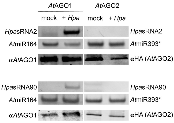
Figure 2—figure supplement 3. Trypan Blue-stained microscopy images presenting the AtmiRNA biogenesis mutants athst-6, athen1-5 and atse-2 did not show any trailing necrosis at 7 dpi.

Figure 2—figure supplement 3.

Scale bars in microscopy images indicate 50 µm and numbers in the micrographs represent observed leaves with necrosis per total inspected leaves.
Figure 2—figure supplement 4. Common defence-related marker gene induction was not enhanced in atago1-27 mutants.

Figure 2—figure supplement 4.

(a) Expression analysis of AtPR1 by RT-PCR in WT and atago1-27 did not show obvious differences at 6 and 12 h post inoculation with H. arabidopsidis. AtActin was used as reference gene with four biological replicates. (b and c) Relative expression of AtPR1 and AtPDF1.2 determined by qRT-PCR using AtActin as reference. The bar represents the average of n ≥ three biological replicates, each comprising two technical replicates.
Figure 2—figure supplement 5. Relative mRNA expression of AtRBOHD and AtRBOHF determined by qRT-PCR using AtActin as reference in WT and atago1-27 in H. arabidopsidis and mock treated plants.

Figure 2—figure supplement 5.

The bars represent the average of n ≥ three biological replicates, each comprising two technical replicates. Letters indicate groups of statistically significant difference within one time point by ANOVA followed by TukeyHSD with p≤0.05.
Figure 2—figure supplement 6. Susceptibility of atago1 mutants to infection with the biotrophic fungus E. cruciferarum and the oomycete A. laibachii remained unaltered.

Figure 2—figure supplement 6.

(a) Trypan Blue-stained microscopy images of WT or atago1-27 leaves infected with Erysiphe cruciferarum did not show necrosis at 8 dpi. Scale bars in microscopy images indicate 50 µm and numbers represent observed leaves with necrosis per total inspected leaves. (b) E. cruciferarum genomic DNA content in WT and atago1-27 was not significantly different at 4 dpi relative to plant genomic DNA as measured by qPCR in n ≥ three biological replicates. (c) Macroscopic infection phenotype of the white rust Albugo laibachii remained unaltered in atago1-27 and atago1-46 mutants at 3 weeks post inoculation. (d) Trypan Blue-stained microscopy images of WT, atago1-27 or atago1-46 leaves infected with A. laibachii did not show necrosis at 7 dpi. Scale bars in microscopy images indicate 50 µm and numbers represent observed leaves with necrosis per total inspected leaves. Scale bars in the total leaf pictures indicate 50 mm. Significance was determined by one tailed Student’s t-test with p≤0.05.

Taken together, these data strongly suggested that translocated HpasRNAs act mainly through AtAGO1 to suppress plant genes for infection. Nevertheless, increased disease resistance of atago1 plants could have been caused by impaired function of plant endogenous sRNAs. For instance, atago1 mutant plants as well as other miRNA pathway mutants, such as atdcl1, athua enhancer(hen)1 athasty(hst) or atserrate(se) show pleiotropic developmental defects because of impaired plant sRNA function (Li and Zhang, 2016; Vaucheret, 2008). To test whether other miRNA pathway mutants also revealed enhanced disease resistance similar to atago1 plants, we inoculated the atdcl1-11 mutant line with H. arabidopsidis. We did not detect any trailing necrosis or reduced pathogen biomass, but in contrast a significantly increased number of conidiospores (Figure 2d–f) indicating a positive role of A. thaliana miRNAs in immune response against H. arabidopsidis. These results provided evidence that necrotic trailing and reduced pathogen susceptibility found in atago1 was not due to the loss of a functional plant miRNA pathway. In support, we did also not observe trailing necrosis upon infection in the atse-2, athen1-5 and athst-6 mutants (Figure 2—figure supplement 3).

Since atago1 exhibited trailing necrosis and reduced susceptibility to H. arabidopsidis, we wanted to examine if constant activation of defence-related marker genes corresponded with enhanced disease resistance. We profiled gene expression of the A. thaliana immunity marker gene AtPATHOGENESIS-RELATED (PR)1. AtPR1 was neither faster nor stronger induced at 6, 12 or 18 h post inoculation in atago1-27 compared to WT (Figure 2—figure supplement 4a). AtPR1 and another immunity marker AtPLANT-DEFENSIN (PDF)1.2 were not higher expressed in atago1-27 at 1, 4 or 7 dpi compared to WT before or after infection (Figure 2—figure supplement 4b–c). To examine plant gene expression related to induced plant cell death, as observed in ago1 mutants, we measured transcript levels of the two NADPH oxidases At REACTIVE BURST OXIDASE HOMOLOG (AtRBOH)D and AtRBOHF. Both genes are required for accumulation of reactive oxygen intermediates to suppress spread of cell death during plant defence (Torres et al., 2005). Moreover, the atrbohd and atrbohf knockout mutant plants previously revealed increased plant cell death after H. arabidopsidis infection and were more resistant against this pathogen (Torres et al., 2002). In consistence, we found that AtRBOHD and AtRBOHF were induced in WT plants at 7 dpi and were significantly higher expressed than in atago1-27 (Figure 2—figure supplement 5). These results gave a first hint of a host defence pathway that might be affected due to AtAGO1-associated HpasRNAs.

Plant miRNAs can initiate the production of secondary phased siRNAs (phasiRNAs), which negatively control the expression of NLR (NOD-like receptor) class Resistance (R) genes (Li et al., 2012; Shivaprasad et al., 2012). Constitutive expression of NLR genes promotes immune responses such as spontaneous plant cell death resembling a hypersensitive response (Lai and Eulgem, 2018). Therefore, lack of phasiRNAs in atago1 could cause enhanced expression of NLRs leading to resistance against H. arabidopsidis. To examine R gene-based enhanced resistance due to lack of phasiRNAs, we inoculated the atrdr6-15 and atdcl2dcl3dcl4 mutants with H. arabidopsidis Noco2. The production of phasiRNAs depends on AtRDR6 and AtDCL2/AtDCL3/AtDCL4 (Fei et al., 2013). Both mutants did not exhibit trailing necrosis (Figure 2g), but in contrast highlighted increased pathogen biomass upon inoculation with H. arabidopsidis (Figure 2h). Higher susceptibility of atrdr6-15 and atdcl2dcl3dcl4 to H. arabidopsidis was also in line with a previous report suggesting a role of Arabidopsis phasiRNAs in silencing of Phytophothora genes for host plant defence (Hou et al., 2019).

In order to further explore whether atago1-27 was more resistant to other biotrophic fungi or oomycetes, we performed infection assays with the powdery mildew fungus Erysiphe cruciferarum and the white rust oomycete Albugo laibachii. We did not observe any plant cell necrosis in neither pathogen. Moreover, there was neither a reduction in the pustules for A. laibachii nor in pathogen biomass of E. cruciferarum (Figure 2—figure supplement 6a–d). Taken together, the observed disease resistance of atago1 plants against H. arabidopsidis was probably neither based on increased basal plant immunity nor on R gene-mediated resistance.

HpasRNAs are crucial for virulence

As we realized that HpasRNAs were associated with the host AtAGO1-RISC, silenced plant target genes, and that Arabidopsis atago1 mutants displayed reduced susceptibility towards H. arabidopsidis infection, we wanted to understand how important HpasRNAs were for H. arabidopsidis virulence. To shed light on the relevance of HpasRNAs for infection, we cloned and expressed a short-tandem-target-mimic (STTM) RNA in Arabidopsis to sequester HpasRNAs. The STTM strategy has been previously used to scavenge endogenous plant sRNAs and to prevent gene silencing of native target genes (Tang et al., 2012). We designed a triple STTM transgene to simultaneously bind the pathogen sRNAs HpasRNA2, HpasRNA30, and HpasRNA90 by RNA base-pairing. A non-complementary 3-base loop structure at the position 10/11 counted from the 5’ end of the HpasRNAs was deliberately incorporated to block potential cleavage by plant AGO/RISCs, as previously described (Tang et al., 2012; Figure 3a). We included the AtAGO1-associated HpasRNA30 in the triple STTM, because it was predicted to silence AtWNK5 (Supplementary file 2), a homolog of AtWNK2, thus we presumed that HpasRNA30-induced AtWNK5 suppression might also be important for virulence. The HpasRNA30 sequence mapped only to the H. arabidopsidis, but not the Arabidopsis genome, and we detected this HpasRNA in infected plants at 4 and 7 dpi by sRNA-seq and stem-loop RT-PCR (Figure 1—figure supplement 2, Supplementary file 2). Remarkably, seven out of eleven individual STTM T1 transgenic lines resembled partially the trailing necrosis phenotype of atago1 (Figure 3b). We isolated two stable STTM T2 lines (#4, #5). The STTM #4 line showed target de-repression of AtAED3 at 7 dpi and of AtWNK2 at 4 dpi upon H. arabidopsidis inoculation when compared to plants expressing an empty vector control (Figure 3—figure supplement 1a). These time points corresponded to target gene suppression as found by qRT-PCR analysis before (Figure 1—figure supplement 3). Moreover, both STTM T2 lines exhibited reduced pathogen biomass (Figure 3—figure supplement 1b) and allowed significantly lower production of pathogen conidiospores (Figure 3c). We also cloned STTMs against an rRNA-derived HpasRNA as well as against a random scrambled sequence for expression in Arabidopsis. These two types of control STTMs did not exhibit trailing necrosis in at least five independent T1 transgenic lines upon H. arabidopsidis inoculation (Figure 3d). Furthermore, we also did not observe disease resistance in transgenic plants expressing the STTM against HpasRNA2/HpasRNA30/HpasRNA90 when inoculated with the unrelated bacterial pathogen Pseudomonas syringae DC3000 (Figure 3—figure supplement 1c). These experiments provided evidence that the expression of anti-HpasRNA STTMs in Arabidopsis blocked HpasRNAs activity that resulted in reduced virulence of H. arabidopsidis.

Figure 3. Translocated HpasRNAs were crucial for virulence.

(a) A triple STTM construct was designed to target the three HpasRNAs HpasRNA2, HpasRNA30 and HpasRNA90 in Arabidopsis. (b) A. thaliana T1 plants expressing the triple STTM to scavenge HpasRNA2, HpasRNA30 and HpasRNA90 exhibited trailing necrosis at 7 dpi. (c) Number of conidiospores per gram FW was significantly reduced in two independent STTM-expressing Arabidopsis T2 lines (#4, #5) compared to WT. (d) Transgenic Arabidopsis plants in T1 expressing a STTM complementary to a rRNA-derived HpasRNA (STTMrRNA) or to a random scrambled (STTMscrRNA) sequence did not exhibit trailing necrosis at 7 dpi. The scale bars indicate 50 µm and numbers represent observed leaves with necrosis per total inspected leaves.

Figure 3.

Figure 3—figure supplement 1. STTM plants revealed higher expression of target genes and lower H. arabidopsidis abundance.

Figure 3—figure supplement 1.

(a) Relative expression of AtAED3 at 7 dpi and AtWNK2 at 4 dpi was determined for STTM or empty vector (ev) expressing plants upon H. arabidopsidis inoculation at 7 and 4 dpi, respectively, by qRT-PCR. One biological replicate represented three leaves, the bars represent the average of n ≥ three biological replicates. The differences of the average were not statistically significant as determined by Student’s t-test. (b) H. arabidopsidis genomic DNA content in leaves was increased in STTM #4 and STTM #5 plants compared to empty vector (ev) expressing WT plants at 4 dpi with n ≥ three biological replicates. The differences were not statistically significant as determined by ANOVA followed by TukeyHSD. (c) STTM-expressing Arabidopsis plants did not exhibit increased resistance against bacterial infection. The bacterial growth was determined by counting colony-forming units (cfu) at 3 days post inoculation. One biological replicate represents bacteria from three leaf discs. Letters in c) indicate significant difference (p≤0.05) according to one site ANOVA followed by TukeyHSD including three biological replicates.

Arabidopsis target genes of HpasRNAs contribute to plant defence

Upon uncovering the importance of HpasRNAs for virulence, we wanted to assess the contribution of Arabidopsis target genes to plant defence. We obtained three T-DNA insertion lines for the identified target genes AtWNK2 and AtAED3, namely atwnk2-2, atwnk2-3, and ataed3-1 (Figure 4—figure supplement 1a). While atwnk2-2 and ataed3-1 are two SALK/SAIL lines (Alonso et al., 2003; Sessions et al., 2002) that carry a T-DNA insertion in their coding sequence, respectively, we now re-located the T-DNA insertion of the atwnk2-3 plant line from the last exon into the 3’ UTR, based on sequencing the T-DNA flanking sites (Figure 4—figure supplement 1a). To study infection phenotypes, we stained H. arabidopsidis-infected leaves with Trypan Blue, and all T-DNA insertion lines resembled pathogen infection structures like in WT plants. However, haustorial density, indicated by the number of haustoria formed per intercellular hyphal distance, was significantly increased in atwnk2-2 (Figure 4—figure supplement 1b). Intensified haustoria formation was previously interpreted as a sign of enhanced susceptibility in other plant/downy mildew pathogen interactions (Hooftman et al., 2007; Unger et al., 2007). Moreover, the pathogen DNA content was slightly but not significantly increased in atwnk2-2 and ataed3-1 compared to WT plants, but this was not the case for atwnk2-3 (Figure 4a). Nevertheless, a significantly increased number of conidiospores (Figure 4b) and sporangiophores (Figure 4c) was observed in all the tested atwnk2 and ataed3 mutant lines upon H. arabidopsidis infection compared to WT plants.

Figure 4. Arabidopsis target genes of HpasRNAs contributed to plant defence.

(a) H. arabidopsidis genomic DNA content in leaves was slightly but not significantly enhanced in atwnk2-2 and ataed3-1 compared to WT, but not in atwnk2-3, at 4 dpi with n ≥ four biological replicates. (b) T-DNA insertion lines of HpasRNA target genes ataed3-1, atwnk2-2, and atwnk2-3 showed significantly higher number of sporangiophores per cotyledon upon infection compared to WT at 5 dpi. (c) ataed3-1, atwnk2-2, and atwnk2-3 showed significantly higher numbers of conidiospores per gram leaf FW upon infection compared to WT at 5 dpi. (d) Number of conidiospores was significantly reduced in gene-complemented mutant lines using the corresponding native promoters proAtEWNK2 or proAtAED3 with native gene sequence, AtAED3 and AtWNK2, or with target site resistant versions, AtAED3r and AtWNK2r compared to the knockout mutant background expressing an empty vector (ev), respectively. Asterisks indicate significant difference by one tailed Student’s t-test with p≤0.05. Letters indicate significant difference by one-site ANOVA test.

Figure 4.

Figure 4—figure supplement 1. Further details on sRNA target gene mutants.

Figure 4—figure supplement 1.

(a) Gene models of AtWNK2 and AtAED3. The insertion site of the T-DNA is marked by the triangles and the genotyping primer binding sites are shown with arrows. T-DNA insertion of HpasRNA target gene mutant lines atwnk2-2, atwnk2-3 and ataed3-1 were verified by genomic DNA PCR. (b) Trypan Blue-stained microscopy images revealed a higher number of haustoria in the first 200 µm of hyphae (indicated by white bar alongside the hyphae) from the spore germination site in atwnk2-2 compared to WT with n ≥ eight leaves. Asterisk indicates significant difference by one tailed Student’s t-test with p≤0.05. Similar results were obtained in two independent experiments.
Figure 4—figure supplement 2. Target sequence-resistant versions of AtAED3 (AtAED3r) and AtWNK2 (AtWNK2r) were created by introducing synonymous nucleotide substitutions indicated by red letters.

Figure 4—figure supplement 2.

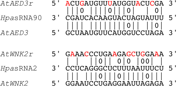
Figure 4—figure supplement 3. Transgenic A. thaliana atwnk2-2 was complemented with proWNK2:WNK2 or proWNK2:WNK2r that resulted in a WT-like flowering time point, while empty vector (ev) exhibited early flowing phenotype, as reported for atwnk2-2 (Wang et al., 2008).

Figure 4—figure supplement 3.

Figure 4—figure supplement 4. A. thaliana plants overexpressing proLjUBI1:AtWNK2r in the atwnk2-2 background revealed local necrosis without pathogen infection (a) and aberrant hyphae and haustoria swellings (b).

Figure 4—figure supplement 4.

Scale bars in microscopy images indicate 50 µm and the numbers represent observed leaves with necrosis or swellings respectively per total inspected leaves.
Figure 4—figure supplement 5. Sequence diversity of HpasRNAs and their predicted Arabidopsis target mRNAs.

Figure 4—figure supplement 5.

(a) HpasRNAs were conserved among the three H. arabidopsidis isolates, Noco2, Cala2, Emoy2. (b) Sequence variations were found in the predicted Arabidopsis mRNAs analysing 1135 A. thaliana accessions. Three categories (colour coded) were considered to possibly prevent target silencing: indels causing bulges that block the HpasRNA/AtmRNA base pairing (red), SNPs at the position 10/11 interfering with RISC-mediated cleavage (orange), and SNPs in the HpasRNA seed region (account to RNA nucleotide positions from 2 to 12) loosing HpasRNA/AtmRNA base pairing (green).
Figure 4—figure supplement 6. The pathogen sRNA2 and its target are conserved across different plant pathogenic oomycetes and hosts.

Figure 4—figure supplement 6.

(a) Oomycete SRNA2 genomic loci are conserved among different plant pathogenic oomycete species of the genera Hyaloperonospora, Phytophthora, and Pythium (Hpa = Hyaloperonospora arabidopsidis, Pcap = Phytophthora capsici, Pso = Phytophthora sojae, Pan = Pythium aphanidermatum, Pinf = Phytophthora infestans, Ppa = Phytophthora parasitica). Blue box at the consensus sequence indicates the region of sRNA transcription as identified by sRNA-seq analysis and red box marks the consensus of the mature 21 nt HpasRNA2 region. (b) Target prediction alignment of sRNA2 homologs from different oomycete species with the target sequences of homolog WNK2s from respective host plant species (At = Arabidopsis thaliana, Gm = Glycine max, St = Solanum tuberosum, Nt = Nicotiana tabacum).

We wanted to investigate in more detail the effect of target gene silencing by HpasRNAs on plant defence. For this, we cloned AtWNK2 and AtAED3 target genes either as native versions or artificially introduced synonymous point mutations in the target sites of HpasRNAs to generate the target gene-resistant versions AtAED3r and AtWNK2r (Figure 4—figure supplement 2). We transformed these gene versions into the respective mutant background ataed3-1 and atwnk2-2 expressing them under the control of their native promoters. Transgenic AtWNK2 and AtWNK2r expressing plants reverted from previously described early flowering of atwnk2-2 (Wang et al., 2008) into the WT phenotype validating successful complementation of atwnk2-2 (Figure 4—figure supplement 3). If AtWNK2 and AtAED3 silencing through HpasRNA2 or HpasRNA90 was relevant to plant defence, we would expect that AtWNK2r and AtAED3r expressing plants become more resistant against H. arabidopsidis. Both, the native gene versions and the target site resistant versions, exhibited reduced number of conidiospores compared to T-DNA mutant plants transformed with an empty expression vector, respectively (Figure 4d). To further explore the role of target genes in plant immunity, we attempted to generate overexpression lines of resistant target gene versions by using the strong Lotus japonicus Ubiquitin1 promoter (proLjUbi1) (Maekawa et al., 2008). We obtained an overexpressor line of the AtWNK2r version (AtWNK2r-OE) in the atwnk2-2 background. These AtWNK2r-OE plants showed ectopic cell death in distance from infection sites (Figure 4—figure supplement 4a), as previously described for overexpression lines of other immunity factors, such as AtBAK1 (Domínguez-Ferreras et al., 2015). Moreover, infection structures frequently displayed aberrant swelling-like structures and extensive branching of hyphae instead of the regular pyriform haustoria formed in atwnk-2–2 (Figure 4—figure supplement 4b), further indicating a role for AtWNK2 in immune reaction.

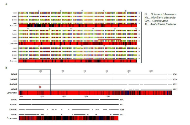
To gain more information on the conservation of the 34 identified AtAGO1-associated HpasRNAs (Supplementary file 2), we analysed RNA sequence diversity using the H. arabidopsidis sequenced genomes of the Noco2, Cala2 and Emoy2 isolates (NCBI BioProject IDs: PRJNA298674; PRJNA297499, PRJNA30969). In a complementary approach, we investigated the variation of the 49 predicted plant target sites among 1135 A. thaliana genome sequenced accessions published by the 1001 genome project (1001 Genomes Consortium, 2016). Interestingly, all HpasRNA were found by BLASTn search in the three H. arabidopsidis isolates with only three allelic variations identified in Emoy2 (Figure 4—figure supplement 5a). On the Arabidopsis target site, we found single nucleotide polymorphisms (SNPs) and indels in 70% of all target genes (Supplementary file 2), many of those might impair in the predicted HpasRNA-induced silencing (Figure 4—figure supplement 5b). Of note, the HpasRNA2 sequence was deeper conserved in other pathogenic oomycete species, compared to other HpasRNAs described in this study (Figure 4—figure supplement 6a). Moreover, the predicted target sites of the pathogen siR2 homologs lie within a conserved region of other plant WNK2 orthologs, with the lowest number of base pair mismatches occurring in the highly-adapted A. thaliana/H. arabidopsidis interaction (Figure 4—figure supplement 6b). Whether RNA sequence diversity in HpasRNAs and A. thaliana target mRNAs drives co-evolution in this co-adapted plant-pathogen system, remains to be further investigated.

Discussion

In this study, we discovered that ck-RNAi happened during H. arabidopsidis host infection and contributed to the virulence of this pathogen. Sequencing sRNAs associated with Arabidopsis AGO1 revealed at least 34 HpasRNAs that entered the host RNAi machinery and potentially targeted multiple plant genes for silencing. These deep sequencing data offered first insights into the H. arabidopsidis sRNA transcriptome during host infection. Total read numbers of AtAGO1-bound HpasRNAs were in the ratio of around 1/1000 compared to AtAGO1-bound Arabidopsis sRNAs, raising the concern that concentration of pathogen sRNAs might not be sufficient to be functional. Nevertheless, our and other studies found genetic and phenotypic evidence for pathogen oomycete sRNA function despite read numbers being in the range of ten per million or lower (Jahan et al., 2015; Qutob et al., 2013). By designing a novel Csy4/GUS repressor reporter system, we demonstrated that HpasRNAs have the capacity to translocate into plant cells and suppress host target genes. This new reporter system was capable of visualizing local gene silencing alongside the H. arabidopsidis hyphae. Therefore, the relatively small proportion of HpasRNAs counted in AtAGO1 sRNA-seq experiment could be explained by strong dilution with AtAGO1 molecules purified from non-colonized tissue. For the same reason, we measured moderate AtWNK2 and AtAED3 target gene suppression due to dilution effects coming from non-infected leaf lamina.

We assumed that diverse HpasRNAs were translocated into Arabidopsis during infection and AtAGO1 was a major hub of HpasRNAs, as detected by AtAGO1 pull down and sRNA-seq analysis. By which pathways and mechanisms HpasRNAs move into plant cells remains an open question. Transport via the extrahaustorial matrix could be a realistic cross-point, as many other biomolecules are exchanged via this route from pathogen to plant cells and vice versa (Judelson and Ah-Fong, 2019). It is noteworthy that accumulation of vesicle-like structures was visualized by electron microscopy at the perihaustorial matrix (Mims et al., 2004). In this regard, transfer of plant sRNAs into pathogen cells via exosomal vesicles was reported to induce ck-RNAi (Cai et al., 2018; Hou et al., 2019), making extracellular vesicles a prime suspect for HpasRNA transport into plant cells.

Plant RISC-associated HpasRNAs were crucial for successful infection, because transgenic Arabidopsis generated to block the suppressive function of the three candidate HpasRNA2, HpasRNA30 and HpasRNA90 via STTM target mimics diminished H. arabidopsidis virulence. As we identified 34 AtAGO1-associated HpasRNAs with 49 predicted plant target genes, we suggest that many HpasRNAs collaboratively sabotage gene expression of the plant immune response. Such a collaborative function was also suggested for proteinaceous pathogen effectors (Cunnac et al., 2011).

Regarding the role of identified HpasRNA target genes in host defence, our data supported quantitative contributions of AtAED3 and AtWNK2 to plant immunity. AtAED3 encodes a putative apoplastic aspartyl protease and has been suggested to be involved in systemic immunity (Breitenbach et al., 2014). AtWNK2 contributes to flowering time regulation in A. thaliana, while other members of the plant WNK family have been linked to the abiotic stress response (Cao-Pham et al., 2018). What is the particular function of these target genes against H. arabidopsidis infection and whether these also play a role against other pathogens, still needs to be explored.

The fact that Arabidopsis siRNA biogenesis mutants like atrdr6-15 and atdcl2dcl3dcl4 displayed increased H. arabidopsidis growth is an indication for the important role of secondary phasiRNAs in plant immunity, that was already observed against fungal pathogens like Verticillium dahliae and Magnaporthe oryzae (Ellendorff et al., 2009; Wagh et al., 2016). This is likely due to the regulatory function of phasiRNAs on endogenous plant immunity genes including the NLRs (Li et al., 2012; Shivaprasad et al., 2012). Two recent studies suggested suppressive roles of secreted plant phasiRNAs in ck-RNAi by silencing fungal B. cinerea and oomycete P. capsici virulence genes (Cai et al., 2018; Hou et al., 2019). Interestingly, exogenously applied sRNAs targeting the Cellulose synthase 3A gene of H. arabidopsidis can lead to pathogen developmental changes and spore germination inhibition, suggesting functional RNA uptake by this pathogen (Bilir et al., 2019). Together with our data, we think that ck-RNAi in H. arabidopsidis/Arabidopsis interaction is bidirectional, as already described in fungal-plant interactions (Cai et al., 2018; Wang et al., 2016).

This study provides evidence that ck-RNAi, originally discovered in the fungal plant pathogen B. cinerea (Weiberg et al., 2013), is part of virulence in the oomycete biotrophic pathogen H. arabidopsidis. The phenomenon of plant-pathogen ck-RNAi is further proposed in the cereal fungal pathogens Puccinia striiformis (Wang et al., 2017) and Blumeria graminis (Kusch et al., 2018). We did not notice any enhanced resistance in an Arabidopsis atago1 mutant against the biotrophic fungus E. cruciferarum and the oomycete A. laibachii, making ck-RNAi via AtAGO1 unlikely. Further experiments are needed to rule out any importance of ck-RNAi for virulence of these two pathogens via alternative plant AGO-RISCs. The fungal wheat pathogen Zymoseptoria tritici was reported to not induce ck-RNAi (Kettles et al., 2019; Ma et al., 2020), while the corn smut pathogen Ustilago maydis has lost its canonical RNAi machinery (Kämper et al., 2006; Laurie et al., 2008). It will be interesting to elucidate why some pathogens have evolved ck-RNAi, while some others not.

Materials and methods

Key resources table.

Reagent type (species) or resource Designation Source or reference Identifiers Additional information
Gene (Arabidopsis thaliana) AtWNK2 arabidopsis.org AT3G22420
Gene (Arabidopsis thaliana) AtAED3 arabidopsis.org AT1G09750
Gene (Arabidopsis thaliana) AtPR1 arabidopsis.org AT2G14610
Gene (Arabidopsis thaliana) AtPDF1.2 arabidopsis.org AT5G44420
Gene (Arabidopsis thaliana) AtAGO1 arabidopsis.org AT1G48410
Gene (Arabidopsis thaliana) AtAGO2 arabidopsis.org AT1G31280
Strain, strain background (Hyaloperonospora arabidopsidis) Noco2 isolated originally in Norwich, UK
Strain, strain background (Albugo laibachii) Nc14 Kemen et al., 2011
DOI:10.1371/journal.pbio.1001094
Strain, strain background (Pseudomonas syringae pv tomato) DC3000 Whalen et al., 1991
DOI:10.1105/tpc.3.1.49
Strain, strain background (Phytophthora capsici) LT263 Hurtado-Gonzales and Lamour, 2009
DOI: 10.1111/j.1365–3059.2009.02059.x
Genetic reagent (Arabidopsis thaliana) atago1-27 Morel et al., 2002
PMID:11910010
Genetic reagent (Arabidopsis thaliana) atago1-45 Nottingham Arabidopsis stock center (NASC) N67861
Genetic reagent (Arabidopsis thaliana) atago1-46 (Nottingham Arabidopsis stock center (NASC) N67862
Genetic reagent (Arabidopsis thaliana) atago2-1 Takeda et al., 2008
DOI: 10.1093/pcp/pcn043
Genetic reagent (Arabidopsis thaliana) atago4-2 Agorio and Vera, 2007
DOI: 10.1093/pcp/pcn043
Genetic reagent (Arabidopsis thaliana) atdcl1-11 Zhang et al., 2008
DOI: 10.1111/j.1365–3040.2008.01786.x
Genetic reagent (Arabidopsis thaliana) atdcl2dcl3dcl4 Deleris et al., 2006
DOI: 10.1126/science.1128214
triple mutant
Genetic reagent (Arabidopsis thaliana) athen1-5 Vazquez et al., 2004
DOI: 10.1016/j.cub.2004.01.035
Genetic reagent (Arabidopsis thaliana) athst-6 Bollman et al., 2003
PMID:12620976
Genetic reagent (Arabidopsis thaliana) atrdr6-15 Allen et al., 2004
DOI: 10.1038/ng1478
Genetic reagent (Arabidopsis thaliana) atse-2 Grigg et al., 2005
DOI: 10.1038/nature04052
Genetic reagent (Arabidopsis thaliana) proAGO2:HA-AGO2 Montgomery et al., 2008
DOI:10.1016/j.cell.2008.02.033
Genetic reagent (Arabidopsis thaliana) atwnk2-2
(SALK_121042)
Nottingham Arabidopsis stock center (NASC) N663846
Genetic reagent (Arabidopsis thaliana) atwnk2-3 (SALK_206118) Nottingham Arabidopsis stock center (NASC) N695550
Genetic reagent (Arabidopsis thaliana) ataed3-1
(SAIL_722_G02C1)
Nottingham Arabidopsis stock center (NASC) N867202
Genetic reagent (Arabidopsis thaliana) proLjUBI:STTMHasR2:
STTMHasR30:STTMHasR90
this study stable triple STTM overexpressor line (maintained in the Weiberg lab)
Genetic reagent (Arabidopsis thaliana) proAtWNK2:HasRNA2/90ts:Csy4:HasRNA2/90ts; proEF1:Csy4ts:GUS this study stable silencing reporter line (maintained in the Weiberg lab)
Genetic reagent (Arabidopsis thaliana) proAtWNK2:AtmiR164ts:Csy4:AtmiR164ts; proEF1:Csy4ts:GUS this study stable silencing reporter line (maintained in the Weiberg lab)
Genetic reagent (Arabidopsis thaliana) proAtWNK2:scrambled:Csy4:scrambled; proEF1:Csy4ts:GUS this study stable silencing reporter line (maintained in the Weiberg lab)
Genetic reagent (Arabidopsis thaliana) atwnk2-2 (proAtWNK2:AtWNK2-GFP) this study stable WNK2 complementation line (maintained in the Weiberg lab)
Genetic reagent (Arabidopsis thaliana) atwnk2-2 (proAtWNK2:AtWNK2r-GFP) this study stable, sRNA resistant WNK2 complementation line (maintained in the Weiberg lab)
Genetic reagent (Arabidopsis thaliana) atwnk2-2 (proAtWNK2:GFP) this study stable plant line as empty vector control (maintained in the Weiberg lab)
Genetic reagent (Arabidopsis thaliana) ataed3-1 (proAtAED3:AtAED3-GFP) this study stable AED3 complementation line (maintained in the Weiberg lab)
Genetic reagent (Arabidopsis thaliana) ataed3-1 (proAtAED3:AtAED3r-GFP) this study stable, sRNA resistant AED3 complementation line (maintained in the Weiberg lab)
Genetic reagent (Arabidopsis thaliana) ataed3-1 (proAtAED3: GFP) this study stable plant line as empty vector control (maintained
in the Weiberg lab)
Antibody anti-AtAGO1 (rabbit polyclonal) Agrisera AS09 527; RRID:AB_2224930 IP(1 µg antibody/g tissue), WB (1:4000)
Antibody anti-HA (3F10; rat monoclonal) Roche Diagnostics Sigma-Aldrich (11867423001); RRID:AB_2314622 IP(0.1 µg antibody/g tissue), WB (1:1000)
Antibody anti-HA (12CA5; mouse monoclonal) provided by Dr. Michael Boshart IP(0.1 µg antibody/g tissue), WB (1:1000), available in the Boshart lab (LMU Munich)
Antibody anti-mouse IRdye800 (goat polyclonal) Li-Cor 926–32210; RRID:AB_2782998 secondary antibody WB (1:15000)
Antibody anti-rat IRdye800 (goat polyclonal) Li-Cor 926–32219; RRID:AB_1850025 secondary antibody WB (1:15000)
Antibody anti-rabbit IRdye800 (goat polyclonal) Li-Cor 926–32211; RRID:AB_621843 secondary antibody WB (1:3000)
Commercial assay or kit NEBNext Multiplex Small RNA Library Prep Set for
Illumina
New England Biolabs (NEB) NEB: E7300
Commercial assay or kit 5′/3′ RACE Kit, 2nd Generation Roche Diagnostics Sigma-Aldrich: 03353621001
Commercial assay or kit sparQ DNA Library Prep Kit Quantabio vwr.com (95191–024)
Software, algorithm Galaxy Server Giardine et al., 2005 hosted by the Gene Center Munich

Plant material

Arabidopsis thaliana (L.) seedlings were grown on soil under long day conditions (16 hr light/8 hr dark, 22°C, 60% relative humidity). The atago1-27, atago1-45, atago1-46, atago2-1, atago4-2, athst-6, athen1-5, atse-2, atdcl1-11, atdcl2dcl3dcl4, atrdr6-15, and proAGO2:HA-AGO2 mutant lines (all in the Col-0 background) were described previously (Agorio and Vera, 2007; Allen et al., 2004; Bollman et al., 2003; Deleris et al., 2006; Grigg et al., 2005; Morel et al., 2002; Smith et al., 2009; Takeda et al., 2008; Vazquez et al., 2004; Zhang et al., 2008Montgomery et al., 2008). The atwnk2-2 (SALK_121042, [Wang et al., 2008]), atwnk2-3 (SALK_206118) and ataed3-1 (SAIL_722_G02C1) lines were verified for the T-DNA insertion by PCR on genomic DNA.

Hyaloperonospora arabidopsidis inoculation

Hyaloperonospora arabidopsidis (Gäum.) isolate Noco2 was maintained on Col-0 plants. Plant inoculation was performed using 2–2.5 × 10⁴ spores/ml and inoculated plants were incubated as described previously (Ried et al., 2019). For atwnk2-2, atwnk2-3, and ataed3-1 pathogen assays inoculum strength was reduced to 1 × 10⁴ spores/ml.

Albugo laibachii (Thines and Y.J. Choi) inoculation

Plants were grown in short-day conditions (10 hr light, 22°C, 65% humidity/14 hr dark, 16°C, 60% humidity, photon flux density 40 μmol m−2 s−1) and inoculated at the age of six weeks. A. laibachii (isolate Nc14; [Kemen et al., 2011]) zoospores obtained from propagation on Arabidopsis accession Ws-0 were suspended in water (105 spores ml−1) and incubated on ice for 30 min. The spore suspension was filtered through Miracloth (Calbiochem, San Diego, CA, USA) and sprayed onto the plants using a spray gun (~700 μl/plant). Plants were incubated at 8°C in a cold room in the dark overnight. Inoculated plants were kept under 10 hr light/14 hr dark cycles with a 20 °C day and 16°C night temperature. Infection rates were determined at 21 dpi for 12 individuals per WT and mutants by visual infection intensity.

Powdery mildew inoculation

Erysiphe cruciferarum (Opiz ex L. Junell) was maintained on highly susceptible Col-0 phytoalexin deficient (pad)4 mutants in a growth chamber at 22°C, a 10 hr photoperiod with 150 µmol m−2s−1, and 60% relative humidity. For pathogen assays 6 week-old Arabidopsis plants were inoculated with E. cruciferarum in a density of 3–5 spores mm−2 and replaced under the same conditions.

Pseudomonas pathogen assay

Pseudomonas syringae pv. tomato DC3000 was streaked from a freezer stock onto LB agar plates with Rifampicin. A single colony was used for inoculation of an overnight culture in liquid LB with Rifampicin. Pseudomonas was resuspended in 10 mM MgCl2 and bacteria concentration was adjusted to OD600 = 0.0002. 5–6 week-old Arabidopsis grown under short day conditions were leaf infiltrated using a needleless syringe, dried for 2 h and incubated under long day conditions. At 3 dpi, three leaf discs per plant (Ø=0.6 cm) were harvested and homogenized in 10 mM MgCl2 for one biological replicate. Bacteria populations were counted as colony forming units using a serial dilution spotted on LB agar plates with Rifampicin.

Phytophthora capsici (Leonian) inoculation

Phytophthora capsici LT263 (Hurtado-Gonzales and Lamour, 2009) was maintained on rye agar plates (Caten and Jinks, 1968). Agar plugs from fresh mycelium (Ø=0.4 cm) were placed on leaves of 5–6 week-old Arabidopsis plants grown under short day conditions. After 24 hr, plugs were removed and leaves were taken for GUS staining at 48 and 72 hpi.

Trypan Blue staining

Infected leaves were stained with Trypan Blue as described previously (Koch and Slusarenko, 1990). Microscopic images were taken with a DFC450 CCD-Camera (Leica) on a CTR 6000 microscope (Leica Microsystems).

GUS staining

Infected leaves were vacuum-infiltrated with GUS staining solution (0.625 mg ml−1 X-Gluc, 100 mM phosphate buffer pH 7.0, 5 mM EDTA pH 7.0, 0.5 mM K3[Fe(CN)6], 0.5 mM K4[Fe(CN)6], 0.1% Triton X-100) and incubated over night at 37°C. Leaves were de-stained with 70% ethanol overnight and microscopic images were taken with the same microscopy set up as Trypan Blue stained samples.

Pathogen quantification

H. arabidopsidis spores were harvested at 7 dpi into 2 ml of water. The spore concentration was determined using a haemocytometer (Neubauer improved, Marienfeld). The sporangiophore number was counted on detached cotyledons using a binocular. For biomass estimation, genomic DNA was isolated using the CTAB method followed by chloroform extraction and isopropanol precipitation (Chen and Ronald, 1999). Four leaves were pooled for one biological replicate and isolated DNA was diluted to a concentration of 5 ng µl−1. H. arabidopsidis and A. thaliana genomic DNA was quantified by qPCR on a qPCR cycler (CFX96, Bio-Rad) using SYBR Green (Invitrogen, Thermo Fischer Scientific) and GoTaq G2 Polymerase (Promega) using species-specific primers (Supplementary file 3). Relative DNA content was calculated using the 2-ΔΔCt method (Livak and Schmittgen, 2001).

A. thaliana gene expression analysis

Total RNA was isolated using a CTAB-based method (Bemm et al., 2016). Genomic DNA was removed using DNase I (Sigma-Aldrich) and cDNA synthesis was performed with 1 µg total RNA using SuperScriptIII RT or Maxima H- RT (Thermo Fisher Scientific). Gene expression was measured by qPCR using a qPCR cycler (Quantstudio5, Thermo Fisher Scientific) and Primaquant low ROX qPCR master mix (Steinbrenner Laborsysteme). Differential expression was calculated using the 2-ΔΔCt method (Livak and Schmittgen, 2001).

Generation of transgene expression vectors

Plasmids for Arabidopsis transformation were constructed using the plant Golden Gate based toolkit (Binder et al., 2014). The coding sequences of AtWNK2 and AtAED3 were amplified by PCR from Arabidopsis cDNA, and silent mutations were introduced by PCR in the target sequence of HpasRNA2 and HpasRNA90, respectively. For overexpression, AtWNK2r was ligated into a binary expression vector with a C-terminal GFP tag under the control of the LjUBQ1 promoter. AtWNK2r and AtAED3r were also ligated into a binary expression vector with a C-terminal GFP tag under the control of their native promoters (~2 kb upstream of the translation start site). Promoter function was tested by fusion to 2xGFP-NLS and fluorescence microscopy of transiently transformed Nicotiana benthamiana leaves. STTM sequences were designed as described previously (Tang et al., 2012), and flanks with BsaI recognition sites were introduced. STTM sequences were synthesized as single stranded DNA oligonucleotides (Sigma Aldrich). The strands were end phosphorylated by T4 polynucleotide kinase (NEB), annealed, and cloned into an expression vector under the control of the pro35S. The final vector with STTMs for HpasRNA2, HpasRNA30, and HpasRNA90 in a row after each other, a rRNA-derived HpasRNA, or a scrambled sequence was assembled, respectively. The coding sequence of Csy4 was synthesized (MWG Eurofins) with codon optimization for expression in plants. Cloned Csy4 was flanked with new overhangs for integration in the Golden Gate toolkit by PCR. A fusion of the target sequences of HpasRNA2 and HpasRNA90, the target sequence of AtmiRNA164a, a scrambled target site, and the target sequence of Csy4 were synthesized as single strands (Sigma Aldrich). The strands were end phosphorylated by T4 polynucleotide kinase (NEB) and annealed. Csy4 was flanked with the respective target sequences and ligated into a vector under the control of the AtWNK2 promoter by BsaI cut ligation. For the reporter, a Csy4 target sequence was inserted between the Kozak sequence and the start codon of the GUS gene and ligated into a vector under the control of the AtEF1α promoter. The final binary expression vector was assembled by combination of the Csy4 and the GUS vectors by BpiI cut ligation. All cloning primers are listed in Supplementary file 3.

Generation of transgenic Arabidopsis plants

Arabidopsis plants of Col-0 (WT), atwnk2-2, and ataed3-1 were transformed with the respective construct using the Agrobacterium tumefaciens strain AGL1 by the floral dip method (Clough and Bent, 1998). Transformed plants were selected on ½ MS + 1% sucrose agar plates containing 50 µg/ml kanamycin, and were subsequently transferred to soil. Experiments were carried out on T1 generation plants representing independent transformants, unless a transformation line number is indicated (e.g. STTM #4). These experiments were carried out using T2 plants.

AGO Western blot analysis and sRNA co-immunopurification

SRNAs bound to A. thaliana AGO1 or HA-tagged AtAGO2 were co-immunopurified (co-IPed) from native proteins without any cross-linking agent and isolated as described previously, with minor modifications (Zhao et al., 2012). In brief, 5 g infected leaf tissue were ground in liquid N2 to fine powder and thawed in 20 ml IP extraction buffer (20 mM Tris-HCl, 300 mM NaCl, 5 mM MgCl2, 0.5% (v/v) NP40, 5 mM, one tablet/50 ml protease inhibitor (Roche Diagnostics), 200 U RNAse inhibitor (RiboLock, Thermo Fisher Scientific)). The cellular debris was removed by centrifugation at 4000 g and 4°C and the supernatant was filtered with two layers or Miracloth (Merck Millipore). 1 µg α-AGO1 antibody (Agrisera)/g leaf tissue or 0.1 µg α-HA antibody (3F10, Roche or 12CA5)/g leaf tissue was incubated on a wheel at 4°C for 30 min. Protein pull down and washing was performed using 400 µl Protein A agarose beads (Roche) as described by Zhao et al., 2012. For Western blot analysis 30% of the co-IP fraction were used, and protein was detected using α-AGO1 antibody (Agrisera) in 1:4000 dilution or α-HA antibody (3F10, Roche or 12CA5) in 1:1000 dilution, respectively. This was followed by an incubation with adequate secondary antibody (α-rabbit IRdye800 (LI-COR, 1:3000 dilution), α-mouse IRdye800 (LI-COR, 1:15000 dilution), and α-rat IRdye800 (LI-COR, 1:15000 dilution)), and protein detection was performed with the Odyssey imaging system (LI-COR). Recovery of the co-IPed sRNAs was achieved as previously described (Carbonell et al., 2012), and was directly used for stem-loop RT-PCR analysis or sRNA library preparation.

Stem-loop RT PCR

SRNAs were detected by stem-loop RT-PCR from 1 µg of total RNA or 5% of the AtAGO co-IPed RNA, as described previously (Varkonyi-Gasic et al., 2007).

5’ RACE-PCR

5’ RACE-PCR was performed on 1 µg of total RNA isolated from Hyaloperonospora-infected Arabidopsis leaves pooled from equal amounts isolated at 4 and 7 dpi, using the 5’/3’ RACE Kit, 2nd Generation (Roche Diagnostics). After the first round of PCR, a gel fraction of the expected size was cut out and a nested PCR was carried out on the eluted DNA. Bands were cut out and DNA was eluted using GeneJet Gel Extraction Kit (Thermo Fisher Scientific). A library was constructed from the eluted PCR fragments using the sparQ DNA Library Prep Kit (Quantabio) and sequenced on an Illumina MiSeq platform.

sRNA cloning, sequencing and target gene prediction

SRNAs were isolated from total RNA for high throughput sequencing as previously described (Weiberg et al., 2013). SRNAs were cloned for Illumina sequencing using the Next Small RNA Prep kit (NEB) and sequenced on an Illumina HiSeq1500 platform. The Illumina sequencing data were analysed using the GALAXY Biostar server (Giardine et al., 2005). Raw data were de-multiplexed (Illumina Demultiplex, Galaxy Version 1.0.0) and adapter sequences were removed (Clip adaptor sequence, Galaxy Version 1.0.0). Sequence raw data are deposited at the NCBI SRA server (BioProject accession: PRJNA395139). Reads were then mapped to a master genome of Hyaloperonospora arabidopsidis comprising the isolates Emoy2 (BioProject PRJNA30969), Cala2 (BioProject PRJNA297499), Noks1 (BioProject PRJNA298674) using the BOWTIE algorithm (Galaxy Version 1.1.0) allowing zero mismatches (-v 0). Subsequently, reads were cleaned from Arabidopsis thaliana sequences (TAIR10 release) with maximal one mismatch. For normalization, ribosomal RNA (rRNA), transfer RNA (tRNA), small nuclear RNAs (snRNAs), and small nucleolar RNA (snoRNA) reads were filtered out using the SortMeRNA program (Galaxy Version 2.1b.1). The remaining reads were counted and normalized on total H. arabidopsidis reads per million (RPM). The HpasRNAs were clustered if their 5’ end position or 3’ end position were within the range of three nucleotides referring to the genomic loci (Weiberg et al., 2013). Target gene prediction of sRNAs was performed with the TAPIR program using a maximal score of 4.5 and a free energy ratio of 0.7 as thresholds (Bonnet et al., 2010). Allelic variation analysis of HpasRNA target sites in A. thaliana mRNAs was done at the 1001Polymorph browser (https://tools.1001genomes.org/polymorph/).

DNA alignment

Search for homologous sequences of HpasRNA was performed by BLASTn search using the genomes of Noco2 (PRJNA298674), Cala2 (PRJNA297499) and Emoy2 (PRJNA30969), or the Ensembl Protists database (http://protists.ensembl.org). Homolog DNA sequences of 100 nucleotides up- and downstream of SRNA2 homologs were aligned using the CLC Main Workbench package.

Statistical analysis

All statistical tests were carried out using R studio (version 1.0.136, rstudio.com). ANOVA tests were performed on log-transformed data. Letters indicate groups of statistically significant difference by ANOVA followed by TukeyHSD with p≤0.05. The dashes on the letters imply an independent ANOVA with TukeyHSD per time point.

Acknowledgements

The authors thank Michaela Pagliara for excellent technical assistance, Alexandra Corduneanu for help with data collection of P. capsici inoculations, Dr. Martin Parniske for critical reading of the manuscript, inspiring scientific discussions, and support, as well as Christopher Alford for reviewing the manuscript as a native English speaker. We want to thank Dr. Aline Banhara and Fang-Yu Hwu for introducing us into the H. arabidopsidis/Arabidopsis pathosystem. We want to thank the Gene Center Munich for Illumina HiSeq sequencing, as well as Gisela Brinkmann and the Genomics Service Unit of the LMU for Illumina MiSeq service. Seeds used in this study were provided by the Nottingham Arabidopsis Stock Centre (NASC) unless otherwise specified. We thank Dr. Hervé Vaucheret, Dr. James Carrington, and Dr. Steven Jacobsen for kindly providing us seeds of the atago1-27, atdcl2dcl3dcl4, atrdr6-15, atse-2, and proHA:HA-AGO2 mutants and Dr. Tino Köster for the atdcl1-11 mutant. We thank Dr. Michael Boshart for providing us αHA (12CA5) antibody. We thank Dr. David Chiasson and Martin Bircheneder for providing Golden Gate entry plasmids and Dr. Dagmar Hann for providing the Pst DC3000 strain. This work was supported by the German Research Foundation (DFG; Grant-ID WE 5707/1–1). The funders had no role in study design, data collection and analysis, decision to publish or in preparation of the manuscript.

Funding Statement

The funders had no role in study design, data collection and interpretation, or the decision to submit the work for publication.

Contributor Information

Arne Weiberg, Email: a.weiberg@lmu.de.

Axel A Brakhage, Hans Knöll Institute, Germany.

Christian S Hardtke, University of Lausanne, Switzerland.

Funding Information

This paper was supported by the following grant:

  • Deutsche Forschungsgemeinschaft WE 5707/1-1 to Arne Weiberg.

Additional information

Competing interests

No competing interests declared.

Author contributions

Conceptualization, Data curation, Formal analysis, Validation, Investigation, Methodology, Writing - original draft, Writing - review and editing.

Formal analysis, Investigation.

Formal analysis, Investigation.

Formal analysis, Investigation.

Methodology.

Resources.

Resources.

Formal analysis, Methodology.

Resources.

Resources.

Conceptualization, Resources, Data curation, Supervision, Funding acquisition, Validation, Investigation, Methodology, Writing - original draft, Project administration, Writing - review and editing.

Additional files

Supplementary file 1. sRNA read numbers.
elife-56096-supp1.xlsx (29KB, xlsx)
Supplementary file 2. Predicted A. thaliana target genes of HpasRNAs.
elife-56096-supp2.xlsx (72.7KB, xlsx)
Supplementary file 3. List of oligonucleotides used in this study.
elife-56096-supp3.xlsx (12.9KB, xlsx)
Transparent reporting form

Data availability

Sequencing data have been deposited in NCBI SRA (PRJNA395139).

The following dataset was generated:

Weiberg A. 2017. Arabidopsis thaliana Col-0 infected with Hyaloperonospora arabidopsidis Noco2 Raw sequence reads. NCBI Sequence Read Archive. PRJNA395139

References

  1. 1001 Genomes Consortium 1,135 genomes reveal the global pattern of polymorphism in Arabidopsis thaliana. Cell. 2016;166:481–491. doi: 10.1016/j.cell.2016.05.063. [DOI] [PMC free article] [PubMed] [Google Scholar]
  2. Agorio A, Vera P. ARGONAUTE4 is required for resistance to Pseudomonas syringae in Arabidopsis. The Plant Cell. 2007;19:3778–3790. doi: 10.1105/tpc.107.054494. [DOI] [PMC free article] [PubMed] [Google Scholar]
  3. Allen E, Xie Z, Gustafson AM, Sung GH, Spatafora JW, Carrington JC. Evolution of microRNA genes by inverted duplication of target gene sequences in Arabidopsis thaliana. Nature Genetics. 2004;36:1282–1290. doi: 10.1038/ng1478. [DOI] [PubMed] [Google Scholar]
  4. Alonso JM, Stepanova AN, Leisse TJ, Kim CJ, Chen H, Shinn P, Stevenson DK, Zimmerman J, Barajas P, Cheuk R, Gadrinab C, Heller C, Jeske A, Koesema E, Meyers CC, Parker H, Prednis L, Ansari Y, Choy N, Deen H, Geralt M, Hazari N, Hom E, Karnes M, Mulholland C, Ndubaku R, Schmidt I, Guzman P, Aguilar-Henonin L, Schmid M, Weigel D, Carter DE, Marchand T, Risseeuw E, Brogden D, Zeko A, Crosby WL, Berry CC, Ecker JR. Genome-wide insertional mutagenesis of Arabidopsis thaliana. Science. 2003;301:653–657. doi: 10.1126/science.1086391. [DOI] [PubMed] [Google Scholar]
  5. Asai S, Rallapalli G, Piquerez SJ, Caillaud MC, Furzer OJ, Ishaque N, Wirthmueller L, Fabro G, Shirasu K, Jones JD. Expression profiling during Arabidopsis/downy mildew interaction reveals a highly-expressed effector that attenuates responses to salicylic acid. PLOS Pathog. 2014;10:e1004443. doi: 10.1371/journal.ppat.1004443. [DOI] [PMC free article] [PubMed] [Google Scholar]
  6. Balakireva A, Zamyatnin A. Indispensable role of proteases in plant innate immunity. International Journal of Molecular Sciences. 2018;19:629. doi: 10.3390/ijms19020629. [DOI] [PMC free article] [PubMed] [Google Scholar]
  7. Bemm F, Becker D, Larisch C, Kreuzer I, Escalante-Perez M, Schulze WX, Ankenbrand M, Van de Weyer AL, Krol E, Al-Rasheid KA, Mithöfer A, Weber AP, Schultz J, Hedrich R. Venus flytrap carnivorous lifestyle builds on herbivore defense strategies. Genome Research. 2016;26:812–825. doi: 10.1101/gr.202200.115. [DOI] [PMC free article] [PubMed] [Google Scholar]
  8. Bilir Ö, Telli O, Norman C, Budak H, Hong Y, Tör M. Small RNA inhibits infection by downy mildew pathogen Hyaloperonospora arabidopsidis. Molecular Plant Pathology. 2019;20:1523–1534. doi: 10.1111/mpp.12863. [DOI] [PMC free article] [PubMed] [Google Scholar]
  9. Binder A, Lambert J, Morbitzer R, Popp C, Ott T, Lahaye T, Parniske M. A modular plasmid assembly kit for multigene expression, gene silencing and silencing rescue in plants. PLOS ONE. 2014;9:e88218. doi: 10.1371/journal.pone.0088218. [DOI] [PMC free article] [PubMed] [Google Scholar]
  10. Bollman KM, Aukerman MJ, Park MY, Hunter C, Berardini TZ, Poethig RS. HASTY, the Arabidopsis ortholog of exportin 5/MSN5, regulates phase change and morphogenesis. Development. 2003;130:1493–1504. doi: 10.1242/dev.00362. [DOI] [PubMed] [Google Scholar]
  11. Bollmann SR, Fang Y, Press CM, Tyler BM, Grünwald NJ. Diverse evolutionary trajectories for small RNA biogenesis genes in the oomycete genus Phytophthora. Frontiers in Plant Science. 2016;7:284. doi: 10.3389/fpls.2016.00284. [DOI] [PMC free article] [PubMed] [Google Scholar]
  12. Bonnet E, He Y, Billiau K, Van de Peer Y. TAPIR, a web server for the prediction of plant microRNA targets, including target mimics. Bioinformatics. 2010;26:1566–1568. doi: 10.1093/bioinformatics/btq233. [DOI] [PubMed] [Google Scholar]
  13. Breitenbach HH, Wenig M, Wittek F, Jordá L, Maldonado-Alconada AM, Sarioglu H, Colby T, Knappe C, Bichlmeier M, Pabst E, Mackey D, Parker JE, Vlot AC. Contrasting roles of the apoplastic aspartyl protease APOPLASTIC, ENHANCED DISEASE SUSCEPTIBILITY1-DEPENDENT1 and LEGUME LECTIN-LIKE PROTEIN1 in Arabidopsis systemic acquired resistance. Plant Physiology. 2014;165:791–809. doi: 10.1104/pp.114.239665. [DOI] [PMC free article] [PubMed] [Google Scholar]
  14. Cai Q, Qiao L, Wang M, He B, Lin FM, Palmquist J, Huang SD, Jin H. Plants send small RNAs in extracellular vesicles to fungal pathogen to silence virulence genes. Science. 2018;360:1126–1129. doi: 10.1126/science.aar4142. [DOI] [PMC free article] [PubMed] [Google Scholar]
  15. Cao-Pham AH, Urano D, Ross-Elliott TJ, Jones AM. Nudge-nudge, WNK-WNK (kinases), say no more? New Phytologist. 2018;220:35–48. doi: 10.1111/nph.15276. [DOI] [PubMed] [Google Scholar]
  16. Carbonell A, Fahlgren N, Garcia-Ruiz H, Gilbert KB, Montgomery TA, Nguyen T, Cuperus JT, Carrington JC. Functional analysis of three Arabidopsis ARGONAUTES using slicer-defective mutants. The Plant Cell. 2012;24:3613–3629. doi: 10.1105/tpc.112.099945. [DOI] [PMC free article] [PubMed] [Google Scholar]
  17. Caten CE, Jinks JL. Spontaneous variability of single isolates of Phytophthora infestans . I. Cultural variation. Canadian Journal of Botany. 1968;46:329–348. doi: 10.1139/b68-055. [DOI] [Google Scholar]
  18. Chen X. Small RNAs and their roles in plant development. Annual Review of Cell and Developmental Biology. 2009;25:21–44. doi: 10.1146/annurev.cellbio.042308.113417. [DOI] [PMC free article] [PubMed] [Google Scholar]
  19. Chen D-H, Ronald PC. A rapid DNA minipreparation method suitable for AFLP and other PCR applications. Plant Molecular Biology Reporter. 1999;17:53–57. doi: 10.1023/A:1007585532036. [DOI] [Google Scholar]
  20. Clough SJ, Bent AF. Floral dip: a simplified method for Agrobacterium-mediated transformation of Arabidopsis thaliana. The Plant Journal. 1998;16:735–743. doi: 10.1046/j.1365-313x.1998.00343.x. [DOI] [PubMed] [Google Scholar]
  21. Coates ME, Beynon JL. Hyaloperonospora arabidopsidis as a pathogen model. Annual Review of Phytopathology. 2010;48:329–345. doi: 10.1146/annurev-phyto-080508-094422. [DOI] [PubMed] [Google Scholar]
  22. Cunnac S, Chakravarthy S, Kvitko BH, Russell AB, Martin GB, Collmer A. Genetic disassembly and combinatorial reassembly identify a minimal functional repertoire of type III effectors in Pseudomonas syringae. PNAS. 2011;108:2975–2980. doi: 10.1073/pnas.1013031108. [DOI] [PMC free article] [PubMed] [Google Scholar]
  23. Deleris A, Gallego-Bartolome J, Bao J, Kasschau KD, Carrington JC, Voinnet O. Hierarchical action and inhibition of plant Dicer-like proteins in antiviral defense. Science. 2006;313:68–71. doi: 10.1126/science.1128214. [DOI] [PubMed] [Google Scholar]
  24. Domínguez-Ferreras A, Kiss-Papp M, Jehle AK, Felix G, Chinchilla D. An overdose of the Arabidopsis coreceptor BRASSINOSTEROID INSENSITIVE1-ASSOCIATED RECEPTOR KINASE1 or its ectodomain causes autoimmunity in a SUPPRESSOR OF BIR1-1-Dependent manner. Plant Physiology. 2015;168:1106–1121. doi: 10.1104/pp.15.00537. [DOI] [PMC free article] [PubMed] [Google Scholar]
  25. Ellendorff U, Fradin EF, de Jonge R, Thomma BP. RNA silencing is required for Arabidopsis defence against verticillium wilt disease. Journal of Experimental Botany. 2009;60:591–602. doi: 10.1093/jxb/ern306. [DOI] [PMC free article] [PubMed] [Google Scholar]
  26. Fahlgren N, Bollmann SR, Kasschau KD, Cuperus JT, Press CM, Sullivan CM, Chapman EJ, Hoyer JS, Gilbert KB, Grünwald NJ, Carrington JC. Phytophthora have distinct endogenous small RNA populations that include short interfering and microRNAs. PLOS ONE. 2013;8:e77181. doi: 10.1371/journal.pone.0077181. [DOI] [PMC free article] [PubMed] [Google Scholar]
  27. Fei Q, Xia R, Meyers BC. Phased, secondary, small interfering RNAs in posttranscriptional regulatory networks. The Plant Cell. 2013;25:2400–2415. doi: 10.1105/tpc.113.114652. [DOI] [PMC free article] [PubMed] [Google Scholar]
  28. Giardine B, Riemer C, Hardison RC, Burhans R, Elnitski L, Shah P, Zhang Y, Blankenberg D, Albert I, Taylor J, Miller W, Kent WJ, Nekrutenko A. Galaxy: a platform for interactive large-scale genome analysis. Genome Research. 2005;15:1451–1455. doi: 10.1101/gr.4086505. [DOI] [PMC free article] [PubMed] [Google Scholar]
  29. Grigg SP, Canales C, Hay A, Tsiantis M. SERRATE coordinates shoot meristem function and leaf axial patterning in Arabidopsis. Nature. 2005;437:1022–1026. doi: 10.1038/nature04052. [DOI] [PubMed] [Google Scholar]
  30. Haurwitz RE, Jinek M, Wiedenheft B, Zhou K, Doudna JA. Sequence- and structure-specific RNA processing by a CRISPR endonuclease. Science. 2010;329:1355–1358. doi: 10.1126/science.1192272. [DOI] [PMC free article] [PubMed] [Google Scholar]
  31. Hooftman DAP, Nieuwenhuis BPS, Posthuma KI, Oostermeijer JGB, den Nijs HCM. Introgression potential of downy mildew resistance from lettuce to Lactuca serriola and its relevance for plant fitness. Basic and Applied Ecology. 2007;8:135–146. doi: 10.1016/j.baae.2006.03.008. [DOI] [Google Scholar]
  32. Hou Y, Zhai Y, Feng L, Karimi HZ, Rutter BD, Zeng L, Choi DS, Zhang B, Gu W, Chen X, Ye W, Innes RW, Zhai J, Ma W. A Phytophthora effector suppresses trans-kingdom RNAi to promote disease susceptibility. Cell Host & Microbe. 2019;25:153–165. doi: 10.1016/j.chom.2018.11.007. [DOI] [PMC free article] [PubMed] [Google Scholar]
  33. Huang J, Yang M, Zhang X. The function of small RNAs in plant biotic stress response. Journal of Integrative Plant Biology. 2016;58:312–327. doi: 10.1111/jipb.12463. [DOI] [PubMed] [Google Scholar]
  34. Hurtado-Gonzales OP, Lamour KH. Evidence for inbreeding and apomixis in close crosses of Phytophthora capsici. Plant Pathology. 2009;58:715–722. doi: 10.1111/j.1365-3059.2009.02059.x. [DOI] [Google Scholar]
  35. Jahan SN, Åsman AK, Corcoran P, Fogelqvist J, Vetukuri RR, Dixelius C. Plant-mediated gene silencing restricts growth of the potato late blight pathogen Phytophthora infestans. Journal of Experimental Botany. 2015;66:2785–2794. doi: 10.1093/jxb/erv094. [DOI] [PMC free article] [PubMed] [Google Scholar]
  36. Jia J, Lu W, Zhong C, Zhou R, Xu J, Liu W, Gou X, Wang Q, Yin J, Xu C, Shan W. The 25-26 nt small RNAs in Phytophthora parasitica are associated with efficient silencing of homologous endogenous genes. Frontiers in Microbiology. 2017;8:773. doi: 10.3389/fmicb.2017.00773. [DOI] [PMC free article] [PubMed] [Google Scholar]
  37. Judelson HS, Ah-Fong AMV. Exchanges at the plant-oomycete interface that influence disease. Plant Physiology. 2019;179:1198–1211. doi: 10.1104/pp.18.00979. [DOI] [PMC free article] [PubMed] [Google Scholar]
  38. Kämper J, Kahmann R, Bölker M, Ma LJ, Brefort T, Saville BJ, Banuett F, Kronstad JW, Gold SE, Müller O, Perlin MH, Wösten HA, de Vries R, Ruiz-Herrera J, Reynaga-Peña CG, Snetselaar K, McCann M, Pérez-Martín J, Feldbrügge M, Basse CW, Steinberg G, Ibeas JI, Holloman W, Guzman P, Farman M, Stajich JE, Sentandreu R, González-Prieto JM, Kennell JC, Molina L, Schirawski J, Mendoza-Mendoza A, Greilinger D, Münch K, Rössel N, Scherer M, Vranes M, Ladendorf O, Vincon V, Fuchs U, Sandrock B, Meng S, Ho EC, Cahill MJ, Boyce KJ, Klose J, Klosterman SJ, Deelstra HJ, Ortiz-Castellanos L, Li W, Sanchez-Alonso P, Schreier PH, Häuser-Hahn I, Vaupel M, Koopmann E, Friedrich G, Voss H, Schlüter T, Margolis J, Platt D, Swimmer C, Gnirke A, Chen F, Vysotskaia V, Mannhaupt G, Güldener U, Münsterkötter M, Haase D, Oesterheld M, Mewes HW, Mauceli EW, DeCaprio D, Wade CM, Butler J, Young S, Jaffe DB, Calvo S, Nusbaum C, Galagan J, Birren BW. Insights from the genome of the biotrophic fungal plant pathogen Ustilago maydis. Nature. 2006;444:97–101. doi: 10.1038/nature05248. [DOI] [PubMed] [Google Scholar]
  39. Kemen E, Gardiner A, Schultz-Larsen T, Kemen AC, Balmuth AL, Robert-Seilaniantz A, Bailey K, Holub E, Studholme DJ, Maclean D, Jones JD. Gene gain and loss during evolution of obligate parasitism in the white rust pathogen of Arabidopsis thaliana. PLOS Biology. 2011;9:e1001094. doi: 10.1371/journal.pbio.1001094. [DOI] [PMC free article] [PubMed] [Google Scholar]
  40. Kettles GJ, Hofinger BJ, Hu P, Bayon C, Rudd JJ, Balmer D, Courbot M, Hammond-Kosack KE, Scalliet G, Kanyuka K. sRNA profiling combined with gene function analysis reveals a lack of evidence for cross-kingdom RNAi in the wheat - Zymoseptoria tritici pathosystem. Frontiers in Plant Science. 2019;10:892. doi: 10.3389/fpls.2019.00892. [DOI] [PMC free article] [PubMed] [Google Scholar]
  41. Khraiwesh B, Zhu J-K, Zhu J. Role of miRNAs and siRNAs in biotic and abiotic stress responses of plants. Biochimica et Biophysica Acta (BBA) - Gene Regulatory Mechanisms. 2012;1819:137–148. doi: 10.1016/j.bbagrm.2011.05.001. [DOI] [PMC free article] [PubMed] [Google Scholar]
  42. Knoth C, Ringler J, Dangl JL, Eulgem T. Arabidopsis WRKY70 is required for full RPP4-mediated disease resistance and basal defense against Hyaloperonospora parasitica. Molecular Plant-Microbe Interactions. 2007;20:120–128. doi: 10.1094/MPMI-20-2-0120. [DOI] [PubMed] [Google Scholar]
  43. Koch E, Slusarenko A. Arabidopsis is susceptible to infection by a downy mildew fungus. The Plant Cell. 1990;2:437–445. doi: 10.1105/tpc.2.5.437. [DOI] [PMC free article] [PubMed] [Google Scholar]
  44. Kusch S, Frantzeskakis L, Thieron H, Panstruga R. Small RNAs from cereal powdery mildew pathogens may target host plant genes. Fungal Biology. 2018;122:1050–1063. doi: 10.1016/j.funbio.2018.08.008. [DOI] [PubMed] [Google Scholar]
  45. Lai Y, Eulgem T. Transcript-level expression control of plant NLR genes. Molecular Plant Pathology. 2018;19:1267–1281. doi: 10.1111/mpp.12607. [DOI] [PMC free article] [PubMed] [Google Scholar]
  46. Laurie JD, Linning R, Bakkeren G. Hallmarks of RNA silencing are found in the smut fungus Ustilago hordei but not in its close relative Ustilago maydis. Current Genetics. 2008;53:49–58. doi: 10.1007/s00294-007-0165-7. [DOI] [PubMed] [Google Scholar]
  47. Li F, Pignatta D, Bendix C, Brunkard JO, Cohn MM, Tung J, Sun H, Kumar P, Baker B. MicroRNA regulation of plant innate immune receptors. PNAS. 2012;109:1790–1795. doi: 10.1073/pnas.1118282109. [DOI] [PMC free article] [PubMed] [Google Scholar]
  48. Li C, Zhang B. MicroRNAs in control of plant development. Journal of Cellular Physiology. 2016;231:303–313. doi: 10.1002/jcp.25125. [DOI] [PubMed] [Google Scholar]
  49. Livak KJ, Schmittgen TD. Analysis of relative gene expression data using real-time quantitative PCR and the 2(-Delta delta C(T)) Method. Methods. 2001;25:402–408. doi: 10.1006/meth.2001.1262. [DOI] [PubMed] [Google Scholar]
  50. Ma X, Wiedmer J, Palma-Guerrero J. Small RNA bidirectional crosstalk during the interaction between wheat and Zymoseptoria tritici. Frontiers in Plant Science. 2020;10:1669. doi: 10.3389/fpls.2019.01669. [DOI] [PMC free article] [PubMed] [Google Scholar]
  51. Maekawa T, Kusakabe M, Shimoda Y, Sato S, Tabata S, Murooka Y, Hayashi M. Polyubiquitin promoter-based binary vectors for overexpression and gene silencing in Lotus japonicus. Molecular Plant-Microbe Interactions. 2008;21:375–382. doi: 10.1094/MPMI-21-4-0375. [DOI] [PubMed] [Google Scholar]
  52. Mallory AC, Bouché N. MicroRNA-directed regulation: to cleave or not to cleave. Trends in Plant Science. 2008;13:359–367. doi: 10.1016/j.tplants.2008.03.007. [DOI] [PubMed] [Google Scholar]
  53. Mi S, Cai T, Hu Y, Chen Y, Hodges E, Ni F, Wu L, Li S, Zhou H, Long C, Chen S, Hannon GJ, Qi Y. Sorting of small RNAs into Arabidopsis argonaute complexes is directed by the 5' terminal nucleotide. Cell. 2008;133:116–127. doi: 10.1016/j.cell.2008.02.034. [DOI] [PMC free article] [PubMed] [Google Scholar]
  54. Mims CW, Richardson EA, Holt III BF, Dangl JL. Ultrastructure of the host–pathogen interface in Arabidopsis thaliana leaves infected by the downy mildew Hyaloperonospora parasitica. Canadian Journal of Botany. 2004;82:1001–1008. doi: 10.1139/b04-073. [DOI] [Google Scholar]
  55. Montgomery TA, Howell MD, Cuperus JT, Li D, Hansen JE, Alexander AL, Chapman EJ, Fahlgren N, Allen E, Carrington JC. Specificity of ARGONAUTE7-miR390 interaction and dual functionality in TAS3 trans-acting siRNA formation. Cell. 2008;133:128–141. doi: 10.1016/j.cell.2008.02.033. [DOI] [PubMed] [Google Scholar]
  56. Morel JB, Godon C, Mourrain P, Béclin C, Boutet S, Feuerbach F, Proux F, Vaucheret H. Fertile hypomorphic ARGONAUTE (ago1) mutants impaired in post-transcriptional gene silencing and virus resistance. The Plant Cell. 2002;14:629–639. doi: 10.1105/tpc.010358. [DOI] [PMC free article] [PubMed] [Google Scholar]
  57. Nikovics K, Blein T, Peaucelle A, Ishida T, Morin H, Aida M, Laufs P. The balance between the MIR164A and CUC2 genes controls leaf margin serration in Arabidopsis. The Plant Cell. 2006;18:2929–2945. doi: 10.1105/tpc.106.045617. [DOI] [PMC free article] [PubMed] [Google Scholar]
  58. Parfrey LW, Lahr DJ, Knoll AH, Katz LA. Estimating the timing of early eukaryotic diversification with multigene molecular clocks. PNAS. 2011;108:13624–13629. doi: 10.1073/pnas.1110633108. [DOI] [PMC free article] [PubMed] [Google Scholar]
  59. Qutob D, Chapman BP, Gijzen M. Transgenerational gene silencing causes gain of virulence in a plant pathogen. Nature Communications. 2013;4:2354. doi: 10.1038/ncomms2354. [DOI] [PMC free article] [PubMed] [Google Scholar]
  60. Ried MK, Banhara A, Hwu FY, Binder A, Gust AA, Höfle C, Hückelhoven R, Nürnberger T, Parniske M. A set of Arabidopsis genes involved in the accommodation of the downy mildew pathogen Hyaloperonospora arabidopsidis. PLOS Pathogens. 2019;15:e1007747. doi: 10.1371/journal.ppat.1007747. [DOI] [PMC free article] [PubMed] [Google Scholar]
  61. Sessions A, Burke E, Presting G, Aux G, McElver J, Patton D, Dietrich B, Ho P, Bacwaden J, Ko C, Clarke JD, Cotton D, Bullis D, Snell J, Miguel T, Hutchison D, Kimmerly B, Mitzel T, Katagiri F, Glazebrook J, Law M, Goff SA. A high-throughput Arabidopsis reverse genetics system. The Plant Cell. 2002;14:2985–2994. doi: 10.1105/tpc.004630. [DOI] [PMC free article] [PubMed] [Google Scholar]
  62. Shivaprasad PV, Chen HM, Patel K, Bond DM, Santos BA, Baulcombe DC. A microRNA superfamily regulates nucleotide binding site-leucine-rich repeats and other mRNAs. The Plant Cell. 2012;24:859–874. doi: 10.1105/tpc.111.095380. [DOI] [PMC free article] [PubMed] [Google Scholar]
  63. Smith MR, Willmann MR, Wu G, Berardini TZ, Möller B, Weijers D, Poethig RS. Cyclophilin 40 is required for microRNA activity in Arabidopsis. PNAS. 2009;106:5424–5429. doi: 10.1073/pnas.0812729106. [DOI] [PMC free article] [PubMed] [Google Scholar]
  64. Takeda A, Iwasaki S, Watanabe T, Utsumi M, Watanabe Y. The mechanism selecting the guide strand from small RNA duplexes is different among argonaute proteins. Plant and Cell Physiology. 2008;49:493–500. doi: 10.1093/pcp/pcn043. [DOI] [PubMed] [Google Scholar]
  65. Tang G, Yan J, Gu Y, Qiao M, Fan R, Mao Y, Tang X. Construction of short tandem target mimic (STTM) to block the functions of plant and animal microRNAs. Methods. 2012;58:118–125. doi: 10.1016/j.ymeth.2012.10.006. [DOI] [PMC free article] [PubMed] [Google Scholar]
  66. Torres MA, Dangl JL, Jones JD. Arabidopsis gp91phox homologues AtrbohD and AtrbohF are required for accumulation of reactive oxygen intermediates in the plant defense response. PNAS. 2002;99:517–522. doi: 10.1073/pnas.012452499. [DOI] [PMC free article] [PubMed] [Google Scholar]
  67. Torres MA, Jones JD, Dangl JL. Pathogen-induced, NADPH oxidase-derived reactive oxygen intermediates suppress spread of cell death in Arabidopsis thaliana. Nature Genetics. 2005;37:1130–1134. doi: 10.1038/ng1639. [DOI] [PubMed] [Google Scholar]
  68. Unger S, Büche C, Boso S, Kassemeyer HH. The course of colonization of two different Vitis genotypes by Plasmopara viticola indicates compatible and incompatible host-pathogen interactions. Phytopathology. 2007;97:780–786. doi: 10.1094/PHYTO-97-7-0780. [DOI] [PubMed] [Google Scholar]
  69. Varkonyi-Gasic E, Wu R, Wood M, Walton EF, Hellens RP. Protocol: a highly sensitive RT-PCR method for detection and quantification of microRNAs. Plant Methods. 2007;3:12. doi: 10.1186/1746-4811-3-12. [DOI] [PMC free article] [PubMed] [Google Scholar]
  70. Vaucheret H. Plant ARGONAUTES. Trends in Plant Science. 2008;13:350–358. doi: 10.1016/j.tplants.2008.04.007. [DOI] [PubMed] [Google Scholar]
  71. Vazquez F, Gasciolli V, Crété P, Vaucheret H. The nuclear dsRNA binding protein HYL1 is required for microRNA accumulation and plant development, but not posttranscriptional transgene silencing. Current Biology. 2004;14:346–351. doi: 10.1016/j.cub.2004.01.035. [DOI] [PubMed] [Google Scholar]
  72. Wagh SG, Alam MM, Kobayashi K, Yaeno T, Yamaoka N, Toriba T, Hirano H-Y, Nishiguchi M. Analysis of rice RNA-dependent RNA polymerase 6 (OsRDR6) gene in response to viral, bacterial and fungal pathogens. Journal of General Plant Pathology. 2016;82:12–17. doi: 10.1007/s10327-015-0630-y. [DOI] [Google Scholar]
  73. Wang Y, Liu K, Liao H, Zhuang C, Ma H, Yan X. The plant WNK gene family and regulation of flowering time in Arabidopsis. Plant Biology. 2008;10:548–562. doi: 10.1111/j.1438-8677.2008.00072.x. [DOI] [PubMed] [Google Scholar]
  74. Wang M, Weiberg A, Lin FM, Thomma BP, Huang HD, Jin H. Bidirectional cross-kingdom RNAi and fungal uptake of external RNAs confer plant protection. Nature Plants. 2016;2:16151. doi: 10.1038/nplants.2016.151. [DOI] [PMC free article] [PubMed] [Google Scholar]
  75. Wang B, Sun Y, Song N, Zhao M, Liu R, Feng H, Wang X, Kang Z. Puccinia striiformis f. sp. tritici microRNA-like RNA 1 (Pst-milR1), an important pathogenicity factor of pst, impairs wheat resistance to pst by suppressing the wheat pathogenesis-related 2 gene. The New Phytologist. 2017;215:338–350. doi: 10.1111/nph.14577. [DOI] [PubMed] [Google Scholar]
  76. Weiberg A, Wang M, Lin FM, Zhao H, Zhang Z, Kaloshian I, Huang HD, Jin H. Fungal small RNAs suppress plant immunity by hijacking host RNA interference pathways. Science. 2013;342:118–123. doi: 10.1126/science.1239705. [DOI] [PMC free article] [PubMed] [Google Scholar]
  77. Weiberg A, Bellinger M, Jin H. Conversations between kingdoms: small RNAs. Current Opinion in Biotechnology. 2015;32:207–215. doi: 10.1016/j.copbio.2014.12.025. [DOI] [PMC free article] [PubMed] [Google Scholar]
  78. Whalen MC, Innes RW, Bent AF, Staskawicz BJ. Identification of Pseudomonas syringae pathogens of Arabidopsis and a bacterial locus determining avirulence on both Arabidopsis and soybean. The Plant Cell. 1991;3:49–59. doi: 10.1105/tpc.3.1.49. [DOI] [PMC free article] [PubMed] [Google Scholar]
  79. Zhang JF, Yuan LJ, Shao Y, Du W, Yan DW, Lu YT. The disturbance of small RNA pathways enhanced abscisic acid response and multiple stress responses in Arabidopsis. Plant, Cell & Environment. 2008;31:562–574. doi: 10.1111/j.1365-3040.2008.01786.x. [DOI] [PubMed] [Google Scholar]
  80. Zhang T, Zhao YL, Zhao JH, Wang S, Jin Y, Chen ZQ, Fang YY, Hua CL, Ding SW, Guo HS. Cotton plants export microRNAs to inhibit virulence gene expression in a fungal pathogen. Nature Plants. 2016;2:16153. doi: 10.1038/nplants.2016.153. [DOI] [PubMed] [Google Scholar]
  81. Zhao H, Lii Y, Zhu P, Jin H. Isolation and profiling of protein-associated small RNAs. Methods in Molecular Biology. 2012;883:165–176. doi: 10.1007/978-1-61779-839-9_13. [DOI] [PMC free article] [PubMed] [Google Scholar]

Decision letter

Editor: Axel A Brakhage1
Reviewed by: Michael J Axtell2

In the interests of transparency, eLife publishes the most substantive revision requests and the accompanying author responses.

Acceptance summary:

The manuscript deals with the transfer of sRNAs between pathogen and plant hosts and vice versa. The authors report that also oomycetes – which are very different from fungi – exploit small RNA mechanisms to suppress plant defence. Further, a novel CRISPR endoribonuclease Csy4/GUS repressor reporter was developed to visualize in situ pathogen-induced target suppression in Arabidopsis thaliana. This first use of an in situ silencing reporter in the context of cross-kingdom RNA interference (ck-RNAi) directly demonstrates the effects of pathogen small RNAs on the host. The author's findings that deletion of the plant dicer-like machinery results in enhanced oomycete burden indicates that the authors discovered a bidirectional ck-RNAi between plants and oomycetes.

Decision letter after peer review:

[Editors’ note: the authors submitted for reconsideration following the decision after peer review. What follows is the decision letter after the first round of review.]

Thank you for submitting your work entitled "Oomycete small RNAs invade the plant RNA-induced silencing complex for virulence" for consideration by eLife. Your article has been reviewed by three peer reviewers, one of whom is a member of our Board of Reviewing Editors, and the evaluation has been overseen by a Senior Editor. The reviewers have opted to remain anonymous.

Our decision has been reached after consultation between the reviewers. Based on these discussions and the individual reviews below, we regret to inform you that your work in its current form will not be considered further for publication in eLife, as we believe that it will be difficult to address all our concerns within two months. Nevertheless, we appreciate the topic and find the work in principle very interesting. Thus, if you choose not to send the work as is elsewhere, but rather revise the study, eLife would be prepared to review the work again. It would, however, be treated as a new submission, although we would try to retain the same reviewers.

Summary:

The manuscript reports that oomycetes have similar mechanisms to suppress the plant defence as previously shown for fungal plant pathogens. A novel CRISPR endoribonuclease Csy4/GUS repressor reporter was developed to visualize in situ pathogen-induced target suppression in Arabidopsis thaliana. This first use of an in situ silencing reporter in the context of cross-kingdom RNA interference (ck-RNAi) directly demonstrates the effect of pathogen small RNAs on the host. The ck-RNAi phenotype is observed only in plant cells near the oomycete and does not appear to spread beyond these neighboring cells. The author's findings that deletion of the plant dicer-like machinery results in enhanced oomycete burden suggest that this might also be another example of bidirectional ck-RNAi. The authors present a corroborating dataset using RNA-seq AtAGO1 pulldown.

Essential revisions:

1) In general, some reviewers were concerned about the conceptual novelty of this study.

2) It needs to be indicated whether the H. arabidopsidis small RNAs were cross-linked to AGO1 before immunoprecipitation in the Materials and methods. Also, were any controls included using an unrelated protein e.g. with a basic domain that unspecifically binds RNAs. This is highly relevant to show the specificity because the authors identified so many RNAs.

3) Adding data indicating whether other infection with other oomycetes also results in trailing necrosis in the ago1-variants is required to increase the scope of this paper.

4) An STTM construct against sRNA that had no predicted targets is a necessary control.

5) The reporter system described is nice, but confirmation by another method is needed to confirm the value of the results. This could be done by FISH or RNA sequencing.

6) Figure 2: The images are unclear, adding propidium iodide staining of the same mutants is needed to confirm the cell death.

7) Figures 1F-1G: The data for AtWNK2 are not convincing. Bands are fuzzy and vague, and the RNA ends cloned are nowhere near the scissile phosphate at position 10-11. We do not consider that target verified. If it's due to "alternative sRNAs" (as suggested in the Results), then these alternatives should be shown, and that hypothesis directly tested. As it stands, that target verification is not well supported.

Reviewer #1:

Transfer of sRNAs between pathogen and plant hosts and vice versa has been impressively (and spectacularly) shown for the fungus Botrytis and its host plants. The corresponding author of the current eLife manuscript was involved in some of this work. The current manuscript is somehow a continuation but also describes novelties. The manuscript reports that oomycetes that are different from fungi have similar mechanisms to suppress the plant defence. Further, a novel CRISPR endoribonuclease Csy4/GUS repressor reporter was developed to visualize in situ pathogen-induced target suppression in Arabidopsis thaliana. This first use of an in situ silencing reporter in the context of cross-kingdom RNA interference (ck-RNAi) directly demonstrates the effect of pathogen small RNAs on the host. The ck-RNAi phenotype is observed only in plant cells near the oomycete and does not appear to spread beyond these neighboring cells.

The author's findings that deletion of the plant dicer-like machinery results in enhanced oomycete burden suggest that this might also be another example of bidirectional ck-RNAi, although this was not further pursued. The authors present a corroborating dataset using RNA-Seq AtAGO1 pulldown.

General comments

The paper is technically sound, but it would benefit from additional editing for grammar and clarification of terms. In particular, for the diverse readership at eLife, the manuscript would greatly benefit from better definition of plant specific tools and terms. Conceptionally, the novelty reported is limited. An important paper describing plant defence mRNAs against Oomycestes and their counter strike has not been considered (Hou et al., 2019).

Specific Comments

1) Introduction: The statement that ck-RNAi has only been observed in fungal-plant interactions is not exactly true. Although other studies may not use the term ck-RNAi, I believe that this phenotype has been observed in other instances across kingdoms (with some contention – See PMID 31018602: Zeng J. Cells. 2019. Cross-Kingdom Small RNAs Among Animals, Plants and Microbes).

2) Results section: The sRNA populations of oomycetes have previously been reported to be 21 and 25 nt as the authors also mention, so the language indicating that this is the first example needs to be refined (for example PMID 28512457).

3) It needs to be indicated whether the H. arabidopsidis small RNAs were cross-linked to AGO1 before immunoprecipitation in the Materials and methods. Also, were any controls included using an unrelated protein e.g. with a basic domain that unspecifically binds RNAs. This is highly relevant to show the specificity because the authors identified so many RNAs.

4) In the experiments of Figure 1—figure supplement 3, why are the AtAED3 levels so different between the" WT mock" and "WT infected" at the "before infection" time point? The levels are so much lower for both conditions after infection that it is hard to understand these data. More explanation needs to be added?

5) Would it be possible to pull down the HpasRNAs and detect AtAGO by western with a tagged STTM construct? The levels might be too low for this to be feasible.

6) In Figure 4—figure supplement 2A, is the "0" symbol indicating wobble pairings? If so, I am not sure if they are all labeled correctly. G-U is generally denoted as a wobble. Also, I would recommend writing U instead of T in the various RNAs shown throughout the text.

7) More detail to plant specific methods and terminology needs to be added to broaden the scope of the manuscript (R genes, T-DNA insertion, "debilitated endogenous RNAs", etc). In addition, the authors need to define the GUS reporter at use (or in the Materials and methods).

8) Adding data indicating whether other infection with other oomycetes also results in trailing necrosis in the ago1-variants is required to increase the scope of this paper.

9) A line further describing the STTM concept for Figure 3B would be beneficial.

10) Are the differences in Figure 3—figure supplement 1 significant? I assume not since all other figures include significance?

11) Did the authors ever try to make STTM constructs for a single HpasRNA instead of three? Is it surprising that an STTM construct against only 3 sRNA can have such drastic impact on infection? Do the authors have examples of sRNA that did not shown an effect on infection? Alternatively, an STTM construct against sRNA that had no predicted targets would be a necessary control.

12) The right panel of Figure 4—figure supplement 1A would benefit from slightly more explanation in the legend.

13) The labeling of significance throughout the paper is confusing (What is the difference between a, b, a', b', ab, etc.?). The figure legends also sometimes define the same statistical abbreviation multiple times, which could be condensed for simplicity.

14) “We expected those plant lines to become more resistant against H. arabidopsidis” should point out that you expect these strains to be more resistant compared to a T-DNA insertion line, right?

15) How do the findings of Figure 4—figure supplement 4A on the overexpression strains fit with the rest of the model?

16) In Figure 4—figure supplement 4, would measuring oomycete DNA be appropriate for comparisons with the rest of the paper?

17) The manuscript has a lot of supplemental material. It seems like some of the supplemental material should be in the main text (parts of Figure 1—figure supplement 3, Figure 2—figure supplement 1 and Figure 4—figure supplement 1 perhaps?)

18) Do the authors have ideas about how the sRNAs are delivered to the plant? Would it be worth a sentence or two postulating in the discussion?

19) Would it be possible to use FISH to stain for the A. thaliana mRNA targets and show that they are decreased in the area around the oomycete? The reporter system described is nice, but confirmation by another method is essential to confirm the results.

20) Can the authors speculate on why atwnk2-3 did not have a phenotype similar to atwnk2-2 in Figure 4A?

21) The authors might consider adding a discussion point on the recently published paper PMID 31333714 describing an example of a fungal pathogen that does not undergo ck-RNAi to the end of the Discussion. This new publication might also partially agree with the findings in Figure 2—figure supplement 6? It seems possible that the fungal pathogen tested may also not be using ck-RNAi (see also Hou et al., 2019). Alternatively, it may just utilize a different AGO protein?

22) It is difficult to see the second band in Figure 1F for AtWNK2. It would also be more convincing to have the WT and resistant target samples all run together on the same blot to observe the minor shift the authors described in the text.

23) Figure 1—figure supplement 1A would be better in color or with a slightly different color scheme.

24) Can the layout of Figure 1—figure supplement 5 be matched to Figure 1E in in terms of inset orientation and inclusion of numbers of observed phenotypes?

25) “HpasRNA30 was detectable in infected plant leaves at 4 and 7 dpi by stem-loop RT-PCR, but not at 0 and 1 dpi supporting that this sRNA was produced by H. arabidopsidis but not by Arabidopsis” – Does this line intend to mean that the sRNA was produced by H. arabidopsidis infection but not by uninfected A. thaliana?

26) More background info on the selected target genes in the beginning of the Results would allow the reader to appreciate their importance for infection earlier than the end of the discussion.

Reviewer #2:

In the paper "Oomycete small RNAs invade the plant RNA-induced silencing complex for virulence" the authors present evidence for a cross-kingdom sRNA transfer and functionality between an oomycete (Hyaloperonospora arabidopsidis) and a plant (Arabidopsis thaliana). In general, this is an interesting work. However, I have few concerns and comments that could improve the paper. I have two particular concerns, (i) the paper is tough to read at some points, making difficult to follow authors' reasoning and conclusions, and (ii) the lack of biological replicates for the small RNA libraries could explain the high number of HpasRNAs targeting Ath genes.

Major comments:

– I'm not very supportive of the "invasion" terminology used in the title and in several places in the manuscript, as it implies a mechanism of AGO loading, and this mechanism is not described in the manuscript. It could be passive loading due to concentration, and a passive movement is not an invasion.

– Throughout the manuscript, there are claims of priority ("first sRNA transcriptome", "data providing first evidence that", etc.) that should be removed as most journals don't allow this.

– It would be interesting to know the genomic origin of the Hpa 21-mers found in the AGO1-IP samples. Do they predominantly derive from un-annotated regions?

– Also, there's no analysis of the genomic sources of the three small RNAs that are used the most in this work, HpasRNA2, HpasRNA30 and HpasRNA90. What are the source loci?

– For the construct with the miR164 target sites flanking Csy4, why is there not constitutive GUS production? miR164 should be expressed in the leaves.

– Could authors specify the sequence of the alternative HpasRNA90/HpasRNA2 found in their libraries and the abundance for each of the libraries? Were they also found in AGO1-IP libraries? Do the authors hypothesize that the HpasRNAs that are cleaving the Ath targets are different from those that they identified in the AGO1-IP libraries, but come from the same "sRNA precursor"? It is not clear in the text what the hypothesis is.

– Several images in figures contain numbers on the upper right corner. However, it is unclear what these numbers are. Maybe the number of images that look like this out of the total? But then why are several zero? Please explain these in the figure legends and the text.

– Figure 1G: Why is the cleavage not between the 10th and 11th nucleotides of the small RNA, in either case? This is atypical, and perhaps indicative that this small RNA is not the cause of the cleavage. Also, the number of captured cleavage events is very low. This is a case in which degradome or PARE sequencing would be far more convincing.

– Figure 2: I think it would be informative if the authors could label the structures observed in the images. What are Hpa haustorium?

– Figure 2: The images are unclear to me. I suggest to adding propidium iodide staining of the same mutants to confirm the cell death.

– Figure 2—figure supplement 4: It would be interesting to have also the ago1-27 mutant in mock conditions, to be able to make a full comparison.

– Figure 3: For clarity, I suggest adding a representation of the alignment of the HpasRNAs and the STTM sRNAs next to Figure 3A.

– Figure 3—figure supplement 1A: I suggest including all the time points in each graph to show that the de-repression only occurs at the mentioned time point. Also, it would be nice to have line #5 for comparison.

– Subsection “Arabidopsis target genes of Hyaloperonospora sRNAs contribute to plant defence”: I do not understand why the mutant lines complemented with the native genes under their own promoter do not behave like WT.

– Discussion: most of the discussion is more a summary of the work presented in the Results section rather than a discussion of the results in the context of the available literature. It is also hard to read at some points (i.e.: "AtAGO was a major RISC that was hijacked by HpasRNAs to success infection, because both blocking HpasRNAs by transgenic target mimics and dysfunctional atago1 mutant alleles displayed a clear disease resistance phenotype").

Reviewer #3:

This manuscript analyzes small RNAs from the oomycete plant pathogen, Hyaloperonospora arabidopsidis, that seem to attack host plant mRNAs. Overall, a convincing case for pathogen to plant small RNA activity is made, with one interaction demonstrated very clearly, with the possibility of several others. Arabidopsis ago1 hypomorphic mutants support increased pathogen growth, while other Arabidopsis mutants in small RNA biogenesis and function do not; this is consistent with the idea that pathogen sRNAs enter the host and use the host's AGO1 protein to do their damage. Transgenic hosts designed to inhibit 3 of the pathogen small RNAs show increased resistance. Knockout mutants of two potential host mRNA targets show increased susceptibility, also consistent with the overall hypothesis. Complementation of one of these mutants (aed3) with a small RNA-resistant allele shows that the presence of the complementary site affects pathogen growth. A clever reporter system, that reports on two pathogen small RNAs at once, also provides convincing evidence of in vivo pathogen sRNA activity against host mRNAs. The work focuses on two host plant targets and corresponding pathogen small RNAs. The data for one of them (AED3) is quite convincing, but less so for the other (WNK2). Small RNA sequencing suggests there may be many more H. arabidopsidis small RNAs that target host mRNAs.

Overall this is an interesting manuscript and a good step forward for the RNAi and plant pathogenesis fields. There are some open questions, some areas that could do with better controls, and a few claims that I feel are unfounded, but these are relatively minor in the context of the overall findings.

Specific Comments:

1) Should there be some kind of control for the AGO1-IP? A no-Ab IP, for instance, or an IP against an irrelevant protein, to assess background levels of contamination? Without this it's quite possible that many of these are non-specific interactions. This doesn't really impact the two small RNAs that were focused on with all of the subsequent focused experiments, but it does seem important to substantiate the claim that there are many of these invading small RNAs that become associated with host AGO1.

2) Figures 1F-1G: The data for AtWNK2 are not convincing to me. Bands are fuzzy and vague, and the RNA ends cloned are nowhere near the scissile phosphate at position 10-11. I do not consider that target verified. If it's due to "alternative sRNAs" (as suggested in the Results), then these alternatives should be shown, and that hypothesis directly tested. As it stands, that target verification is not well supported.

3) More evidence against WNK2: Figure 4D shows no effect of presence or absence of the proposed sRNA target site. And the reporter (Figure 1), and STTM experiments (Figure 3) are designed against multiple pathogen small RNAs, so the contribution, if any, of the single small RNA that might target WNK2, is unknowable.

4) Figure 2H/subsection “Arabidopsis atago1 exhibited enhanced disease resistance against downy mildew”: It seems that there is more pathogen in the rdr mutant and dcl triple mutant. That is consistent with prior work in Phytophthora that showed that plant secondary siRNAs attack oomycete genes. I think that interpretation and that prior work should be mentioned here. This is an alternative interpretation to the secondary siRNA –> R gene connection that is currently discussed by the authors.

5) “By Arabidopsis AtAGO1-IP coupled to sRNA-seq, we identified 34 H. arabidopsidis sRNAs that hijacked the host RNAi machinery to target multiple plant genes for silencing”: This is not true. Only two small RNAs were directly shown to target plant genes, not 34. All of the others remain merely untested predictions.

6) Figure 4—figure supplement 6B and related text (Discussion paragraph three): I disagree. This is in fact very poor evidence that the WNK2 complementary sites to this sRNA are conserved – the bottom three alignments are almost certainly non-functional given current understanding of the base-pairing requirements for plant small RNA function.

7) Introduction “Cross-kingdom RNA interference (ck-RNAi) has been reported so far only in fungal-plant interaction” and Discussion “Our study demonstrates the invasion, function, and significance of Hyaloperonospora sRNAs in virulence, the first natural ck-RNAi case ever reported for an oomycete plant pathogen”: I'm not sure about this claim. I think Wenbo Ma's work has already shown plant-to-oomycete small RNA activity. If true, rephrase please, because I think "cross kingdom" RNAi from plant to oomycete already in the literature. It would be more accurate to say this is the first report of oomycete –> to plant RNAi transfer, as the reverse (plant –> oomycete) has previously been demonstrated. Indeed, I was very surprised that this prior work in Phytophthora was not discussed or cited at all.

[Editors’ note: further revisions were suggested prior to acceptance, as described below.]

Thank you for submitting your article "Oomycete small RNAs bind to the plant RNA-induced silencing complex for virulence" for consideration by eLife. Your article has been reviewed by three peer reviewers, one of whom is a member of our Board of Reviewing Editors, and the evaluation has been overseen by Christian Hardtke as the Senior Editor.

The reviewers have discussed the reviews with one another and the Reviewing Editor has drafted this decision to help you prepare a revised submission. The reviewers feel that your manuscript has been considerably improved and describes a very nice study which gives a lot of new insight. Still, there remains some aspects that need your attention (see below).

In recognition of the fact that revisions may take longer than the two months we typically allow, until the research enterprise restarts in full, we will give authors as much time as they need to submit revised manuscripts.

Summary:

The manuscript reports cross-kingdom RNA interference (ck-RNAi) in the interaction of oomycetes that are very different from fungi and plants which leads to suppression of the plant defence. Further, a novel CRISPR endoribonuclease Csy4/GUS repressor reporter was developed to visualize in situ pathogen-induced target suppression in Arabidopsis thaliana. This first use of an in situ silencing reporter in the context of ck-RNAi demonstrates the presence of pathogen small RNAs in host cells. The ck-RNAi phenotype was only observed in plant cells near the oomycete and was also specific for the oomycete investigated. The author's findings that deletion of the plant dicer-like machinery results in enhanced oomycete burden suggest that this represents an interesting example of oomycete-to-plant ck-RNAi.

Essential revisions:

1) The one item of some significance remains the 5'-RACE data and more specifically the conclusions being drawn from those data. Figure 1F-H and associated text: The reviewers still feel the data shown do not support the conclusions being made. These data are not strong enough to conclude slicing for either mRNA. We are aware that the previous studies cited (Cai et al., 2018; Zhang et al., 2016) have concluded slicing based on 5'-ends that are not at position 10/11. But just because other studies have made such conclusions does not mean that they were correct. We are aware of no biochemical evidence whatsoever that shows that AtAGO1 ever can cut anywhere but position 10/11. One explanation in some cases could be that there are "isomiRs" (positional variants of the sRNAs), but the authors backed off of that claim (correctly, since there's no evidence presented). We really don't think these data can be used to draw any firm conclusions and suggest they be struck from the study or presented/discussed as inconclusive rather than conclusive data. This does not substantially affect the overall conclusions of the whole study, which we feel are very convincing based on all the other data shown.

2) Supplementary file 2 holds the predicted targets. How conserved are these targeted sequences among Arabidopsis accessions?

Other critical points:

1) The PR1 and PDF1.2 expression analysis shows no difference. We think the choice of these genes is not optimal as clearly the plant mounts a defense? response in ago1 mutants visible through the trailing necrosis. So, what is activated then?

2) We are considering the P. capsici experiments as suboptimal. The images show sporangia and at 48-72hpi most host tissue is likely dead and therefore unable to activate GUS.

3) We are also concerned by the choice of the WNK2 promoter to drive the Csy4 reporter. The authors state that transcript levels of WNK2 are altered in compatible vs incompatible interactions. Is it clear that the transcript levels are altered post-transcriptionally and that the promoter itself shows the same responsiveness in different infection scenarios?

4) Have the authors addressed whether the Hpa sRNAs could also target Hpa transcripts or are they exclusive to plant transcripts? I could not find such data.

5) Point 4 in the previous points from reviewers is still valid and, in our view, using bacteria as an additional system is debatable.

eLife. 2020 May 22;9:e56096. doi: 10.7554/eLife.56096.sa2

Author response


[Editors’ note: the authors resubmitted a revised version of the paper for consideration. What follows is the authors’ response to the first round of review.]

Essential revisions:

1) In general, some reviewers were concerned about the conceptual novelty of this study.

We are convinced that our study provides conceptual novel insights into the mechanism of cross-kingdom RNAi. First, we here provided data that expand the concept of cross-kingdom RNAi as a virulence strategy into a new pathogen class, that of the oomycetes. Cross-kingdom RNAi events have been found in fungal, plant, and animal systems. However, oomycetes have been estimated to have diverged from the plant, animal or fungal clades over 1.5 billion years ago (Parfrey et al., 2011). Under this aspect, we suggest that cross-kingdom RNAi is either evolutionary deeply conserved among distinct eukaryotic pathogens, or it has been independently evolved multiple times. Given that, we feel that our manuscript contains clearly conceptual novelty and it is noteworthy that cross-kingdom RNAi occurs in phylogenetic very distinct organismic interactions.

From the technical point of view, we would also like to point out that we provide a novel type of cross-kingdom silencing reporter in this manuscript. This reporter is based on an inverse readout, meaning that our reporter is switched on by the action of a pathogen small RNA entering a host plant cell and RNAi machinery. This is superior to other cross-kingdom RNAi reporters that typically quantify reduction of target gene expression, for instance by fusion to a fluorescence reporter (e.g. GFP). Using our novel and unique cross-kingdom RNAi reporter, we are able to visualize, which plant cells experience pathogen small RNA-induced gene silencing. With this reporter, we provide conceptual new insights into the spatial-temporal effects of cross-kingdom RNAi in the infected host plant tissue during pathogen attack. We foresee that this novel class of cross-kingdom RNAi reporter will be widely used in future research on inter-kingdom RNA communication.

2) It needs to be indicated whether the H. arabidopsidis small RNAs were cross-linked to AGO1 before immunoprecipitation in the Materials and methods. Also, were any controls included using an unrelated protein e.g. with a basic domain that unspecifically binds RNAs. This is highly relevant to show the specificity because the authors identified so many RNAs.

We thank the reviewer for this important suggestion. We have now implemented a new paragraph describing the applied protocol in greater detail in the Materials and methods section. We now state that there was no cross-linking for AtAGO-IP coupled to small RNA analysis. Regarding an independent control experiment that rules out any unspecific small RNA binding to Arabidopsis AtAGO1, we have performed an additional experiment, in which we in parallel pulled down AtAGO1 and AtAGO2 for small RNA deep sequencing using Hyaloperonospora-infected leaf materials. We decided on AtAGO2 as our control, because AtAGO2 is a small RNA binding protein in Arabidopsis, but typically binds types of plant small RNAs different to AtAGO1 (Mi et al., 2008). AGO2 as a control were also used in fungal-plant cross-kingdom RNAi studies (Weiberg et al., 2013, Wang et al., 2013). Indeed, our new data indicate enriched binding of the Hyaloperonospora small RNAs under investigation to AtAGO1 compared to AtAGO2. This is in line with the AtAGO1 and AtAGO2 pull-down coupled to small RNA RT-PCR results, as presented in Figure 2—figure supplement 2. We now have incorporated the new data of Hyaloperonospora small RNA reads found in AtAGO1 or AtAGO2 into the Supplementary file 2. The new sequencing data of AtAGO1 and AtAGO2 co-IP will be added to the NCBI SRA database, upon acceptance of the manuscript.

3) Adding data indicating whether other infection with other oomycetes also results in trailing necrosis in the ago1-variants is required to increase the scope of this paper.

We thank the reviewer for this suggestion. We have now investigated and compared the infection phenotypes of Arabidopsis wild type and ago1 mutant plants after inoculation with another oomycete, white rust pathogen Albugo laibachii. This infection assay was repeated three times. In none of these could we detect any obvious phenotypic differences between wild type and atago1-27 plants, and we also did not observe any trailing necrosis in atago1-27. Based on this result, we believe that trailing necrosis occurring in the Arabidopsis ago1 mutants is rather specific to H. arabidopsidis infection. We would like to emphasize that this result does not exclude the possibility of cross-kingdom RNAi in the Arabidopsis-Albugo interaction. The new data on Albugo infection phenotypes are now described in subsection “Arabidopsis atago1 exhibited enhanced disease resistance against downy mildew” and incorporated into the new Figure 2—figure supplement 6C and D.

4) An STTM construct against sRNA that had no predicted targets is a necessary control.

We understand and share the reviewer’s concern about the possibility that our STTM RNA could have imposed a general immune reaction in plants, and thus this experiment requires a necessary control. In order to minimize the chance of the STTM affecting general immune reaction, we have inoculated our STTM lines with the bacterial pathogen Pseudomonas syringae pv tomato strain DC3000 as a control, that does not possess a canonical RNAi pathway. In this experiment, we did not observe any increased resistance of STTM plants compared to wild type. That means STTM expressing plants confer resistance exclusively to H. arabidopsidis. We now have included description of the bacterial pathogen data in subsection “HpasRNAs are crucial for virulence” and presented them in the new Figure 3D. We decided to not construct additional new STTMs against Hyaloperonospora small RNAs for plant transformation, which were not predicted to target plant genes, because this would not exclude the possibility of plant gene silencing, as we chose stringent criteria for target prediction to minimize the number of false positives.

5) The reporter system described is nice, but confirmation by another method is needed to confirm the value of the results. This could be done by FISH or RNA sequencing.

We thank the reviewer(s) for the comments to confirm our novel reporter system. We have undertaken two additional experiments to further validate the strength of our reporter. First, specific activation of GUS upon H. arabidopsidis infection was confirmed by quantitative RT-PCR. However, we have experienced the same only moderate effects in GUS up-regulation similar to down-regulation of Arabidopsis target mRNAs AtWNK2 and AtAED3 by H. arabidopsidis infection. Thanks to our novel reporter system we now understand that the effect of pathogen small RNAs is restricted to plant cells under direct infection by H. arabidopsidis. We have not included the GUS expression data into the manuscript, because we think that the data do not provide any additional information to the readership. However, we are happy to share GUS expression data as Author response image 1. Upon request, we can include the data in the manuscript. In a second experiment, we have challenged our reporter plants with the hemibiotrophic oomycete pathogen Phytophthora capsici. As displayed in Figure 4—figure supplement 6, HpasRNA2 is conserved among oomycetes, but the additional mismatches in the seed region and the predicted cleavage position make efficient silencing highly unlikely as also pointed out by reviewer 3. We observed GUS activation in few, individual plant cells, but these were independent from infection sites. This result supports the specificity of the reporter system to individual pathogen small RNAs (in our case from H. arabidopsidis) that are functional to enter plant cells and induce cross-kingdom RNAi. We now have incorporated phenotype data of P. capsici infection into the new Figure 1—figure supplement 5B.

Author response image 1. Relative GUS mRNA levels in cross-kingdom RNAi reporter plants upon water treatment or H. arabidopsidis inoculation at 5 dpi.

Author response image 1.

AtActin was used as a reference gene. Expression was measured in four independent biological replicates.

We believe that confirming experiments suggested by one reviewer, such as single cell RNA sequencing and RNA FISH, are beyond the scope of this study, as these experiments are highly challenging and cannot be provided by our laboratory in reasonable time. Just to share some thoughts about this, cell-specific RNA-seq is a very challenging experiment, as H. arabidopsidis is growing mainly through the leaf intercellular space. Laser micro-dissection was indeed successfully used to collect plant epidermis cells under direct fungal infection by e.g. powdery mildew or rust (Chandran et al., 2010; Hacquard et al., 2010). However, we are not aware of any protocol available or any lab in the world that succeeded in collecting the leaf parenchyma cells that are in direct contact with H. arabidopsidis cells, with keeping the RNA intact. Moreover, the most solid visualization of H. arabidopsidis infection is Trypan Blue staining involving leaf fixation with lactophenol that is not compatible with any downstream RNA analysis. Given the fact that cross-kingdom RNAi reporter, AtAGO1-IP, plant ago1 mutant phenotypes, and STTM plants are independent experiments that all contribute to or provide direct evidence for pathogen small RNA-induced plant gene silencing, let us conclude that cross-kingdom RNAi is a virulence strategy of H. arabidopsidis.

6) Figure 2: The images are unclear, adding propidium iodide staining of the same mutants is needed to confirm the cell death.

We appreciate this suggestion. We have now used Propidium Iodide staining of wild type and ago1 mutant plants at 7 dpi with H. arabidopsidis. In summary, microscopic examination revealed fluorescence activity in ago1 mutants, probably indicating dead plant cells, however to us the pictures are less informative than Trypan Blue staining, because it was impossible to localize H. arabidopsidis in the Propidium Iodide assay. To our knowledge, Trypan Blue staining is a well-established method to visualize the pathogen structure and it can detect local plant cell death upon H. arabidopsidis infection in parallel (Knoth and Eulgem, 2008; van Wees, 2008). Representative Propidium Iodide microscopic images are shown in Author response image 2, but have not been included in the manuscript.

Author response image 2. Arabidopsis wild type, atago1-27 and atago1-45 plants were stained with Propidium Iodide at 7 dpi with H. arabidopsidis.

Author response image 2.

Fluorescence signals were detected with a rhodamine filter set and are shown in yellow. A minimum of 5 leaves was inspected per genotype with comparable results.

7) Figures 1F-1G: The data for AtWNK2 are not convincing. Bands are fuzzy and vague, and the RNA ends cloned are nowhere near the scissile phosphate at position 10-11. We do not consider that target verified. If it's due to "alternative sRNAs" (as suggested in the Results), then these alternatives should be shown, and that hypothesis directly tested. As it stands, that target verification is not well supported.

We agree with the reviewer that RACE-PCR bands are not perfect in showing a clear cleavage product for AtWNK2. However, sequencing PCR products revealed that Hpa-infected wild type plants indeed contained mRNA 5` ends that fall into the predicted cleavage site. To make these more obvious, we have repeated the whole experiment by isolating fresh RNA from infected plants and performed another independent RACE-PCR and sequence analysis for AtWNK2. By this, we could identify additional clones confirming cleavage at the predicted target site of HpasRNA2. The numbers have now been incorporated into updated Figure 1H. As pointed out by the reviewer, our sequencing results indicated cleavage of AtWNK2 at position 11/12, 12/13, which is not the exact position of 10/11. We have stated this observation in the manuscript text. We removed our comment that this slight shift might be indicative for alternative HpasRNAs, which we agree with the reviewer, is not supported by the accompanying experiments in this manuscript.

However, we would like to emphasise that unexpected shift of the cleavage position had been also reported in previous plant-fungal cross kingdom RNAi studies (Cai et al., 2018; Zhang et al., 2016).

Reviewer #1:

[…]

Specific Comments

1) Introduction: The statement that ck-RNAi has only been observed in fungal-plant interactions is not exactly true. Although other studies may not use the term ck-RNAi, I believe that this phenotype has been observed in other instances across kingdoms (with some contention – See PMID 31018602: Zeng J. Cells. 2019. Cross-Kingdom Small RNAs Among Animals, Plants and Microbes).

We have corrected this sentence and acknowledge correctly recently published work to this subject. The text now reads: “The exchange of small RNAs between host and pathogen can lead to functional gene silencing in the recipient organism, a mechanism termed cross-kingdom RNAi (ck-RNAi). While fungal small RNAs promoting virulence is relatively well established, it is not clear how conserved and significant ck-RNAi is for virulence of distinct plant pathogens.”

2) Results section: The sRNA populations of oomycetes have previously been reported to be 21 and 25 nt as the authors also mention, so the language indicating that this is the first example needs to be refined (for example PMID 28512457).

We have removed the sentence, claiming a “first report” to avoid confusion. We now have included the suggested reference next to the Fahlgren et al. citation.

3) It needs to be indicated whether the H. arabidopsidis small RNAs were cross-linked to AGO1 before immunoprecipitation in the Materials and methods. Also, were any controls included using an unrelated protein e.g. with a basic domain that unspecifically binds RNAs. This is highly relevant to show the specificity because the authors identified so many RNAs.

We have addressed this comment in the essential revision under point 2. In brief, we now describe that AtAGO pull-down experiments were done without any cross-linking in the Materials and methods section. We performed a new experiment of AtAGO1 and AtAGO2-IP sRNA-seq and we now provide new data (Supplementary file 2) that validate Hyaloperonospora small RNAs under investigation were enriched in binding to AtAGO1.

4) In the experiments of Figure 1—figure supplement 3, why are the AtAED3 levels so different between the" WT mock" and "WT infected" at the "before infection" time point? The levels are so much lower for both conditions after infection that it is hard to understand these data. More explanation needs to be added?

We have now included an additional explanation why expression levels are changed also under mock condition. One explanation might be the high humidity (almost 100%) during the course of infection. We have added a sentence “that might have been caused by the almost 100% relative humidity during the infection assay”.

5) Would it be possible to pull down the HpasRNAs and detect AtAGO1 by western with a tagged STTM construct? The levels might be too low for this to be feasible.

We thank the reviewer for this intriguing idea. However, we agree with the reviewer that this is not feasible.

6) In Figure 4—figure supplement 2A, is the "0" symbol indicating wobble pairings? If so, I am not sure if they are all labeled correctly. G-U is generally denoted as a wobble. Also, I would recommend writing U instead of T in the various RNAs shown throughout the text.

We wish to thank the reviewer for pointing out the mistakes in the alignment. We now have fixed all of them.

7) More detail to plant specific methods and terminology needs to be added to broaden the scope of the manuscript (R genes, T-DNA insertion, "debilitated endogenous RNAs", etc). In addition, the authors need to define the GUS reporter at use (or in the Materials and methods).

We have made significant efforts to make our manuscript easier to read and understandable to the broad readership of eLife. As suggested by this reviewer, we have defined R genes and T-DNA insertion lines as : “While atwnk2-2 and ataed3-1 are two SALK/SAIL lines (Alonso et al., 2003) that carry a T-DNA insertion in their CDS, respectively, we now re-located the T-DNA insertion of the atwnk2-3 plant line from the last exon into the 3´ UTR, based on sequencing the T-DNA flanking sites” We also have changed the term “debilitated endogenous RNAs” to “impaired function of endogenous sRNAs”.

8) Adding data indicating whether other infection with other oomycetes also results in trailing necrosis in the ago1-variants is required to increase the scope of this paper.

We have addressed this comment in the essential revision point 3. In brief, we now have investigated the infection phenotype of Arabidopsis ago1 mutants after inoculation with the white rust pathogen Albugo laibachii. We could not detect any trailing necrosis; thus, it seems that this particular phenotype is specific to H. arabidopsidis infection. The data are now described in subsection “Arabidopsis atago1 exhibited enhanced disease resistance against downy mildew” and in the new Figure 2—figure supplement 6C and D.

9) A line further describing the STTM concept for Figure 3B would be beneficial.

We now have added a sentence that describes the STTM lines, as follows: “The triple STTM RNA was designed to bind the target pathogen small RNAs HpasRNA2, HpasRNA30, and HpasRNA90 by base-pairing. An intended 3-base loop structure at the position 10/11 from the 5` ends of the small RNAs was incorporated to block any potential cleavage by plant AGO. The STTM strategy has been used in plants to scavenge small RNAs to prevent gene silencing of their native target genes (Tang et al., 2012).”

10) Are the differences in Figure 3—figure supplement 1 significant? I assume not since all other figures include significance?

We apologize for being unclear. These data do not indicate statistical significance. We now have stated in the figure legend: “The differences of the average are not statistically significant as determined by Student’s t-test.”

11) Did the authors ever try to make STTM constructs for a single HpasRNA instead of three? Is it surprising that an STTM construct against only 3 sRNA can have such drastic impact on infection? Do the authors have examples of sRNA that did not shown an effect on infection? Alternatively, an STTM construct against sRNA that had no predicted targets would be a necessary control.

We are very excited about the observed reduced infection levels in our triple STTM plants. Yet, we have not tested the contribution of single sRNA STTMs. Nevertheless, we have now performed infection assays with the bacterial pathogen P. syringae in order to test, if STTMs induce a more general immune response. We can now exclude this possibility, because we did not observe any enhanced resistance of STTM plants against P. syringae. These data are now incorporated into the manuscript in Figure 3D.

12) The right panel of Figure 4—figure supplement 1A would benefit from slightly more explanation in the legend.

We have added an explanatory sentence into the legend of Figure 4—figure supplement 1, stating: “Gene models of AtWNK2 and AtAED3. The insertion site of the T-DNA is marked by the triangles and the genotyping primer binding sites are shown with arrows.”

13) The labeling of significance throughout the paper is confusing (What is the difference between a, b, a', b', ab, etc.?). The figure legends also sometimes define the same statistical abbreviation multiple times, which could be condensed for simplicity.

We apologize for the confusion. We thought to use dashes, if ANOVA tests were not to compare between all the treatments, but only between the treatments of a given time point. We have clarified this now in the Materials and methods section. We now also indicate the used statistics at the end of each figure legend in order to avoid repetition. We hope that this brings clarification and simplicity.

14) “We expected those plant lines to become more resistant against H. arabidopsidis” should point out that you expect these strains to be more resistant compared to a T-DNA insertion line, right?

The assumption made by this reviewer is correct. To avoid misunderstandings, we have now clarified this point, which now reads: “We expected those plant lines to become more resistant against H. arabidopsidis, compared to the knockout mutant lines ataed3-1 and atwnk2-2.”

15) How do the findings of Figure 4—figure supplement 4A on the overexpression strains fit with the rest of the model?

We interpret the data as further indicator that AtWNK2 is part in the plant immune system, because overexpression of immunity genes often leads to autoimmune response. We now state in the manuscript text: “AtWNK2r-OE plants showed ectopic cell death in distance from infection sites (Figure 4—figure supplement 4A), as previously described for overexpression lines of other immunity factors, such as AtBAK1 (Domínguez-Ferreras et al., 2015).”

16) In Figure 4—figure supplement 4, would measuring oomycete DNA be appropriate for comparisons with the rest of the paper?

We agree with the reviewer. Nevertheless, given that such data are of minor relevance, we decided to work on the major critics that have been raised by the three reviewers and the editor for a revised version of this manuscript.

17) The manuscript has a lot of supplemental material. It seems like some of the supplemental material should be in the main text (Parts of Figure 1—figure supplement 3, Figure 2—figure supplement 1 and Figure 4—figure supplement 4 perhaps?)

We have carefully chosen most representative and informative data as main figures of the manuscript, and we feel to not overload those with additional data. However, we are totally open for suggestions, which data could be moved from the supplementary section into the main manuscript.

18) Do the authors have ideas about how the sRNAs are delivered to the plant? Would it be worth a sentence or two postulating in the discussion?

We are happy to share our thoughts on the matter of small RNA delivery in the Discussion section. In fact, extracellular vesicles have garnered a lot of attention in the context of small RNA transport during plant-microbe interactions. We therefore discuss about EVs as potential vehicle of small RNAs based on published reports, and now include a statement in the Discussion: “By which pathways and mechanisms H. arabidopsidis small RNAs are translocated into plant cells remains an open question. […] In this regard, transfer of plant small RNA into pathogen cells via exosomal vesicles was reported to induce cross-kingdom RNAi (Cai et al., 2018; Hou et al., 2019), making extracellular vesicles a prime suspect for H. arabidopsidis small RNA transport into plant cells.”

19) Would it be possible to use FISH to stain for the A. thaliana mRNA targets and show that they are decreased in the area around the oomycete? The reporter system described is nice, but confirmation by another method is essential to confirm the results.

We have addressed this comment in the essential revision under point 5.

20) Can the authors speculate on why atwnk2-3 did not have a phenotype similar to atwnk2-2 in Figure 4A?

We believe that the difference might be due to the fact that T-DNA is inserted in the 3’ UTR in atwnk2-3 plants, and thus might represent a weaker knockout allele. We now have stated this in the text, accordingly.

21) The authors might consider adding a discussion point on the recently published paper PMID 31333714 describing an example of a fungal pathogen that does not undergo ck-RNAi to the end of the Discussion. This new publication might also partially agree with the findings in Figure 2—figure supplement 6? It seems possible that the fungal pathogen tested may also not be using ck-RNAi (see also Hou et al., 2019). Alternatively, it may just utilize a different AGO protein?

We agree with the reviewer to include such a possibility in our discussion part by referring to recent publications. However, we do not feel confident to speculate if plant ago1 mutants do not exhibit enhanced disease resistance, as in the cases of Erysiphe and Albugo, those pathogens are not capable to use small RNAs for infection. As already pointed out by the reviewer, this observation could be explained by various reasons, including a balancing effect between the use for pathogen small RNAs and the use of endogenous plant miRNAs and siRNAs to regulate immune activation. Nevertheless, we now discuss on potential absence of cross-kingdom RNAi in individual pathogen systems in the last part of the discussion.

22) It is difficult to see the second band in Figure 1F for AtWNK2. It would also be more convincing to have the WT and resistant target samples all run together on the same blot to observe the minor shift the authors described in the text.

We now provide the original gel pictures in the modified Figure 1F and 1G including a size marker that allow to directly compare band sizes between the wild type plants and plants expressing a target site resistant version in the respective target genes knockout background.

23) Figure 1—figure supplement 1A would be better in color or with a slightly different color scheme.

We now make the color code in Figure 1—figure supplement 1A compatible to Figure 1—figure supplement 1B.

24) Can the layout of Figure 1—figure supplement 5 be matched to Figure 1E in in terms of inset orientation and inclusion of numbers of observed phenotypes?

We now have made the suggested changes in the Figure 1—figure supplement 5, accordingly.

25) “HpasRNA30 was detectable in infected plant leaves at 4 and 7 dpi by stem-loop RT-PCR, but not at 0 and 1 dpi supporting that this sRNA was produced by H. arabidopsidis but not by Arabidopsis” – Does this line intend to mean that the sRNA was produced by H. arabidopsidis infection but not by uninfected A. thaliana?

The assumption of this reviewer is correct. We have clarified this point, as follows: “The HpasRNA30 sequence mapped only to the Hyaloperonospora genome but not the Arabidopsis reference (see Materials and methods), and it was detected only in infected plants at 4 and 7 dpi by stem-loop RT-PCR (Figure 1—figure supplement 2). Thus, we concluded HpasRNA30 was produced by H. arabidopsidis but not by Arabidopsis.”.

26) More background info on the selected target genes in the beginning of the Results would allow the reader to appreciate their importance for infection earlier than the end of the discussion.

We now have added a sentence at the beginning of the result part: “In addition, members of the WNK protein family and extracellular aspartyl proteases have been previously linked to stress responses and immunity (Balakireva and

Zamyatnin, 2018; Cao-Pham et al., 2018).”

Reviewer #2:

[…]

Major comments:

– I'm not very supportive of the "invasion" terminology used in the title and in several places in the manuscript, as it implies a mechanism of AGO loading, and this mechanism is not described in the manuscript. It could be passive loading due to concentration, and a passive movement is not an invasion.

According to the reviewer`s concern, we now have modified the term to avoid any misunderstandings. The title was changed to “Oomycete small RNAs bind to the plant RNA-induced silencing complex for virulence”. Throughout the manuscript text, we exchanged “invade” with either “enter” or “translocate”.

– Throughout the manuscript, there are claims of priority ("first sRNA transcriptome", "data providing first evidence that", etc.) that should be removed as most journals don't allow this.

We thank the reviewer for this hint. We have now changed the respective terminology and removed the claims of priority.

– It would be interesting to know the genomic origin of the Hpa 21-mers found in the AGO1-IP samples. Do they predominantly derive from un-annotated regions?

To provide this valuable information, we now have included a respective column in Supplementary file 2. The data represent all small RNAs mapped to non-annotated regions, with one exception that mapped to an intronic region of a protein coding gene. We have included in the text: “Most of the AtAGO1-bound HpasRNAs with predicted Arabidopsis target genes mapped to non-annotated, intergenic regions (Supplementary file 2).”

– Also, there's no analysis of the genomic sources of the three small RNAs that are used the most in this work, HpasRNA2, HpasRNA30 and HpasRNA90. What are the source loci?

These three small RNAs originate from non-annotated, intergenic genomic regions. We now include this information in the Supplementary file 2.

– For the construct with the miR164 target sites flanking Csy4, why is there not constitutive GUS production? miR164 should be expressed in the leaves.

We appreciate this comment. We now provide additional information in our manuscript. In brief, Arabidopsis miR164 expression is refined to the apical meristem in younger, developing leaves at the serrating leaf margins (Nikovics et al., 2006). Our miR164-specific GUS reporter confirms such an expression pattern around the leaf teeth (see Author response image 3). In addition, we also detected GUS activation in some leaf meristem cells. However, these are unrelated to H. arabidopsis infection, as we cannot find any pathogen hyphae at these spots in the microscopy images. In addition, the miR164-specific GUS reporter was constitutively activated in adult rosette leaves. We have obtained such results through our infection assay with P. capsici (see Author response image 3B). We are happy to include the results into our manuscript, upon request.

Author response image 3. AtmiR164 reporter plants revealed GUS activation at leaf teeth and in mature leaves.

Author response image 3.

a) Plants expressing AtmiR164 target site (ts) reporter displayed GUS activation at the serrated leaf tips and at the leaf tooth. In rare cases (two over all three round of experimental replication), also patchy GUS activity was observed, which was unlinked to pathogen presence. b) In mature plants, GUS activity was visible throughout the leaf in AtmiR164ts reporter plants, here shown with infecting P. capsici. However, GUS activity was independent of the pathogen presence. Scale bars represent 50 μm.

– Could authors specify the sequence of the alternative HpasRNA90/HpasRNA2 found in their libraries and the abundance for each of the libraries? Were they also found in AGO1-IP libraries? Do the authors hypothesize that the HpasRNAs that are cleaving the Ath targets are different from those that they identified in the AGO1-IP libraries, but come from the same "sRNA precursor"? It is not clear in the text what the hypothesis is.

We thank the reviewer for pointing out this unclarity. For target gene prediction, the sRNAs were grouped, if their 5’ end position and 3’ end position were within a 3-nucleotides sliding window, but mapped to the same genomic locus, according to Weiberg et al., 2013. Reads with slight differences at 5` and 3` ends were identified throughout all libraries. Thus, prediction of the slicing position was eventually deviating from the expected position 10/11. We now have clarified small RNA definition for target gene prediction within the Materials and methods part. Moreover, we have now removed our comment that this slight shift might be indicative for alternative HpasRNAs, which we agree with the reviewer #3 is not supported by the accompanying experiments in this manuscript.

– Several images in figures contain numbers on the upper right corner. However, it is unclear what these numbers are. Maybe the number of images that look like this out of the total? But then why are several zero? Please explain these in the figure legends and the text.

We apologize for this confusion and we have now clarified this aspect in our figures and manuscript. As already assumed by the reviewer, the digits incorporated into the figures refer to the case number of a certain event, e.g. trailing necrosis or GUS activity, in relation to the total number of the inspected leaves. In case of a zero value, it means that not a single leaf displayed the event. For example, (0/34) in Figure 2A means that not a single leaf out of 34 inspected leaves showed trailing necrosis. We have now specified an explanation at the end of each figure legend that comes together with the applied statistical test in order to improve the understanding of each figure. However, we believe that additional explanation on this subject in the main manuscript would introduce lots of redundancy, in that case we decided to not make changes in the manuscript text.

– Figure 1G: Why is the cleavage not between the 10th and 11th nucleotides of the small RNA, in either case? This is atypical, and perhaps indicative that this small RNA is not the cause of the cleavage. Also, the number of captured cleavage events is very low. This is a case in which degradome or PARE sequencing would be far more convincing.

We noticed this unexpected shift in the expected cleavage sites. One explanation that is already mentioned in the manuscript text, is alternative H. arabidopsidis small RNA sequences that we detected in our small RNA-seq data could be responsible for alternative mRNA cleavage position. Slight shifts away from the expected cleavage position were also reported in other studies describing cross kingdom RNAi (Cai et al., 2018; Zhang et al., 2016). Thus, this seems to be a common observation that could be also partially explained by rapid degradation of 5` uncapped mRNA cleavage products. We believe that this circumstance makes it so hard to detect the expected, precise AtWNK2 mRNA cleavage product in our study. In any case, we have run an independent RACE-PCR experiments extracting fresh RNA from H. arabidopsis-infected wild type plants. From this, we sequenced additional clones that confirmed cleavage sites in the predicted target sequence of HpasiR2. The data are now incorporated into the updated Figure 1h. In theory, PARE sequencing could give additional information on global mRNA cleavage profiles, however, in this study, our goal was to confirm predicted target mRNA slicing of two Arabidopsis genes.

– Figure 2: I think it would be informative if the authors could label the structures observed in the images. What are Hpa haustorium?

We now have included arrow symbols to point H. arabidopsidis haustoria in microscopic images in Figure 2. A respective introduction of the arrow symbols is included in the figure legend, accordingly.

– Figure 2: The images are unclear to me. I suggest to adding propidium iodide staining of the same mutants to confirm the cell death.

We have addressed this comment in the essential revision under point 6. In brief, we now have performed Propidium Iodide staining experiments upon H. arabidopsidis inoculation. Results can be found in the Author response image 2.

– Figure 2—figure supplement 4: It would be interesting to have also the ago1-27 mutant in mock conditions, to be able to make a full comparison.

Indeed, it would be interesting to include an atago1-27 mock condition in this experiment. In our opinion, this condition contributes to a lesser extent to the major scope of this study, and therefore was not included in this revision.

– Figure 3: For clarity, I suggest adding a representation of the alignment of the HpasRNAs and the STTM sRNAs next to Figure 3A.

We have now added the respective alignments as part of a modified Figure 3A.

– Figure 3—figure supplement 1A: I suggest including all the time points in each graph to show that the de-repression only occurs at the mentioned time point. Also, it would be nice to have line #5 for comparison.

We here have focused on respective time points to investigate the effect of STTMs. Since the two data sets come from two independent experiments with the respective time points taken, it is unfortunately not possible to display the entire time course. It would mix up data of two different experimental sets in one graph, which we believe is misleading. We agree that data for STTM #5 would be nice for comparison, but we had to make priorities for the experiments to be feasible in the given time frame and therefore selected only one line for these measurements.

– Subsection “Arabidopsis target genes of Hyaloperonospora sRNAs contribute to plant defence”: I do not understand why the mutant lines complemented with the native genes under their own promoter do not behave like WT.

We apologize for this misunderstanding. In this experiment, we did not use wild type plants as our control, but the respective T-DNA insertion knockout line that we transformed with an empty vector. We believe that this is a more appropriate control than wild type plants, as these also represent transformed individuals. To clarify this point, we now state in: “We expected those plant lines to become more resistant against H. arabidopsidis, compared to the knockout mutant lines ataed3-1 and atwnk2-2.”

– Discussion: most of the discussion is more a summary of the work presented in the Results section rather than a discussion of the results in the context of the available literature. It is also hard to read at some points (i.e.: "AtAGO1 was a major RISC that was hijacked by HpasRNAs to success infection, because both blocking HpasRNAs by transgenic target mimics and dysfunctional atago1 mutant alleles displayed a clear disease resistance phenotype").

We thank the reviewer for this comment. We have revised the Discussion section, accordingly, and now cite additional literature to set our findings into the context of current knowledge in the field. Thereby, we hope to provide the readership a valuable discussion and conclusion of our findings.

Reviewer #3:

[…]

Specific Comments:

1) Should there be some kind of control for the AGO1-IP? A no-Ab IP, for instance, or an IP against an irrelevant protein, to assess background levels of contamination? Without this it's quite possible that many of these are non-specific interactions. This doesn't really impact the two small RNAs that were focused on with all of the subsequent focused experiments, but it does seem important to substantiate the claim that there are many of these invading small RNAs that become associated with host AGO1.

We have addressed this comment in the essential revision under point 2. In brief, we now have implemented a new dataset of AtAGO1 and AtAGO2-IP RNA-seq at 4 dpi with H. arabidopsidis, with AtAGO2-IP as a control sample. Our data indicates that most H. arabidopsidis small RNAs under investigation are enriched in binding to AtAGO1. We think AtAGO2 is a valuable control, as it also binds to small RNAs, but to different small RNA populations compared to AtAGO1 (Mi et al., 2008). Our new AtAGO sRNA-seq data are now available in Supplementary file 2.

2) Figures 1F-1G: The data for AtWNK2 are not convincing to me. Bands are fuzzy and vague, and the RNA ends cloned are nowhere near the scissile phosphate at position 10-11. I do not consider that target verified. If it's due to "alternative sRNAs" (as suggested in the Results), then these alternatives should be shown, and that hypothesis directly tested. As it stands, that target verification is not well supported.

We have addressed this comment in the essential revision under point 7. We agree with the reviewer that RACE-PCR bands are not perfect in showing a clear cleavage product for AtWNK2. However, sequencing PCR products revealed that Hpa-infected wild type plants indeed contained mRNA 5` ends that fall into the predicted cleavage site. To make these more obvious, we have repeated the whole experiment by isolating fresh RNA from infected plants and performed another independent RACE-PCR and sequence analysis for AtWNK2. By this, we could identify additional clones confirming cleavage at the predicted target site of HpasRNA2. The numbers have now been incorporated into updated Figure 1H. As pointed out by the reviewer, our sequencing results indicated cleavage of AtWNK2 at position 11/12, 12/13, which is not the exact position of 10/11. We have stated this observation in the manuscript text. We removed our comment that this slight shift might be indicative for alternative HpasRNAs, which we agree with the reviewer is not supported by the accompanying experiments in this manuscript. However, we would like to emphasise that unexpected shift of the cleavage position had been also reported in previous plant-fungal cross kingdom RNAi studies (Cai et al., 2018; Zhang et al., 2016).

3) More evidence against WNK2: Figure 4D shows no effect of presence or absence of the proposed sRNA target site. And the reporter (Figure 1), and STTM experiments (Figure 3) are designed against multiple pathogen small RNAs, so the contribution, if any, of the single small RNA that might target WNK2, is unknowable.

We acknowledge the concern of this reviewer. We have now weakened our statement about AtWNK2 being a target of HpasRNA2 by RACE-PCR, accordingly. Nevertheless, STTM plants exhibited enhanced mRNA levels of AtWNK2 compared to EV control plants, strongly suggesting that HpasRNA2 is able to suppress AtWNK2. Moreover, atwnk2 knockout plants were more susceptible to Hyaloperonospora (Figure 4B-C), which was compensated by expressing AtWNK2 in the mutant background.

4) Figure 2H/subsection “Arabidopsis atago1 exhibited enhanced disease resistance against downy mildew”: It seems that there is more pathogen in the rdr mutant and dcl triple mutant. That is consistent with prior work in Phytophthora that showed that plant secondary siRNAs attack oomycete genes. I think that interpretation and that prior work should be mentioned here. This is an alternative interpretation to the secondary siRNA –> R gene connection that is currently discussed by the authors.

We now have extended the respective paragraph to: “Both mutants did not exhibit either trailing necrosis (Figure 2G) or reduced, but even increased, pathogen biomass (Figure 2H) upon inoculation with H. arabidopsidis. This higher susceptibility was also in line with the previously described role of secondary siRNAs in anti-oomycete defence silencing pathogen genes (Hou et al., 2019).” In the light of recently published literature, we have added also a new paragraph in the Discussion about the possibility of this as the first indication for bi-directional cross kingdom RNAi in the Arabidopsis/H. arabidopsidis interaction.

5) “By Arabidopsis AtAGO1-IP coupled to sRNA-seq, we identified 34 H. arabidopsidis sRNAs that hijacked the host RNAi machinery to target multiple plant genes for silencing”: This is not true. Only two small RNAs were directly shown to target plant genes, not 34. All of the others remain merely untested predictions.

We now have reworded this statement, accordingly, and it now reads: “Sequencing small RNAs isolated from Arabidopsis AGO1 revealed at least 34 H. arabidopsidis sRNAs that entered the host RNAi machinery and potentially target multiple plant genes for silencing.”

6) Figure 4—figure supplement 6B and related text (Discussion paragraph three): I disagree. This is in fact very poor evidence that the WNK2 complementary sites to this sRNA are conserved – the bottom three alignments are almost certainly non-functional given current understanding of the base-pairing requirements for plant small RNA function.

We now have revised our statement, accordingly. We hope the reviewer can agree that the siR2 sequence is rather conserved in oomycete pathogens, as depicted in Figure 4—figure supplement 6A. The WNK2 target region and sites of siR2 lies towards the Nterminal part of the gene that is also more conserved than the C-terminus (see Author response image 4). However, whether individual base pairing between oomycete siR2 and target sites can still result in gene silencing is speculative, and as rightfully pointed out by reviewer #3 unlikely for the last three alignments shown in the Figure 4—figure supplement 6B. We have therefore rephrased the entire paragraph: “Moreover, the target site of the pathogen siR2 homologs lies within a conserved region of other plant WNK2 orthologs, with the lowest number of base pair mismatches occurring in the highly-adapted A. thaliana/H. arabidopsidis interaction (Figure 4—figure supplement 6B). Whether this is a sign of pathogen adaptation and whether siR2 plays any role in other oomycete-plant interactions, remains to be further investigated.”

Author response image 4. Alignment of the WNK2 orthologs from four distinct plant species, Solanum lycopersicum, Nicotiana benthamiana, Glycine max and Arabidopsis thaliana.

Author response image 4.

a) N-terminal section, b) full-length gene alignment indicating different levels of nucleotide sequence conservation with black (low) to red (high) sequence identity. The red arrow indicates the siR2 target site.

7) Introduction “Cross-kingdom RNA interference (ck-RNAi) has been reported so far only in fungal-plant interaction” and Discussion “Our study demonstrates the invasion, function, and significance of Hyaloperonospora sRNAs in virulence, the first natural ck-RNAi case ever reported for an oomycete plant pathogen”: I'm not sure about this claim. I think Wenbo Ma's work has already shown plant-to-oomycete small RNA activity. If true, rephrase please, because I think "cross kingdom" RNAi from plant to oomycete already in the literature. It would be more accurate to say this is the first report of oomycete –> to plant RNAi transfer, as the reverse (plant –> oomycete) has previously been demonstrated. Indeed, I was very surprised that this prior work in Phytophthora was not discussed or cited at all.

We agree that the work by Hou et al., 2019 should be cited, and we now have cited this paper in our Results and Discussion parts, stating that plant defense against oomycetes with small RNAs has been suggested (but not proven) previously. Additionally, we now have stated without a claim of priority: “Our study demonstrates that ck-RNAi occurs during Hyaloperonospora host infection, which contributes to the virulence of this pathogen.” Moreover, we have now revised the first two sentences of our Abstract, accordingly, to avoid confusion as suggested by this and the other reviewers. It reads now: The exchange of small RNAs between host and pathogen can lead to functional gene silencing in the recipient organism, a mechanism termed cross-kingdom RNAi (ck-RNAi). While fungal small RNAs promoting virulence is relatively well established, it is not clear how conserved and significant ck-RNAi is for virulence of distinct plant pathogens.

[Editors’ note: what follows is the authors’ response to the second round of review.]

Essential revisions:

1) The one item of some significance remains the 5'-RACE data and more specifically the conclusions being drawn from those data. Figure 1F-H and associated text: The reviewers still feel the data shown do not support the conclusions being made. These data are not strong enough to conclude slicing for either mRNA. We are aware that the previous studies cited (Cai et al, 2018; Zhang et al., 2016) have concluded slicing based on 5'-ends that are not at position 10/11. But just because other studies have made such conclusions does not mean that they were correct. We are aware of no biochemical evidence whatsoever that shows that AtAGO1 ever can cut anywhere but position 10/11. One explanation in some cases could be that there are "isomiRs" (positional variants of the sRNAs), but the authors backed off of that claim (correctly, since there's no evidence presented). We really don't think these data can be used to draw any firm conclusions and suggest they be struck from the study or presented/discussed as inconclusive rather than conclusive data. This does not substantially affect the overall conclusions of the whole study, which we feel are very convincing based on all the other data shown.

We understand the remaining significance regarding the 5’RACE-PCR results. We agree that sequencing a few single clones to clarify the cleavage site is not sufficient, and we want to thank for the insightful comments regarding shift of cleavage sites. We now have performed a new 5’RACE-PCR experiment using Hyaloperonospora-infected Arabidopsis thaliana Col-0 plants and re-analyze cleavage sites of AtWNK2 and AtAED3 by next generation high-throughput sequencing. For both genes, sequencing data revealed only very few 5` ends of mRNAs at the predicted cleavage site, while several orders of magnitude more reads mapped further 3’ downstream of the target sites, indicating rapid degradation of the transcripts. The few remaining 5` ends of mRNAs that mapped at the predicted target sites did not display a prominent peak at the expected position of 10/11. Therefore, we agree with the reviewers that the 5’RACE-PCR does not provide any direct evidence for Hyaloperonospora small RNA-directed transcript cleavage. Although being a negative result, we suggest to report these new data in our manuscript in Figure 1—figure supplement 4 for transparency reason, however we have removed all other RACE-PCR data from the Figure 1. Moreover, the RACE-PCR results are now clearly described as inconclusive in the manuscript text as follows:

“We isolated PCR products at the predicted cleavage sizes (Figure 1—figure supplement 4A) for next generation sequencing analysis. […] Therefore, RACE-PCR did not support HpaRNA-guided cleavage of the Arabidopsis target mRNAs.”

We have removed a respective statement from the Abstract section of our manuscript text, accordingly.

2) Supplementary file 2 holds the predicted targets. How conserved are these targeted sequences among Arabidopsis accessions?

We are happy to address this question. We now have analyzed sequence variations at the predicted target sites given in Supplementary file 2 among the sequenced Arabidopsis accessions of the "1001 genome project". Indeed, we detected several SNPs and indels in HpasRNA target sites in at least one A. thaliana accession for 34 out of 49 target genes, and some of the detected mutations most likely impair HpasRNA-induced silencing. We have now included the information of the target site position within Arabidopsis genes (5`/3` UTRs, CDS), and target site conservation by giving the types and numbers of mutations for each case in the Supplementary file 2. We now further providing a summary of allelic variations comprising mutations that likely disturb target silencing in a new Figure 4—figure supplement 5. We have now described these new findings in the manuscript text as follows: “To gain more information on the conservation of the 34 identified AtAGO1-associated HpasRNAs and their 49 predicted plant target sites (Supplementary file 2) we analysed RNA sequence diversity using the H. arabidopsidis sequenced genomes of the Noco2, Cala2 and Emoy2 isolates (NCBI BioProject IDs: PRJNA298674; PRJNA297499, PRJNA30969) as well as in 1135 A. thaliana accessions published by the 1001 genome project (1001 Genomes Consortium, 2016). Interestingly, all HpasRNA genes were found by BLASTn search in the three H. arabidopsidis isolates with only three allelic variations identified in Emoy2 (Figure 4—figure supplement 5A). On the Arabidopsis target site, we found SNPs and indels in 70 % of all target genes (Supplementary file 2), many of those might impair in the predicted HpasRNA-induced silencing (Figure 4—figure supplement 5B).” We have joined results of our analysis on sRNA2 conservation among different oomycete species, moving it from the discussion to the result part in the manuscript text.

Other critical points:

1) The PR1 and PDF1.2 expression analysis shows no difference. We think the choice of these genes is not optimal as clearly the plant mounts a defense? response in ago1 mutants visible through the trailing necrosis. So, what is activated then?

We understand the reviewers’ concern about expression analysis on AtPR1 and AtPDF1.2. We share the surprise that strong resistance found in atago1-27 did not result in increased expression of these two defense marker genes. To gain more insights into potential candidate genes associated with the phenotype of trailing necrosis in atago1-27, we have now profiled the expression of two reactive burst oxidases, AtRBOHD and AtRBOHF, in A. thaliana Col-0 wild type and atago1-27 upon infection with H. arabidopsidis. We chose these two genes, because enhanced resistance against H. arabidopsidis infection accompanied with spread of cell death was previously reported for atrbohdand atrbohf knockout mutant plants (Torres et al., 2002). Moreover, a role in limiting salicylic acid-triggered cell death was assigned to AtRBOHD and AtRBOHF (Torres et al., 2005). In consistence to published data, AtRBOHD and AtRBOHF are significantly higher expressed in wild type plants compared to atago1-27 at 7 days post inoculation. These new data provide a first hint that ROS production pathway might be involved in trailing necrosis in atago1-27. We have now included the new data of AtRBOHD and AtRBOHF gene expression in the new Figure 2—figure supplement 5 and have described these new findings in the manuscript text, as follows:

"To examine plant gene expression related to induced plant cell death, as observed in ago1 mutants, we measured transcript levels of the two NADPH oxidases At REACTIVE BURST OXIDASE HOMOLOG (AtRBOH)B and AtRBOHF. […] These results gave a first hint of a host defence pathway that might be affected due to AtAGO1-associated HpasRNAs."

Finding all unknown genes associated to the observed trailing necrosis in atago1 mutant plants would however require a holistic approach such as an RNA-seq experiment, which we feel goes beyond the scope of this study.

2) We are considering the P. capsici experiments as suboptimal. The images show sporangia and at 48-72hpi most host tissue is likely dead and therefore unable to activate GUS.

We understand the concern of the reviewers that P. capsici as a pathogen is rather suboptimal, because it might have killed infected host tissue disabling GUS activation. We would like to explain why we have chosen P. capsici in this assay. a) To our understanding, P. capsici is – after H. arabidopsidis – the second best established oomycete leaf-infecting pathogen of A. thaliana with standardized pathogen cultivation and plant inoculation protocols (Wang et al., 2013). b) Although a hemibiotrophic pathogen, P. capsici has elongated biotrophic phase of at least two days post inoculation (Wang et al., 2013). We think that images of P. capsici-infected A. thaliana reporter plants taken at 2-3 days post inoculation (dpi) were at this stage. Supporting this assumption, another Arabidopsis wild type leaf that we infected with P. capsici exhibited only few dead plant cells at 2 dpi, while the majority of plant cells remained viable (Author response image 5A). The images of HpasRNA90/HpasRNA2 ts:Csy4 plants depict not only pathogen sporangia but also hyphae. To make this clearer, we have inserted an arrow that point at hyphae in the improved image in Figure 1—figure supplement 5. We further would like to guide your attention to the Author response image 5B, which shows a leaf of the miR164 ts:Csy4 reporter line infected by P. capsici at 2 dpi. In this control image, sporangia and hyphae are visible; however, the leaf was stained completely blue due to AtmiR164-triggered GUS activation. Therefore, we are convinced that GUS activity in plant cells that are under infection threat by P. capsici at 2 dpi is possible. Based on these observations, we would like to support the results of our Csy4/GUS reporter plants with the inoculation experiments with P. capsici.

Author response image 5. Proof of the viability of Arabidopsis cells colonized by Phytophthora capsici under inoculation condition used in the Csy4/GUS ck-RNAi reporter-based assays.

Author response image 5.

a) Two representative microscopy images showing Trypan-blue stained Arabidopsis leaves infected with P. capsici hyphae (arrows) and oospores (triangle) at 3 dpi. Local plant cell death was started eliciting at infection sites (asterisk); however, most plant cells yet appeared to be still alive. b) A mature leaf of the Csy4/GUS reporter plants including the AtmiR164 target site expressed GUS. The arrow indicates the infection structures of P. capsici. GUS activity was stained at 2 dpi. The scale bars represent 50 μm.

3) We are also concerned by the choice of the WNK2 promoter to drive the Csy4 reporter. The authors state that transcript levels of WNK2 are altered in compatible vs incompatible interactions. Is it clear that the transcript levels are altered post-transcriptionally and that the promoter itself shows the same responsiveness in different infection scenarios?

We thank the reviewers for pointing out the remaining ambiguity of the intentional choice of the AtWNK2 promoter in the reporter construct. To our opinion, Hyaloperonospora infection-specific GUS activation must be attributed to posttranscriptional regulation in the Arabidopsis reporter line, as the same AtWNK2 promoter was used for all reporter constructs including the negative controls. Important to note, all transgenic plant reporter lines displayed full compatibility with H. arabidopsidis, we therefore exclude the possibility that differential promoter activation due to incompatible interaction could result in GUS activation only in the native target site version, but not in versions carrying the AtmiR164 target site or scrambled sequences. We have now included an explanation in the manuscript text that all reporter constructs were under the control of the same proAtWNK2 promoter and that all transgenic reporter plant lines were fully compatible with H. arabidopsidis. Therefore, we concluded that the observed GUS activity induced by H. arabidopsidis in plants expressing Csy4 fused to HpasRNA2/HpasRNA90 target sites was neither due to target sequence-unspecific regulation of Csy4 or GUS nor pathogen-triggered regulation of the AtWNK2 promoter.

4) Have the authors addressed whether the Hpa sRNAs could also target Hpa transcripts or are they exclusive to plant transcripts? I could not find such data.

HpasRNAs listed in Supplementary file 2 are derived from non-coding regions of the Hyaloperonospora genome. Indeed, it is possible that they could also be capable of regulating endogenous Hyaloperonospora mRNAs in trans. However, sRNA-induced gene silencing in oomycetes has, to our knowledge, only been suggested in cis for instance in co-expressional silencing of transposons and effector genes in Phytophthora (Jia et al., 2017; Qutob et al., 2013). We would here be only able to provide in silico prediction of Hyaloperonospora mRNA targets based on the assumption that oomycete small RNAs function like plant small RNAs. We decided to not include this analysis in our study, because we feel that such analysis would be premature and would not provide any significantly novel insights into HpasRNA-induced endogenous gene regulation to the readership. However, if reviewers and the editor feel that such data could be useful to report, we are happy to provide an analysis on this subject.

5) Point 4 in the previous points from reviewers is still valid and, in our view, using bacteria as an additional system is debatable.

We understand that using a bacterial pathogen in this assay is debatable. We have shifted these data into the figure supplement. To strengthen the result of this assay, we now provide additional data that based on two new transgenic Arabidopsis plant lines expressing either a STTM complementary to a random, scrambled sRNA sequence or complementary to a Hyaloperonospora rRNA-derived sRNA sequence. For both constructs, at least 5 independent T1 lines were challenged with H. arabidopsidis, and we did not detect any case of trailing necrosis. These results further support that expression of STTMs did not stimulate plant immunity per se, but blocked HpasRNA2, HpasRNA30 and HpasRNA90 activity, which resulted in reduced virulence of H. arabidopsidis. The new data are now included in Figure 3D and are described in the manuscript text as follows:

"We also cloned STTMs against an rRNA-derived HpasRNA as well as against a random scrambled sequence for expression in Arabidopsis. These two types of control STTMs did not exhibit trailing necrosis in at least 5 independent T1 transgenic lines upon H. arabidopsidis inoculation (Figure 3D)."

References:

Cai Q, He B, Weiberg A, Buck AH, Jin H. 2019. Small RNAs and extracellular vesicles: New mechanisms of cross-species communication and innovative tools for disease control. PLoS Pathog 15:e1008090. doi:10.1371/journal.ppat.1008090

Chandran D, Inada N, Hather G, Kleindt CK, Wildermuth MC. 2010. Laser microdissection of Arabidopsis cells at the powdery mildew infection site reveals site-specific processes and regulators. Proc Natl Acad Sci 107:460–465. doi:10.1073/pnas.0912492107

Hacquard S, Delaruelle C, Legué V, Tisserant E, Kohler A, Frey P, Martin F, Duplessis S. 2010. Laser capture microdissection of uredinia formed by Melampsora laricipopulina revealed a transcriptional switch between biotrophy and sporulation. Mol Plant Microbe Interact 23:1275–1286. doi:10.1094/MPMI-05-10-0111

Knoth C, Eulgem T. 2008. The oomycete response gene LURP1 is required for defense against Hyaloperonospora parasitica in Arabidopsis thaliana. Plant J 55:53–64. doi:10.1111/j.1365-313X.2008.03486.x

van Wees S. 2008. Phenotypic analysis of Arabidopsis mutants: Trypan blue stain for fungi, oomycetes, and dead plant cells. Cold Spring Harb Protoc 2008:pdb.prot4982pdb.prot4982. doi:10.1101/pdb.prot4982

Wang Y, Bouwmeester K, van de Mortel JE, Shan W, Govers F. 2013. A novel Arabidopsis-oomycete pathosystem: differential interactions with Phytophthora capsici reveal a role for camalexin, indole glucosinolates and salicylic acid in defence. Plant Cell Environ 36:1192–1203. doi:10.1111/pce.12052

Associated Data

    This section collects any data citations, data availability statements, or supplementary materials included in this article.

    Data Citations

    1. Weiberg A. 2017. Arabidopsis thaliana Col-0 infected with Hyaloperonospora arabidopsidis Noco2 Raw sequence reads. NCBI Sequence Read Archive. PRJNA395139

    Supplementary Materials

    Supplementary file 1. sRNA read numbers.
    elife-56096-supp1.xlsx (29KB, xlsx)
    Supplementary file 2. Predicted A. thaliana target genes of HpasRNAs.
    elife-56096-supp2.xlsx (72.7KB, xlsx)
    Supplementary file 3. List of oligonucleotides used in this study.
    elife-56096-supp3.xlsx (12.9KB, xlsx)
    Transparent reporting form

    Data Availability Statement

    Sequencing data have been deposited in NCBI SRA (PRJNA395139).

    The following dataset was generated:

    Weiberg A. 2017. Arabidopsis thaliana Col-0 infected with Hyaloperonospora arabidopsidis Noco2 Raw sequence reads. NCBI Sequence Read Archive. PRJNA395139


    Articles from eLife are provided here courtesy of eLife Sciences Publications, Ltd

    RESOURCES