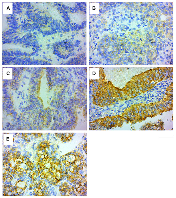
Figure 4. GRB7 expression in ovarian carcinoma cells.
Representative images of high grade ovarian cancer tissues stained with the GRB7 antibody (brown) and Hematoxylin (blue). (A) No GRB7 expression (0). (B) Weak cytoplasmic GRB7 expression (1+). (C) Medium cytoplasmic GRB7 expression (2+). (D) Strong cytoplasmic GRB7 expression (3+). (E) Strong cytoplasmic GRB7 expression with membranous accentuation (3+). Images were taken with a 40 × objective, scale bar = 50 μm.

