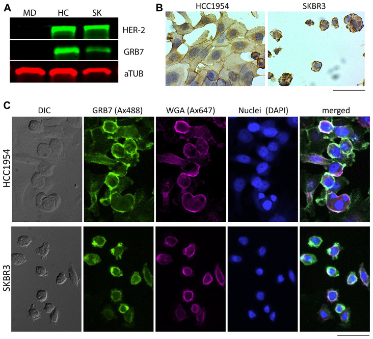
Figure 6. GRB7 expression levels and localization of breast cancer cell lines (MDA-MB-231, HCC1954, and SKBR3).
(A) Western blot showing GRB7 expression levels (middle) panel using total protein lysates (10 μg/lane). Alpha-tubulin (aTUB, bottom panel) was used as internal reference, HER-2 was used to show co-amplification of HER-2 and GRB7 as expected (top panel). MD = MDA-MB-231, HC = HCC1954, SK = SKBR3. (B) Representative images of breast cancer (HCC1954, SKBR3) cell lines stained (IHC) with the GRB7 antibody (brown, developed using HRP-DAB) and nuclei (Hematoxylin, blue). Images show cells with strong cytoplasmic GRB7 expression with membranous accentuation. (C) Representative images of breast cancer (HCC1954, SKBR3) cell lines stained (IF) with the GRB7 antibody (green), WGA cell membrane marker (magenta), nuclei (blue), and a merged image from all three channels, showing GRB7 and WGA co-localization in white (most right). A DIC image is shown on the most left. Images were taken with a 20 × objective, scale bar = 100 μm.

