PAC1 receptor blockade using a monoclonal antibody inhibits nociceptive neuronal activity in the trigeminocervical complex.
Keywords: Pituitary adenylate cyclase activating peptide, Sumatriptan, Headache, Migraine, Cluster headache, Trigeminal activation, Monoclonal antibody, PAC1 receptor
Abstract
Pituitary adenylate cyclase activating polypeptide-38 (PACAP38) may play an important role in primary headaches. Preclinical evidence suggests that PACAP38 modulates trigeminal nociceptive activity mainly through PAC1 receptors while clinical studies report that plasma concentrations of PACAP38 are elevated in spontaneous attacks of cluster headache and migraine and normalize after treatment with sumatriptan. Intravenous infusion of PACAP38 induces migraine-like attacks in migraineurs and cluster-like attacks in cluster headache patients. A rodent-specific PAC1 receptor antibody Ab181 was developed, and its effect on nociceptive neuronal activity in the trigeminocervical complex was investigated in vivo in an electrophysiological model relevant to primary headaches. Ab181 is potent and selective at the rat PAC1 receptor and provides near-maximum target coverage at 10 mg/kg for more than 48 hours. Without affecting spontaneous neuronal activity, Ab181 effectively inhibits stimulus-evoked activity in the trigeminocervical complex. Immunohistochemical analysis revealed its binding in the trigeminal ganglion and sphenopalatine ganglion but not within the central nervous system suggesting a peripheral site of action. The pharmacological approach using a specific PAC1 receptor antibody could provide a novel mechanism with a potential clinical efficacy in the treatment of primary headaches.
1. Introduction
Migraine and cluster headache are highly disabling disorders50 involving activation of the trigeminovascular system, which includes the perivascular meningeal afferents and the trigeminal ganglion in the peripheral nervous system, and the trigeminocervical complex (TCC) as well as higher brain structures including the hypothalamus, thalamus, and periaqueductal gray in the central nervous system (CNS).18,33
Pituitary adenylate cyclase activating polypeptide-38 (PACAP38) has received increasing attention in the context of migraine and cluster headache. PACAP38 is a 38-amino acid neuropeptide that is structurally and functionally related to vasoactive intestinal peptide (VIP).26,39 Both neuropeptides act on the same set of receptors, namely PAC1, VPAC1, and VPAC2 receptors, which belong to the G-protein-coupled receptors of the secretin family.26 PACAP3843 and VIP22 have vasodilatory properties and play a major role in parasympathetic communication.7,26,35 In cluster headache, both neuropeptides are released during a spontaneous attack.15,49 In migraine, despite some shared biology, both molecules have important differences. During a spontaneous migraine attack, PACAP38 is released into the cranial circulation regardless of the presence of autonomic symptoms48,52 while VIP is only released if cranial autonomic symptoms accompany the attack.17 The infusion of PACAP38,2,25,43 but less reliably VIP,42 induces migraine-like attacks, which can be effectively treated with sumatriptan. Sumatriptan normalizes elevated levels of PACAP38 during migraine.52 These findings strikingly resemble the preclinical and clinical observations made with calcitonin gene-related peptide (CGRP),14,16,17,36,54 which plays a prominent role in the pathophysiology of migraine and cluster headache and has been proven to be a validated target in the treatment of both disorders.13,18,23,29,38,44
A large body of preclinical evidence has supported and begun to dissect the mechanisms behind these clinical observations. The fact that PACAP38 and VIP share similar affinities to the VPAC1 and VPAC2 receptors but that the PAC1 receptor has a 100- to 1000-fold higher affinity to PACAP389 has led to the conclusion that the relevant action of PACAP38 is likely to be mediated mainly through the PAC1 receptor.52 These findings are supported by in vivo studies that demonstrate the release of PACAP38 into the cranial circulation upon peripheral trigeminal activation52,53 and the facilitation of nociceptive neuronal transmission in the TCC that is reversible by the administration of a PAC1 receptor antagonist.1
These observations suggest that targeting the PAC1 receptor may offer an effective and highly selective approach to reduce trigeminal activation and offer a novel target for the treatment of primary headaches. We therefore developed a potent and selective monoclonal mouse anti-rat PAC1 antibody (Ab181), fully characterized the pharmacologic and pharmacokinetic properties of the agent, and studied its effect on nociceptive neuronal activity within the TCC in an in vivo model that has been proven to be highly predictive for clinical efficacy in primary headaches.19–21,47 Preliminary results have been presented at the fifth European Headache and Migraine Trust International Congress31 and the International Headache Congress.32
2. Methods
The generation and in vitro characterization of Ab181, the pharmacodynamic and pharmacokinetic analyses as well as the immunohistochemistry were conducted between 2009 and 2013 in the Amgen laboratories, Thousand Oaks, CA. The electrophysiological studies were conducted between 2012 and 2013 in the laboratory of the Headache Group at the Department of Neurology, University of California, San Francisco, CA.
2.1. The generation of the Ab181
Mice anti-rat PAC1-specific monoclonal antibodies were generated using a conventional immunization method. Five 4- to 6-week-old hybrid 129xC57BL/6 mice (Charles River Laboratories, Hollister, CA) received 3 rounds of immunizations with soluble rat PAC1, chemically conjugated to Padre Peptides (Pan DR Helper T-cell epitopes). Mice were immunized SQ/IP with up to 50 µg of antigen-mixed or emulsified in either Freund's complete adjuvant (Cat# 77140; Pierce, Thermo Fisher Scientific, Waltham, MA), RIBI (Cat# S6322; Sigma-Aldrich, St. Louis, MO), or Poly I: C/CpG. Soluble PAC1 receptor polypeptides containing the N-terminal extracellular domains of rat PAC1 (amino acids 1-135 of GenBank accession no. NM133511.1) were generated by transiently cotransfecting 293-6E cells. All mice were maintained according to the regulations of the Amgen Institutional Animal Care and Use Committees in Thousand Oaks, CA.
Mice with the highest detected FACS titer to rat PAC1 expressed on CHO AMID cells were selected for fusion. Four days before spleen harvest, mice selected for fusion were given a final IP boost of 50-µg soluble rat PAC1 in phosphate-buffered saline (PBS) (Cat# 14040; GIBCO, Thermo Fisher Scientific, Waltham, MA). B-cell hybridomas were obtained by fusing immune splenocytes with nonsecreting murine myeloma cells, Sp2/0-Ag14 (American Type Culture Collection), at a ratio of 2.5:1 by electrofusion. Ab181 is identified through screening assays including binding competition, functional blocking, and receptor selectivity against the rat PAC1 receptor.
2.2. In vitro characterization of Ab181
Potency and selectivity of Ab181 were analyzed in vitro cell-based functional assay.
2.2.1. Cell culture
In house developed stable rPAC1/CHO cells were grown in Ham's F12 nutrient mixture (Cat# 11765; GIBCO, Thermo Fisher Scientific), 10% fetal bovine serum (Cat# 10099; GIBCO, Thermo Fisher Scientific), 1X Penicillin–Streptomycin–Glutamine (Cat# 10378; GIBCO, Thermo Fisher Scientific), 400-µg/mL G418 (Cat# 10131; GIBCO, Thermo Fisher Scientific), and 250-µg/mL Zeocin (Cat# R250-01; Invitrogen, Thermo Fisher Scientific). All cell flasks were maintained in incubators at 37°C with 5% CO2. U2OS (Cat# HTB-96; ATCC, Manassas, VA) cells were grown in the medium of McCoy's 5A (Cat# 16600; GIBCO, Thermo Fisher Scientific) containing 10% fetal bovine serum (Cat# 10099; GIBCO, Thermo Fisher Scientific), 1X L-glutamine (Cat# 25030; GIBCO, Thermo Fisher Scientific), 1X Penicillin–Streptomycin–Glutamine, and 1X MEM nonessential amino acids (Cat# 11140, GIBCO; Thermo Fisher Scientific). All cell flasks were maintained in incubator at 37°C with 5% CO2.
2.2.2. BacMam virus construct
The rVPAC1 and rVPAC2 BacMam virus constructs were prepared in house. The titer of rVPAC1 BacMam virus is 7.01 × 10⁸ IU/mL, and the titer of rVPAC1 BacMam virus is 5.75 X 10⁸ IU/mL.
2.2.3. Preparation of BacMam virus transduced rVPAC1 and rVAPC2 cells
U2OS cells were cultured in T-75 flask until the cell density reaches to 70% to 80% confluent before transduction. Cell medium was removed from flask, and the cells were rinsed with 1X PBS (Cat# 14040; GIBCO, Thermo Fisher Scientific) once, and then Versene (Cat# 15040; GIBCO, Thermo Fisher Scientific) was added to detach the cells. 3 × 10⁶ U2OS cells were resuspended with culture medium and mixed with rVPAC1 or rVPAC2 BacMam virus at a concentration of 50 multiplicity of infection/cell. The cell mixture was further incubated overnight for assay.
2.2.4. Cell-based functional assay
The cAMP assay was performed by using LANCE cAMP ultra assay kit (Cat # TRF0263; PerkinElmer, Inc, Waltham, MA) to determine the activity of Ab181. Assay buffer contain Ham's F12 nutrient mixture (Cat# 11765; GIBCO, Thermo Fisher Scientific), 0.1% bovine serum albumin (Cat# CR84-100; PerkinElmer, Inc, Waltham, MA), and 1 mM IBMX (Cat# I5879, Sigma-Aldrich, St. Louis, MO). Agonist dose–response curve was first run to determine the appropriate concentration to be used in subsequent antagonism studies (data not shown) (Table 1).
Table 1.
Antagonist activities (IC50) of Ab181.
The antagonist activity of Ab181 was performed by using EC80 of PACAP38 (Cat# H-8430; Bachem, Bubendorf, Switzerland) or maxadilan (Cat# H-6734; Bachem, Bubendorf, Switzerland). PACAP6-38 (Cat# H-2734; Bachem), a PAC1 receptor antagonist, was used as a positive control. Ab181 (0.5 pM-1 μM) was preincubated with the rPAC1-CHO cell suspension (2000 cells/well) at room temperature for 30 minutes before the addition of agonists. PACAP38 or maxadilan was then added and incubated with the mixture for additional 15 minutes at room temperature. The reaction was stopped by adding detection mix of Eu-cAMP tracer and ULight-anti-cAMP (Cat# TRF0263; PerkinElmer, Inc) to all wells. After a 45-minute incubation at room temperature, the assay plate was read on an EnVision instrument (Cat# 2105-0100; PerkinElmer, Inc).
Same assay method was used to determine the activity of Ab181 on rVPAC1 and rVPAC2 receptors. The activity of VIP (Cat#: H-3775; Bachem), a rVPAC1 and rVPAC2 receptor agonist, was first evaluated, and the EC80 concentration of VIP was used in the assay to determine the activity of Ab181 on these receptors.
All results were analyzed using GraphPad Prism software's nonlinear regression curve fit (GraphPad Software, Inc, La Jolla, CA), and data are presented as mean ± SD. Data from the agonist dose–response curves were used to calculate the half maximal effective concentration (EC50) and the half maximal inhibitory concentration (IC50) values for agonist and antagonist studies, respectively.
2.3. Pharmacokinetic and pharmacodynamic analysis
Male naïve Sprague–Dawley rats, 6 to 12 weeks from either Taconic Farms, Inc (Oxnard, CA) or Harlan Laboratories (Indianapolis, IN), at the average age of initiation of treatment were used, and all procedures in this report were conducted in compliance with the Animal Welfare Act, the Guide for the Care and Use of Laboratory Animals, and the Office of Laboratory Animal Welfare. Animals were group-housed in nonsterile, ventilated microisolator housing on corn cob bedding in Amgen's Assessment and Accreditation of Laboratory Animal Committee (AAALAC)-accredited facility with controlled temperature (70 ± 5° F), relative humidity (50 ± 20%), and 12-hour light/dark cycles (06:00-18:00). Animals had ad libitum access to pelleted feed (Harlan Teklad 2020X, Indianapolis, IN) and water (onsite-generated reverse osmosis) through automatic watering system.
2.3.1. Test and control materials
Ab181 was generated from Amgen and diluted to a series of concentrations in A5Su (10 mM sodium acetate, 9% sucrose, pH = 5.0). An isotype control was used as a dummy antibody in A5Su at a concentration of 5 mg/mL. Maxadilan (trifluoroacetate salt, Bachem) was used, and a dosing solution was freshly prepared daily by dissolving maxadilan in 1X Dulbecco's PBS (DPBS) (Sigma-Aldrich) at a final concentration of 0.5 µg/mL.
2.3.2. Laser Doppler imaging
A laser Doppler imager (Moor Instruments, Ltd, Wilmington, DE) was used to measure dermal blood flow (DBF) on the shaved skin of the rat abdomen.
After anesthetic with propofol on the test day, the rat's abdominal area was shaved, and each animal was placed in a supine position on a temperature-controlled circulating warm-water heating pad to help maintain a stable body temperature during the study. After a 10- to 15-minute stabilization period, a black rubber O-ring (0.925-cm inner diameter; O-Rings West, Seattle, WA) was placed on the rat abdomen without directly positioning it over a visible blood vessel. After placement of an O-ring on the selected area, a baseline (BL) DBF measurement was taken. After the BL scan, the maxadilan solution prepared fresh daily in 20-µL vehicle (DPBS) was injected intradermally at the center of the O-ring. The postmaxadilan DBF was measured either every 15 minutes over a 60-minute period or at specified time points such as 15 and 30 minutes. The O-ring serves as an area of interest in which the DBF will be analyzed within the O-ring. Ab181 was prepared in A5Su at different concentrations depending on the dose range and given in a single bolus intravenous injection. In this report, MIIBF was measured and expressed as % change from the baseline [100 × (individual postagonist flux − individual baseline flux)/individual baseline flux] or as % inhibition [(mean of % change from BL from vehicle-treated animals − individual % change from BL from drug-treated animals)/mean % change from BL from vehicle-treated animals].
2.3.2.1. Dose–response study at 48 hours after Ab181 treatment
Ab181 was administered through penile vein or tail vein at various doses (0.1, 0.3, 1, and 10 mg/kg). Forty-eight hours later, rats received an intradermal maxadilan injection (10 ng in 20-µL DPBS) followed by postmaxadilan DBF scans every 15 minutes over a 60-minute period. After the final DBF scan, 3 rats that had been pretreated with 10 mg/kg of Ab181 were allowed to recover from the propofol anesthesia, returned to their home cages, and underwent a second DBF measurement at 168 hours after drug administration. Serum PK samples were taken through tail vein immediately before the first maxadilan challenge at 48 and 168 hours after Ab181 treatment. The post-MIIBF scan time point for the later studies was determined based on the postmaxadilan DBF response from 15 to 60 minutes.
2.3.2.2. Dose–response study at 3.25 hours after Ab181 treatment
Ab181 was administered through the penile vein at various doses (0.1, 0.3, 1, 3, and 10 mg/kg). Three hours later, rats received an intradermal maxadilan injection (10 ng of maxadilan in 20-µL DPBS) followed by a post-MIIBF scan 15 minutes later. Serum PK samples were taken through tail vein immediately after the DBF scan (ie, 3.25 hours after Ab181 treatment). The time point was chosen to confirm that the receptor occupancy of Ab181 had reached and stayed at the desired pharmacodynamic measures when performing the electrophysiological studies.
2.3.2.3. Time-course study of Ab181 at a dose of 10 mg/kg
Ab181 was dosed intravenously at 10 mg/kg through either rat tail vein or penile vein at various pretreatment times (0.5, 3, 6.25 hours) before maxadilan challenge. The full time-course study comprised several experiments, including the 48 and 168 hours after Ab181 treatment studies described above.
2.3.2.4. Statistical analysis of laser Doppler flow experiments
All DBF results were expressed as the mean ± SEM. A one-way analysis of variance followed by Dunnett's multiple comparison test was used to assess the statistical significance of Ab181 effects relative to either vehicle or the control antibody. A P < 0.05 was used to determine significance between any 2 groups. ED50 values were calculated in GraphPad Prism after logarithmic transformation and nonlinear fit of data using a sigmoidal dose–response model with variable slope and with the top constrained to the resulting mean percent change in the corresponding control group (vehicle or control antibody-treated group) and bottom constrained to zero.
2.4. Electrophysiological recordings
All experiments were approved by the Institutional Animal Care and Use Committee of the University of California, San Francisco. Experiments were conducted in accordance with the United States Public Health Service's Policy on Humane Care and Use of Laboratory Animals, the ARRIVE guidelines, and the guidelines of the Committee for Research and Ethical Issues of the International Association of the Study of Pain.
2.4.1. General surgical preparation
Twenty-four male Sprague–Dawley rats (Charles River Laboratories) were used in the experiments. In each animal, only one experiment was conducted, and recordings were performed from one site. The animals were anesthetized by an induction with a single dose of pentobarbital (60 mg/kg intraperitoneally; Nembutal, Lundbeck, Deerfield, IL) followed by a continuous infusion of propofol (20-25 mg/kg/h intravenously; Propoflo, Abbott, Abbott Park, IL) for maintenance throughout the entire experiment. For the administration of the anesthetic and drugs, both femoral veins were cannulated. The left femoral artery was cannulated for the continuous monitoring of arterial blood pressure.
2.4.2. Physiological monitoring
Rats were placed on a self-regulating homeothermic blanket system with a rectal probe (Harvard Apparatus, Holliston, MA), and core body temperature was maintained at 37 ± 0.5°C. Arterial blood pressure was monitored from the femoral artery using a transducer (DTX Plus DT-XX; Becton Dickinson, Sandy, UT) connected to an amplifier (PM-1000; CWE, Ardmore, PA). Following a tracheostomy, animals were mechanically ventilated (3-5 mL/min, 75-90 strokes/min; 7025, Ugo Basile, Comerio, VA, Italy) with oxygen-enriched air, and end-expiratory CO2 was kept between 3.5% and 4.5%. Data on arterial blood pressure and CO2 concentration were continuously displayed and fed into a data acquisition system (Power 1401; Cambridge Electronic Design-CED, Hertfordshire, Cambridge, United Kingdom) and saved on a hard disk.
2.4.3. Recording preparation
The rat's heads were fixed in a stereotaxic frame (Kopf Instruments, Tujunga, CA). A craniotomy was performed over the parietal cortex with a dental burr using constant irrigation to reduce heat production. With this procedure, the middle meningeal artery (MMA) was exposed without lesioning the dura mater. For electrical stimulation, a bipolar stimulating electrode (NE200; Rhodes Medical Instruments, Summerland, CA) was placed above the MMA touching the dura mater at either side of the blood vessel. The electrode was connected to a stimulus isolation unit (SIU5A; Grass Instruments, Quincy, MA).
For the extracellular recording of neuronal activity in the TCC, a C1 partial hemilaminectomy was performed, and the spinal dura mater was removed. A tungsten electrode with a nominal impedance of 1 MΩ (TM31A10; World Precision Instruments, Sarasota, FL) was then introduced in the TCC near the dorsal root entry zone. For the localization of optimal site for the extracellular recording, the electrode was advanced or retracted in 5-μm steps with a piezoelectric motor-driven micromanipulator. Wide dynamic range neurons with convergent input from the dura mater and the facial skin were identified by their responsiveness to electrical stimulation of the perivascular afferents surrounding the MMA as well as innocuous brush and noxious pinch of the skin innervated by the first branch of the trigeminal nerve.
2.4.4. Stimulation of meningeal afferents and recording in the trigeminocervical complex
Electrical stimulation of the perivascular meningeal afferents was performed applying electrical square wave pulses (10-18 V, 0.1-0.2 ms, 0.5 Hz, 20 sweeps) (S88; Grass Instruments).
The stimulus-evoked neuronal signal and the neuronal background activity were acquired by the recording electrode placed in the TCC. The electrical signal was fed into a headstage amplifier (NL100AK; Neurolog, Digitimer, Welwyn Garden City, Hertfordshire, United Kingdom) and passed to an AC preamplifier (NL104, Neurolog, Digitimer) set to a gain of 1000×. The signal was then passed through a band-pass filter (bandwidth 300 Hz-10 kHz) (NL125/126; Neurolog, Digitimer) and a 60-Hz noise eliminator (Humbug; Quest Scientific, Vancouver, BC, Canada) before further amplification by an AC-DC amplifier (NL106; Neurolog, Digitimer). This signal was fed to a gated amplitude discriminator (NL201; Neurolog, Digitimer) and a data acquisition system (Power 1401; Cambridge Electronic Design-CED). Data were collected, analyzed, and stored using Spike 5.2 software (Cambridge Electronic Design-CED). The output of the gated amplitude discriminator was also fed into an audio amplifier (NL120; Neurolog, Digitimer) and loudspeaker as well as an oscilloscope to assist spike discrimination from background activity. For the analysis of stimulus-evoked neuronal activity, poststimulus histograms were produced online. Background activity gated through the amplitude discriminator was collected into successive bins.
2.4.5. Experimental protocol and drug administration
After completing the surgical procedure, the animals had a resting period of 30 minutes. After this period, baseline recordings were obtained. These were obtained by calculating the mean of 3 series of poststimulus histograms, each consisting of 20 electrical stimuli.
After the assessment of the baseline values, Ab181 (10 mg/kg) or its vehicle (A5Su) were administered intravenously over 1 minute. Because of the pharmacological properties of the antibody, the animals underwent a second resting period of 2.5 hours to allow sufficient time for the antibody to bind its target. After this second resting, sumatriptan (10 mg/kg) or vehicle was administered intravenously over 1 minute followed by another resting period of 30 minutes. Poststimulus histograms were then established 180, 185, 190, 195, 200, 205, 210, and 225 minutes after the administration of the first pharmacological intervention (Ab181 or vehicle) (Fig. 1). Based on the treatments described above, animals were divided in 3 treatment groups, group 1 receiving the Ab181 (intervention 1) and sumatriptan vehicle (intervention 2), group 2 receiving vehicle (intervention 1) and sumatriptan (intervention 2), and group 3 receiving vehicle at both interventions (Fig. 1).
Figure 1.

Timeline of the electrophysiological in vivo experiments. After the surgical procedure and the recording of baseline activity, either Ab181 (10 mg/kg) or vehicle (A5Su) was injected intravenously. After a resting period of 2.5 hours to allow for the distribution of the antibody throughout the circulation and the relevant tissues, sumatriptan or its vehicle was administered intravenously. Following another resting period of 30 minutes to allow sumatriptan to act, stimulus-evoked and spontaneous neuronal activity was recorded in the TCC. TCC, trigeminocervical complex.
2.4.6. Statistical analysis
Statistical analysis was performed using SPSS 22.0 software (IBM Corporation, Armonk, NY). Data are expressed as percentages of baseline values with SEs of the mean (±SEM). Effects within a treatment group were calculated using the analysis of variance with repeated measures applying the Greenhouse–Geisser correction of the assumption of sphericity was violated. Bonferroni correction was applied for multiple comparisons. Statistical significance was assumed at P < 0.05. For a detailed comparison of individual data points with the baseline value within one treatment group, the dependent t test was used.
2.5. Immunohistochemistry
Adult male Sprague–Dawley rats (n = 3 per group) were injected intravenously with Ab181 or a monoclonal mouse isotype control antibody against an unrelated target or vehicle. After 3.5 hours, animals were terminally anesthesized by FatalPlus (Vortech Pharmaceuticals, Dearborn, MI) and perfused with ice-cold PBS at pH 7.4 (Cat# 14040; GIBCO, Thermo Fisher Scientific) followed by 4% paraformaldehyde (Cat# P6148; Sigma-Aldrich) in PBS (Cat# 14040; GIBCO, Thermo Fisher Scientific). The sphenopalatine ganglia, TG, and brains were dissected and cryoprotected with 30% sucrose in PBS. Twelve-micrometer sections were cut, washed in PBS, and incubated in 3% normal goat serum and 0.3% TritonX-100 in PBS for 1 hour at room temperature. Sections were then washed in PBS and incubated with AlexaFluor488 goat anti-mouse IgG (Invitrogen, Thermo Fisher Scientific) for 1 hour at room temperature. After final washes in PBS (Cat# 14040; GIBCO, Thermo Fisher Scientific), sections were cover slipped with Vectashield mounting medium with DAPI (Cat# H-1200; Vector Labs, Burlingame, CA). The following additional controls were conducted: A set of sections from Ab181-dosed rats was incubated with secondary antibodies against rabbit (AlexaFluor488 goat anti-rabbit IgG; Invitrogen, Thermo Fisher Scientific) as negative control. As positive control for PAC1 staining, a set of vehicle sections was incubated with 10-µg/mL Ab181 in blocking solution at 4°C for 48 hours before further processing with AlexaFluor488 goat anti-mouse IgG (Invitrogen, Thermo Fisher Scientific) as described above.
3. Results
3.1. In vitro potency of the PAC1 receptor antibody Ab181
Ab181 is a full antagonist of the rat PAC1 receptor. It dose-dependently inhibited PAC1 receptor agonist PACAP38 or maxadilan, a PAC1 selective receptor agonist, induced cAMP accumulation in CHO cells expressing rat PAC1 receptors with IC50 of 20 ± 3.3 nM (n = 3) and 4.5 ± 0.1 nM (n = 2), respectively (Fig. 2 and Table 1). Ab181 is selective to the PAC1 receptor. IC50 at rat VPAC1 and rat VPAC2 receptors against the agonist VIP is greater than 100 nM, the highest concentration tested.
Figure 2.
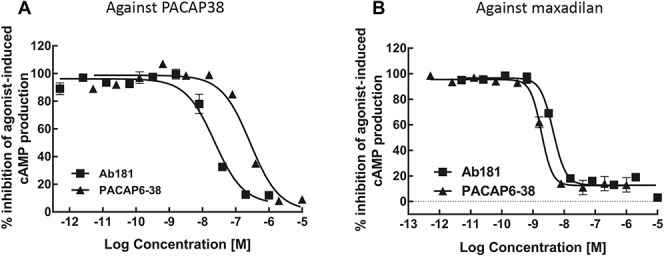
Antagonist activity of Ab181 at rat rPAC1 CHO cell (rPAC1-CHO) against PACAP38 or maxadilan. The antagonist activity of Ab181 at the rat PAC1 receptor was performed by measuring the potency of Ab181 in inhibiting EC80 of agonist PACAP38 or maxadilan-stimulated cAMP production. PACAP6-38, a PAC1 receptor antagonist, was used as a positive control. Full antagonist activity of Ab181 against both PACAP38 (A) and maxadilan (B) induced cAMP accumulation was observed in rPAC1-CHO cells with IC50 of 20 ± 3.3 nM (n = 3) and 4.5 ± 0.1 nM (n = 2) against respective agonist.
3.2. Pharmacokinetic and pharmacodynamic effect of Ab181
3.2.1. Dose–response study at 48 hours after Ab181 treatment
Pretreatment of Ab181 48 hours before maxadilan challenge prevented the maxadilan-induced increase in DBF (MIIDBF). The change in DBF over 60 minutes after maxadilan is depicted in Figure 3A. There was a statistically significant difference (P < 0.001) between the vehicle-treated group and each of 3 Ab181 dose groups (0.3, 1, and 10 mg/kg) at 15 minutes after maxadilan injection with a calculated ED50 of 0.16 mg/kg (CI95% = 0.10-0.26 mg/kg). Serum concentration of Ab181 is plotted on the Y-axis of Figure 3B.
Figure 3.
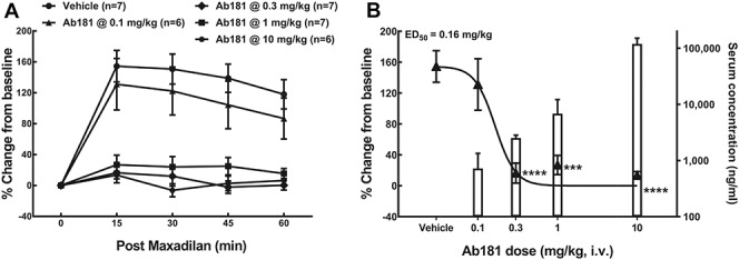
Prophylactic effect of Ab181 on maxadilan-induced increase in DBF in rats 48 hours after drug treatment. Rats were given intravenous bolus injection of Ab181 (0.1, 0.3, 1, and 10 mg/kg, n = 6-7 rats/group). Forty-eight hours later, after obtaining a postdrug baseline (BL), maxadilan at a dose of 10 ng (in 20-µL DPBS) was injected intradermally. The dermal blood flow (DBF) was measured by a laser Doppler imager. (A) The increase in DBF (change from BL) caused by maxadilan injection over 60 minutes was expressed as % change from BL = [100 × (individual postmaxadilan flux − individual BL flux)/individual BL flux]. (B) % change from BL (left-axis) vs resulting serum concentrations (right axis) at 15 minutes after maxadilan; solid triangle represents % change from BL at 48 hours after drug treatment (left axis, mean ± SEM), and open bar represents serum concentration at 48 hours after drug treatment (right axis, mean ± SD). ***P < 0.001, ****P < 0.0001 compared with vehicle-treated group by one-way ANOVA followed by Dunnett's MCT. ANOVA, analysis of variance; DBPS, Dulbecco's phosphate-buffered saline; MCT, multiple comparison test.
3.2.2. Dose–response study at 3.25 hours after Ab181 treatment
A shorter pretreatment of Ab181 at various doses was also evaluated. A 15-minute postmaxadilan measurement was used in this study. The change in MIIDBF after 3.25 hours after Ab181 is depicted in Figure 4. There was a statistically significant inhibition of the MIIDBF following dose of 1, 3, and 10 mg/kg at 3.25 hours after treatment (P < 0.01), resulting in a calculated ED50 of 0.95 mg/kg (CI95% = 0.59-1.51 mg/kg). Serum concentration of Ab181 is plotted on the Y-axis of Figure 4.
Figure 4.
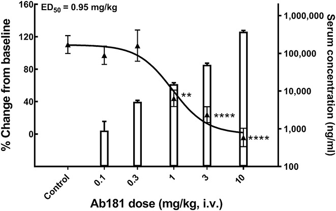
Effect of Ab181 on a maxadilan-induced increase in DBF in rats 3.25 hours after treatment. Rats were given intravenous (i.v.) bolus injection of Ab181 (0.1, 0.3, 1, 3, and 10 mg/kg, n = 7-8 rats/group). Three hours later, after obtaining a postdrug baseline (BL), maxadilan at a dose of 10 ng (in 20-µL DPBS) was injected intradermally. The DBF was measured by a laser Doppler imager. Solid triangle represents % change from baseline (mean ± SEM, left axis), and open bar represents resulting serum concentration (mean ± SD, right axis). Control: Dummy antibody + vehicle (n = 13 rats). **P < 0.01, ****P < 0.0001 compared with the control group by one-way ANOVA followed by Dunnett's MCT. ANOVA, analysis of variance; DBF, dermal blood flow; DBPS, Dulbecco's phosphate-buffered saline; MCT, multiple comparison test.
3.2.3. Time-course study of Ab181 at a dose of 10 mg/kg
A 15-minute postmaxadilan measurement was used in this study. Ab181 at 10 mg/kg produced a statistically significant inhibition of the MIIDBF starting (P < 0.05) from a pretreatment time of 0.75 hours and lasting through the last measured 168-hour time point (Fig. 5). The resulting mean serum concentration at 1, 3, 6, 48, and 168 hours after drug treatment is plotted on the Y-axis in Figure 5.
Figure 5.
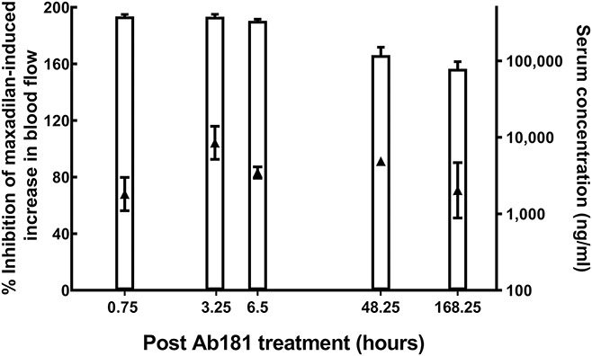
Time course of effect of Ab181 (at 10 mg/kg) on a maxadilan-induced increase in DBF in rats. Rats were given intravenous bolus injection of Ab181 at 10 mg/kg with pretreatment time at 0.5, 3, 6.25, 48, and 168 hours before maxadilan challenge (n = 3-8 rats/group). After obtaining a postdrug baseline (BL), maxadilan at a dose of 10 ng (in 20 µL DPBS) was injected intradermally. The DBF was measured by a laser Doppler imager. Solid triangle represents % inhibition (mean ± SEM, left axis), and open bar represents resulting serum concentration (mean ± SD, right axis). DBF, dermal blood flow; DBPS, Dulbecco's phosphate-buffered saline.
3.3. Electrophysiological recordings
3.3.1. Ab181 inhibits stimulus-evoked responses in the trigeminocervical complex
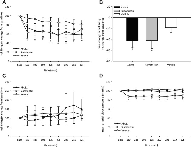
The intravenous administration of Ab181 (Fig. 1) induced a long-lasting inhibition of stimulus-evoked nociceptive neuronal activity within the TCC (F1.48, 5.91 = 8.43, P = 0.022). Compared with the baseline value, the effect was significant at all investigated time points throughout the entire observational period. Maximum inhibition reached −43 ± 13% (t7 = 3.41, P = 0.011) compared with baseline.
Sumatriptan, which was administered as a positive control, significantly reduced stimulus-evoked responses in the TCC (F2.21, 15.49 = 4.97, P = 0.019). Similar to the Ab181 group, the inhibiting effect of sumatriptan was significant at all investigated time points reaching a maximum inhibition of −42 ± 15% (t7 = 2.78, P = 0.027) compared to baseline without recovering until the end of the observational period.
By contrast, the intravenous administration of the vehicle did not affect stimulus-evoked neuronal activity (F2.21, 15.48 = 2.45, P = 0.115; Figs. 6A, B).
Figure 6.
Effect of Ab181 on neuronal activity in the TCC. Ab181 effectively inhibited stimulus-evoked neuronal activity in the TCC (A). The maximum inhibition was very similar to the one observed after the intravenous administration of sumatriptan (B). By contrast, an inhibiting effect on spontaneous background activity was not observed (C). Ab181 did not affect arterial blood pressure throughout the entire course of the experiment (D). TCC, trigeminocervical complex.
3.3.2. Neuronal background activity in the trigeminocervical complex is not affected by Ab181
In contrast to the observed effect on stimulus-evoked neuronal activity, intravenous administration of Ab181 does not attenuate unspecific neuronal background activity within the TCC (F1.32, 5.27 = 0.767, P = 0.456). The same lack of effect on background activity was observed in animals treated with sumatriptan (F2.33, 16.28 = 0.821, P = 0.474) and those treated with vehicle (F1.68, 11.78 = 0.99, P = 0.385; Fig. 6C).
3.3.3. Arterial blood pressure is unaffected by Ab181
Intravenous administration of Ab181 does not affect arterial blood pressure (F1.69, 6.78 = 0.656, P = 0.525). By contrast, sumatriptan induced a significant decrease in arterial blood pressure (F2.55, 17.84 = 5.82, P = 0.008) throughout the entire observational period, reaching a maximum reduction of −13 ± 3% (t7 = 4.89, P = 0.002) compared to baseline without recovering until the end of the experiment. In the vehicle control group, arterial blood pressure was unaffected (F2.29, 16.02 = 0.93, P = 0.426; Fig. 6D).
3.4. Immunohistochemistry
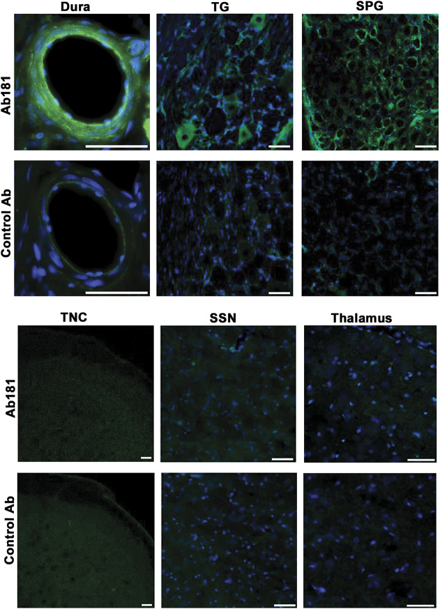
To investigate whether Ab181 or the control antibody distributed into tissues of interest after intravenous administration, we treated satellite rats from the electrophysiology study but dissected and fixed tissues of interest at the 3.5-hour time point after injection. Using fluorescently labeled secondary antibodies against the Fc portion of the antibodies, we were able to detect immunoreactivity in dura, trigeminal ganglion (TG), and SPG of Ab181-dosed rats, but not in rats dosed with control antibody (Fig. 7). By contrast, no labeling above background was detected in spinal trigeminal nucleus or superior salivary nucleus of the brainstem as well as in the hypothalamus and thalamus indicating that Ab181 did not cross the blood–brain barrier, or the amount of antibodies that entered the CNS after intravenous injection was below the detection threshold of the immunohistochemical method (Fig. 7).
Figure 7.
Distribution of Ab181 in tissues after intravenous dosing. Ex vivo detection of the IgG portion of Ab181 or control antibody after intravenous dosing in dural vessels, trigeminal ganglion (TG), and sphenopalatine ganglion (SPG) as well as spinal trigeminal nucleus caudalis (TNC), superior salivatory nucleus (SSN), and thalamus. Staining of neuronal and glial structures is detected in dura, TG, and SPG of Ab181-dosed rats, but not in TNC, SSN, or thalamus. Only background staining is detected after intravenous dosing with control antibody (lower panels). Scale bars 50 µm.
4. Discussion
PACAP38, first described in 1989,39 is one of the key neurotransmitters of the parasympathetic system. Soon after its discovery, preclinical evidence suggested effects beyond its parasympathetic role, in particular in specific pain pathways related to headache.53 Neuroanatomical and functional data obtained since strongly suggest a prominent role in primary headaches including cluster headache and migraine. Our data may provide a plausible underpinning biology for a potential clinical effect of PAC1 blockade in these disorders.
First, PACAP receptors (PAC1, VPAC1, and VPAC2) are located in several key areas along the trigeminovascular system. Within the central part of the pain pathways processing trigeminal pain, they have been identified in the thalamus,27 hypothalamus,27 and the TCC,34 while in the peripheral part, they are located in the TG34 and the meningeal vasculature.5,6 Beyond that, they are found in the SPG8 highlighting the relevance of PACAP38 in the parasympathetic system, as well as its potential role in the functional interaction between the trigeminal and parasympathetic systems, namely the trigeminoautonomic reflex.
Second, from a functional perspective, PACAP38 may play a role in cluster headache and migraine since its plasma concentration is elevated in spontaneous attacks49,52 and normalizes after effective attack abortion with sumatriptan.52 In addition, PACAP38 triggers migraine-like attacks in migraineurs,25,43 most likely through an activation of the PAC1 receptor.52 In the context of migraine, the ability to trigger attacks clearly distinguishes PACAP38 from VIP. These clinical observations can now be explored in specific animal models. For example, electrical stimulation of the superior sagittal sinus of the cat induces the release of PACAP38 into the cranial circulation.52,53 In line, the administration of PACAP38 facilitates nociceptive neuronal activity in the TCC.1 These functional features of PACAP38 strikingly resemble the preclinical47 and clinical observations16,17 made with CGRP, which predicted clinical efficacy of 6 small-molecule CGRP-receptor antagonists10,28,30,37,41,51 and CGRP/CGRP-receptor antibodies11,23,45 in the acute and preventive treatment of migraine as well as a CGRP antibody in the preventive treatment of episodic cluster headache.13
Third, the parasympathetic and trigeminal sensory systems connect and interact with each other. From an anatomic perspective, CGRP-positive sensory fibers project from the TG to the SPG,8 and in vivo data demonstrates that PACAP38 induces the release of CGRP from the TCC.34 Nevertheless, the detailed molecular mechanisms behind the attack-triggering capability of PACAP38, and in particular, the clinical relevance of the above-described mechanism remain to be fully elucidated. Interestingly, the administration of PACAP38, despite creating an activation of the trigeminovascular system with the clinical picture of a migraine-like attack, does not induce the release of CGRP in a human model of migraine.25 These results suggest that the PACAP38-induced effects on trigeminal activation are likely to be the result of a direct effect on nociceptive trigeminal neurons rather than a CGRP-mediated mechanism, although both mechanisms do not exclude each other. In the case of a direct activation of either of the PACAP receptors, PACAP38 stimulates the cAMP/PKA pathway thereby increasing neuronal excitability.46 In addition, all PACAP receptors may be modulated in their expression profile under chronic pain conditions.55 It may be speculated that these upregulating and downregulating properties and the ability to produce neuronal sensitization may even play a role in the chronification of migraine. Based on this large body of evidence, we set out to develop a specific antibody against the PAC1 receptor because this receptor, as outlined above, is most likely the most relevant PACAP receptor in mediating the attack-triggering effect of PACAP38 and to test its effects on a model system that represents significant elements of cluster headache and migraine pathophysiology.4
PACAP38 is a nonselective agonist at all the PACAP receptors, and VIP is only mildly selective to the VPAC1 and VPAC2 receptors.26 Maxadilan, on the other hand, is the most selective agonist at the PAC1 receptor,40 which allowed us to measure pharmacodynamic activities specifically mediated through the PAC1 receptor. Although some years have passed since PACAP38 was discovered,39 selective antagonists to the individual receptors in the PACAP receptor family are still lacking. Several peptide antagonists have been reported with mild selectivity between different receptors,5,9,24 but their pharmacokinetic properties, especially their very short plasma half-life, hindered their utility in in vivo pharmacology studies. Therefore, Ab181 was developed. As demonstrated in the Results section, it is a potent and selective antagonist at the PAC1 receptor, with no activity at the VPAC receptors. It is also a potent and selective inhibitor of maxadilan-induced increases in DBF in a time- and dose-dependent manner. Through the in vitro and in vivo profiling, Ab181 demonstrated full-target coverage of the PAC1 receptor at 3.5 hours after intravenous administration and maintained sustained plasma concentration throughout the study duration. It is therefore the first ideal tool for the studying of selective PAC1 pharmacology in preclinical species.
The new results of the study show that the intravenous administration of Ab181 inhibits stimulus-evoked nociceptive activity in the TCC. Unlike conventional small molecular agents whose molecule weight (MW) are normally <500, the much larger sized antibodies (MW > 145KD) generally have a slower tissue distribution rate. As a result, it requires a substantial equilibrium period between the administration of the antibody and the initiation of the experiment before the assessment of its influence on stimulus-evoked activity. Therefore, the experimental design resembles more of a short-term preventive scenario than an acute reversal of nociceptive trigeminal activation. The results show that the extent of neuronal inhibition was almost identical to that observed with sumatriptan, suggesting a robust effect. The fact that we did not observe an effect on nonspecific background activity reflects the specificity of the effect on nociceptive neuronal transmission.
The exact site of action of PAC1 remains speculative. Literature suggested that agonist PACAP38, with MW >4000, triggers migraine-like attacks without crossing the blood–brain barrier.12 However, passage of PACAP27 and PACAP38 across the blood–brain barrier has been suggested.3 Here, immunohistochemical analysis of Ab181 found a minimum presence in CNS, which suggests that the site of the modulating action on nociceptive activity is likely to be located outside of the CNS. This would be in line with the results obtained from the electrophysiological experiments showing no effect on neuronal background activity and the immunohistochemical analysis in which Ab181 was detected in the relevant peripheral structures but not within the CNS. These findings suggest that Ab181 does not cross the blood–brain barrier in a significant concentration, a finding that is expected given the molecular size of the antibody. It cannot be excluded that a small amount of antibody reaches the CNS but remains below the immunohistochemical detection limit; however, target coverage with such small amount is unlikely sufficient for a full antagonism effect. Therefore, the current study with a selective PAC1 antagonist antibody supports the hypothesis that peripheral PAC1 receptor inhibition can be sufficient to abort or prevent attacks of cluster headache and migraine.
Recently, a randomized, placebo-controlled trial (phase 2) with a monoclonal PAC1 receptor antibody (AMG 301) was reported in abstract form to be ineffective in the preventive treatment of migraine. In the context of our data, some comments are relevant. First, in our experiments, we used an antibody that was developed for rodents and differs significantly in its pharmacological properties, including its affinity to the PAC1 receptor, for which it is more potent than the one used in the human phase 2 trial. Second, although the available evidence suggests that the PAC1 receptor plays a relevant role in trigeminal activation, the possibility that VPAC receptors are clinically significant is an open issue. The fact that VIP may trigger migraine attacks in subset of migraineurs may support this hypothesis.2 Finally, in the clinical trial, patients were not stratified by the presence or absence of cranial autonomic symptoms. Therefore, it may be hypothesized that the ability of PACAP38 to induce migraine attacks may require an action on PAC1 receptors on trigeminal and parasympathetic neurons thereby increasing the activation of the trigeminoautonomic reflex. If this would be the case, one could speculate of a higher relevance of this potentially therapeutic mechanism in migraine with cranial autonomic symptoms or in cluster headache.
Taken together, the findings of our studies suggest that the PAC1 antibody Ab181 can inhibit nociceptive neuronal traffic, so that this pharmacological approach may offer a new strategy for the preventive treatment of primary headache disorders, including cluster headache and migraine.
Conflict of interest statement
J. Hoffmann is consulting for and/or serves on advisory boards of Allergan, Autonomic Technologies, Inc (ATI), Chordate Medical AB, Eli Lilly, Hormosan Pharma, Novartis and Teva. He has received honoraria for speaking from Allergan, Chordate Medical AB, Novartis and Teva. He received personal fees for MedicoLegal Work as well as from Sage Publishing, Springer Healthcare and Quintessence Publishing. S. Miller is an employee of Amgen, Inc. S. Akerman reports an unrestricted grant, honoraria and travel reimbursements from electroCore LLC, unrelated to the submitted work. H. Sun is an employee of Amgen, Inc. L. Shi is an employee of Amgen, Inc. D. Zhu is an employee of Amgen, Inc. S. Lehto is an employee of Amgen, Inc. H. Liu is an employee of Amgen, Inc. R. Yin is an employee of Amgen, Inc. B.D. Moyer is an employee of Amgen, Inc. C. Xu is an employee of Amgen, Inc. P.J. Goadsby reports grants and personal fees from Amgen and Eli-Lilly and Company, and personal fees from Alder Biopharmaceuticals, Allergan, Autonomic Technologies, Inc, Biohaven Pharmaceuticals, Inc, Electrocore LLC, eNeura, Impel Neuropharma, MundiPharma, Novartis, Teva Pharmaceuticals, and Trigemina, Inc. WL Gore, and personal fees from MedicoLegal work, Massachusetts Medical Society, Up-to-Date, Oxford University Press, and Wolters Kluwer; and a patent Magnetic stimulation for headache assigned to eNeura without fee. The remaining authors have no conflicts interest to declare.
Acknowledgments
The study was funded by Amgen, Inc, Thousand Oaks, CA, USA.
Footnotes
Sponsorships or competing interests that may be relevant to content are disclosed at the end of this article.
C. Xu and P.J. Goadsby contributed equally to this work.
References
- [1].Akerman S, Goadsby PJ. Neuronal PAC1 receptors mediate delayed activation and sensitization of trigeminocervical neurons: relevance to migraine. Sci Transl Med 2015;7:1–11. [DOI] [PubMed] [Google Scholar]
- [2].Amin FM, Hougaard A, Schytz HW, Asghar MS, Lundholm E, Parvaiz AI, de Koning PJH, Andersen MR, Larsson HBW, Fahrenkrug J, Olesen J, Ashina M. Investigation of the pathophysiological mechanisms of migraine attacks induced by pituitary adenylate cyclase-activating polypeptide-38. Brain 2014;137:779–94. [DOI] [PubMed] [Google Scholar]
- [3].Banks WA, Kastin AJ, Komaki G, Arimura A. Pituitary adenylate cyclase activating polypeptide (PACAP) can cross the vascular component of the blood-testis barrier in the mouse. J Androl 1993;14:170–3. [PubMed] [Google Scholar]
- [4].Bergerot A, Holland PR, Akerman S, Bartsch T, Ahn AH, MaassenVanDenBrink A, Reuter U, Tassorelli C, Schoenen J, Mitsikostas DD, van den Maagdenberg AMJM, Goadsby PJ. Animal models of migraine. Looking at the component parts of a complex disorder. Eur J Neurosci 2006;24:1517–34. [DOI] [PubMed] [Google Scholar]
- [5].Boni LJ, Ploug KB, Olesen J, Jansen-Olesen I, Gupta S. The in vivo effect of VIP, PACAP-38 and PACAP-27 and mRNA expression of their receptors in rat middle meningeal artery. Cephalalgia 2009;29:837–47. [DOI] [PubMed] [Google Scholar]
- [6].Chan KY, Baun M, de Vries R, van den Boogaert AJ, Dirven CM, Danser AH, Jansen Olesen I, Olesen J, Villalon CM, Maassen Van Den Brink A, Gupta S. Pharmacological characterization of VIP and PACAP receptors in the human meningeal and coronary artery. Cephalalgia 2011;31:181–9. [DOI] [PubMed] [Google Scholar]
- [7].Couvineau A, Laburthe M. VPAC receptors: structure, molecular pharmacology and interaction with accessory proteins. Br J Pharmacol 2012;166:42–50. [DOI] [PMC free article] [PubMed] [Google Scholar]
- [8].Csati A, Tajti J, Kuris A, Tuka B, Edvinsson L, Warfvinge K. Distribution of vasoactive intestinal polypeptide, pituitary adenylate cyclase activating peptide, nitric oxide synthase and their receptors in human and rat sphenopalatine ganglion. Neuroscience 2012;202:158–68. [DOI] [PubMed] [Google Scholar]
- [9].Dickson L, Finlayson K. VPAC and PAC receptors: from ligands to function. Pharmacol Ther 2009;121:294–316. [DOI] [PubMed] [Google Scholar]
- [10].Diener HC, Barbanti P, Dahlof C, Reuter U, Habeck J, Podhorna J. BI 44370 TA, an oral CGRP antagonist for the acute treatment of migraine attacks: results from a phase II study. Cephalalgia 2011;31:573–84. [DOI] [PubMed] [Google Scholar]
- [11].Dodick DW, Goadsby PJ, Silberstein SD, Lipton RB, Olesen J, Ashina M, Wilks K, Kudrow D, Kroll R, Kohrman B, Bargar R, Hirman J, Smith J. Randomized, Double-blind, Placebo-controlled, Phase II Trial of ALD403, an anti-CGRP peptide antibody in the prevention of frequent episodic migraine. Lancet Neurol 2014;13:1100–7. [DOI] [PubMed] [Google Scholar]
- [12].Erdling A, Sheykhzade M, Maddahi A, Bari F, Edvinsson L. VIP/PACAP receptors in cerebral arteries of rat: characterization, localization and relation to intracellular calcium. Neuropeptides 2013;47:85–92. [DOI] [PubMed] [Google Scholar]
- [13].Goadsby PJ, Dodick DW, Leone M, Bardos JN, Oakes TM, Millen BA, Zhou C, Dowsett SA, Aurora SK, Ahn AH, Yang JY, Conley RR, Martinez JM. Trial of galcanezumab in prevention of episodic cluster headache. N Engl J Med 2019;381:132–41. [DOI] [PubMed] [Google Scholar]
- [14].Goadsby PJ, Edvinsson L. The trigeminovascular system and migraine: studies characterizing cerebrovascular and neuropeptide changes seen in humans and cats. Ann Neurol 1993;33:48–56. [DOI] [PubMed] [Google Scholar]
- [15].Goadsby PJ, Edvinsson L. Human in vivo evidence for trigeminovascular activation in cluster headache. Brain 1994;117:427–34. [DOI] [PubMed] [Google Scholar]
- [16].Goadsby PJ, Edvinsson L, Ekman R. Release of vasoactive peptides in the extracerebral circulation of man and the cat during activation of the trigeminovascular system. Ann Neurol 1988;23:193–6. [DOI] [PubMed] [Google Scholar]
- [17].Goadsby PJ, Edvinsson L, Ekman R. Vasoactive peptide release in the extracerebral circulation of humans during migraine headache. Ann Neurol 1990;28:183–7. [DOI] [PubMed] [Google Scholar]
- [18].Goadsby PJ, Holland PR, Martins-Oliveira M, Hoffmann J, Schankin C, Akerman S. Pathophysiology of Migraine—a disorder of sensory processing. Physiol Rev 2017;97:553–622. [DOI] [PMC free article] [PubMed] [Google Scholar]
- [19].Goadsby PJ, Hoskin KL. Inhibition of trigeminal neurons by intravenous administration of the serotonin (5HT)1B/D receptor agonist zolmitriptan (311C90): are brain stem sites a therapeutic target in migraine? PAIN 1996;67:355–9. [DOI] [PubMed] [Google Scholar]
- [20].Goadsby PJ, Hoskin KL. Serotonin inhibits trigeminal nucleus activity evoked by craniovascular stimulation through a 5-HT1B/1D receptor: a central action in migraine? Ann Neurol 1998;43:711–18. [DOI] [PubMed] [Google Scholar]
- [21].Goadsby PJ, Hoskin KL, Knight YE. Substance P blockade with the potent and centrally acting antagonist GR205171 does not effect central trigeminal activity with superior sagittal sinus stimulation. Neuroscience 1998;86:337–43. [DOI] [PubMed] [Google Scholar]
- [22].Goadsby PJ, Macdonald GJ. Extracranial vasodilatation mediated by VIP (vasoactive intestinal polypeptide). Brain Res 1985;329:285–8. [DOI] [PubMed] [Google Scholar]
- [23].Goadsby PJ, Reuter U, Hallstrom Y, Broessner G, Bonner JH, Zhang F, Sapra S, Picard H, Mikol DD, Lenz RA. A controlled trial of erenumab for episodic migraine. N Engl J Med 2017;377:2123–32. [DOI] [PubMed] [Google Scholar]
- [24].Gourlet P, Vandermeers A, Vandermeers-Piret MC, Rathe J, De Neef P, Robberecht P. Fragments of pituitary adenylate cyclase activating polypeptide discriminate between type I and II recombinant receptors. Eur J Pharmacol 1995;287:7–11. [DOI] [PubMed] [Google Scholar]
- [25].Guo S, Vollesen AL, Hansen RD, Esserlind AL, Amin FM, Christensen AF, Olesen J, Ashina M. Part I: pituitary adenylate cyclase-activating polypeptide-38 induced migraine-like attacks in patients with and without familial aggregation of migraine. Cephalalgia 2017;37:125–35. [DOI] [PubMed] [Google Scholar]
- [26].Harmar AJ, Fahrenkrug J, Gozes I, Laburthe M, May V, Pisegna JR, Vaudry D, Vaudry H, Waschek JA, Said SI. IUPHAR reviews 1: pharmacology and functions of receptors for vasoactive intestinal peptide and pituitary adenylate cyclase-activating polypeptide. Br J Pharmacol 2012;166:4–17. [DOI] [PMC free article] [PubMed] [Google Scholar]
- [27].Hashimoto H, Nogi H, Mori K, Ohishi H, Shigemoto R, Yamamoto K, Matsuda T, Mizuno N, Nagata S, Baba A. Distribution of the mRNA for a pituitary adenylate cyclase-activating polypeptide receptor in the rat brain: an in situ hybridization study. J Comp Neurol 1996;371:567–77. [DOI] [PubMed] [Google Scholar]
- [28].Hewitt DJ, Aurora SK, Dodick DW, Goadsby PJ, Ge J, Bachman R, Taraborelli D, Fan X, Assaid C, Lines C, Ho TW. Randomized controlled trial of the CGRP receptor antagonist, MK-3207, in the acute treatment of migraine. Cephalalgia 2011;31:712–22. [DOI] [PubMed] [Google Scholar]
- [29].Ho AP, Dahlof CG, Silberstein SD, Saper JR, Ashina M, Kost JT, Froman S, Leibensperger H, Lines CR, Ho TW. Randomized, controlled trial of telcagepant over four migraine attacks. Cephalalgia 2010;30:1443–57. [DOI] [PubMed] [Google Scholar]
- [30].Ho TW, Ferrari MD, Dodick DW, Galet V, Kost J, Fan X, Leibensperger H, Froman S, Assaid C, Lines C, Koppen H, Winner PK. Efficacy and tolerability of MK-0974 (telcagepant), a new oral antagonist of calcitonin gene-related peptide receptor, compared with zolmitriptan for acute migraine: a randomised, placebo-controlled, parallel-treatment trial. Lancet 2008;372:2115–23. [DOI] [PubMed] [Google Scholar]
- [31].Hoffmann J, Martins-Oliveira M, Akerman S, Supronsinchai W, Xu C, Goadsby PJ. PAC-1 receptor antibody modulates nociceptive trigeminal activity in rat. Cephalalgia 2016;36:141. [Google Scholar]
- [32].Hoffmann J, Martins-Oliveira M, Akerman S, Supronsinchai W, Xu C, Goadsby PJ. Nociceptive trigeminal neurotransmission is inhibited by a PAC-1 receptor antibody in an in vivo model relevant to migraine. Cephalagia 2017;31:3–4. [Google Scholar]
- [33].Hoffmann J, May A. Diagnosis, pathophysiology, and management of cluster headache. Lancet Neurol 2018;17:75–83. [DOI] [PubMed] [Google Scholar]
- [34].Jansen-Olesen I, Baun M, Amrutkar DV, Ramachandran R, Christophersen DV, Olesen J. PACAP-38 but not VIP induces release of CGRP from trigeminal nucleus caudalis via a receptor distinct from the PAC1 receptor. Neuropeptides 2014;48:53–64. [DOI] [PubMed] [Google Scholar]
- [35].Laburthe M, Couvineau A, Tan V. Class II G protein-coupled receptors for VIP and PACAP: structure, models of activation and pharmacology. Peptides 2007;28:1631–9. [DOI] [PubMed] [Google Scholar]
- [36].Lassen LH, Haderslev PA, Jacobsen VB, Iversen HK, Sperling B, Olesen J. CGRP may play a causative role in migraine. Cephalalgia 2002;22:54–61. [DOI] [PubMed] [Google Scholar]
- [37].Marcus R, Goadsby PJ, Dodick D, Stock D, Manos G, Fischer TZ. BMS-927711 for the acute treatment of migraine: a double-blind, randomized, placebo controlled, dose-ranging trial. Cephalalgia 2014;34:114–25. [DOI] [PubMed] [Google Scholar]
- [38].Martinez JM, Goadsby PJ, Dodick DW, Bardos JN, Oakes TMM, Millen BA, Zhou C, Dowsett SA, Aurora S, Yang JY, Conley RR. Study CGAL: a placebo-controlled study of galcanezumabin patients with eisodic cluster headache: results from the 8-week double-blind treatment phase. Cephalalgia 2018;38:145–6. [Google Scholar]
- [39].Miyata A, Arimura A, Dahl RR, Minamino N, Uehara A, Jiang L, Culler MD, Coy DH. Isolation of a novel 38 residue-hypothalamic polypeptide which stimulates adenylate cyclase in pituitary cells. Biochem Biophys Res Commun 1989;164:567–74. [DOI] [PubMed] [Google Scholar]
- [40].Moro O, Lerner EA. Maxadilan, the vasodilator from sand flies, is a specific pituitary adenylate cyclase activating peptide type I receptor agonist. J Biol Chem 1997;272:966–70. [DOI] [PubMed] [Google Scholar]
- [41].Olesen J, Diener HC, Husstedt IW, Goadsby PJ, Hall D, Meier U, Pollentier S, Lesko LM. Calcitonin gene-related peptide receptor antagonist BIBN 4096 BS for the acute treatment of migraine. N Engl J Med 2004;350:1104–10. [DOI] [PubMed] [Google Scholar]
- [42].Rahmann A, Wienecke T, Hansen JM, Fahrenkrug J, Olesen J, Ashina M. Vasoactive intestinal peptide causes marked cephalic vasodilatation but does not induce migraine. Cephalalgia 2008;28:226–36. [DOI] [PubMed] [Google Scholar]
- [43].Schytz HW, Birk S, Wienecke T, Kruuse C, Olesen J, Ashina M. PACAP38 induces migraine-like attacks in patients with migraine without aura. Brain 2009;132:16–25. [DOI] [PubMed] [Google Scholar]
- [44].Silberstein SD, Dodick DW, Bigal ME, Yeung PP, Goadsby PJ, Blankenbiller T, Grozinski-Wolff M, Yang R, Ma Y, Aycardi E. Fremanezumab for the preventive treatment of chronic migraine. N Engl J Med 2017;377:2113–22. [DOI] [PubMed] [Google Scholar]
- [45].Skljarevski V, Oakes TM, Zhang Q, Ferguson MB, Martinez J, Camporeale A, Johnson KW, Shan Q, Carter J, Schacht A, Goadsby PJ, Dodick DW. Galcanezumab for episodic migraine prevention: a randomized phase 2b placebo-controlled dose-ranging clinical trial. JAMA Neurol 2018;75:187–93. [DOI] [PMC free article] [PubMed] [Google Scholar]
- [46].Spengler D, Waeber C, Pantaloni C, Holsboer F, Bockaert J, Seeburg PH, Journot L. Differential signal transduction by five splice variants of the PACAP receptor. Nature 1993;365:170–5. [DOI] [PubMed] [Google Scholar]
- [47].Storer RJ, Akerman S, Goadsby PJ. Calcitonin gene-related peptide (CGRP) modulates nociceptive trigeminovascular transmission in the cat. Br J Pharmacol 2004;142:1171–81. [DOI] [PMC free article] [PubMed] [Google Scholar]
- [48].Tuka B, Helyes Z, Markovics A, Bagoly T, Szolcsanyi J, Szabo N, Toth E, Kincses ZT, Vecsei L, Tajti J. Alterations in PACAP-38-like immunoreactivity in the plasma during ictal and interictal periods of migraine patients. Cephalalgia 2013;33:1085–95. [DOI] [PubMed] [Google Scholar]
- [49].Tuka B, Szabo N, Toth E, Kincses ZT, Pardutz A, Szok D, Kortesi T, Bagoly T, Helyes Z, Edvinsson L, Vecsei L, Tajti J. Release of PACAP-38 in episodic cluster headache patients—an exploratory study. J Headache Pain 2016;17:69. [DOI] [PMC free article] [PubMed] [Google Scholar]
- [50].Vos T, Abajobir AA, Abate KH, Abbafati C, Abbas KM, Abd-Allah F, Abdulkader RS, Abdulle AM, Abebo TA, Abera SF, Aboyans V, Abu-Raddad LJ, Ackerman IN, Adamu AA, Adetokunboh O, Afarideh M, Afshin A, Agarwal SK, Aggarwal R, Agrawal A, Agrawal S, Ahmadieh H, Ahmed MB, Aichour MTE, Aichour AN, Aichour I, Aiyar S, Akinyemi RO, Akseer N, Al Lami FH, Alahdab F, Al-Aly Z, Alam K, Alam N, Alam T, Alasfoor D, Alene KA, Ali R, Alizadeh-Navaei R, Alkerwi Aa, Alla F, Allebeck P, Allen C, Al-Maskari F, Al-Raddadi R, Alsharif U, Alsowaidi S, Altirkawi KA, Amare AT, Amini E, Ammar W, Amoako YA, Andersen HH, Antonio CAT, Anwari P, Ärnlöv J, Artaman A, Aryal KK, Asayesh H, Asgedom SW, Assadi R, Atey TM, Atnafu NT, Atre SR, Avila-Burgos L, Avokphako EFGA, Awasthi A, Bacha U, Badawi A, Balakrishnan K, Banerjee A, Bannick MS, Barac A, Barber RM, Barker-Collo SL, Bärnighausen T, Barquera S, Barregard L, Barrero LH, Basu S, Battista B, Battle KE, Baune BT, Bazargan-Hejazi S, Beardsley J, Bedi N, Beghi E, Béjot Y, Bekele BB, Bell ML, Bennett DA, Bensenor IM, Benson J, Berhane A, Berhe DF, Bernabé E, Betsu BD, Beuran M, Beyene AS, Bhala N, Bhansali A, Bhatt S, Bhutta ZA, Biadgilign S, Bicer BK, Bienhoff K, Bikbov B, Birungi C, Biryukov S, Bisanzio D, Bizuayehu HM, Boneya DJ, Boufous S, Bourne RRA, Brazinova A, Brugha TS, Buchbinder R, Bulto LNB, Bumgarner BR, Butt ZA, Cahuana-Hurtado L, Cameron E, Car M, Carabin H, Carapetis JR, Cárdenas R, Carpenter DO, Carrero JJ, Carter A, Carvalho F, Casey DC, Caso V, Castañeda-Orjuela CA, Castle CD, Catalá-López F, Chang HY, Chang JC, Charlson FJ, Chen H, Chibalabala M, Chibueze CE, Chisumpa VH, Chitheer AA, Christopher DJ, Ciobanu LG, Cirillo M, Colombara D, Cooper C, Cortesi PA, Criqui MH, Crump JA, Dadi AF, Dalal K, Dandona L, Dandona R, das Neves J, Davitoiu DV, de Courten B, De Leo DD, Defo BK, Degenhardt L, Deiparine S, Dellavalle RP, Deribe K, Des Jarlais DC, Dey S, Dharmaratne SD, Dhillon PK, Dicker D, Ding EL, Djalalinia S, Do HP, Dorsey ER, dos Santos KPB, Douwes-Schultz D, Doyle KE, Driscoll TR, Dubey M, Duncan BB, El-Khatib ZZ, Ellerstrand J, Enayati A, Endries AY, Ermakov SP, Erskine HE, Eshrati B, Eskandarieh S, Esteghamati A, Estep K, Fanuel FBB, Farinha CSES, Faro A, Farzadfar F, Fazeli MS, Feigin VL, Fereshtehnejad SM, Fernandes JC, Ferrari AJ, Feyissa TR, Filip I, Fischer F, Fitzmaurice C, Flaxman AD, Flor LS, Foigt N, Foreman KJ, Franklin RC, Fullman N, Fürst T, Furtado JM, Futran ND, Gakidou E, Ganji M, Garcia-Basteiro AL, Gebre T, Gebrehiwot TT, Geleto A, Gemechu BL, Gesesew HA, Gething PW, Ghajar A, Gibney KB, Gill PS, Gillum RF, Ginawi IAM, Giref AZ, Gishu MD, Giussani G, Godwin WW, Gold AL, Goldberg EM, Gona PN, Goodridge A, Gopalani SV, Goto A, Goulart AC, Griswold M, Gugnani HC, Gupta R, Gupta R, Gupta T, Gupta V, Hafezi-Nejad N, Hailu GB, Hailu AD, Hamadeh RR, Hamidi S, Handal AJ, Hankey GJ, Hanson SW, Hao Y, Harb HL, Hareri HA, Haro JM, Harvey J, Hassanvand MS, Havmoeller R, Hawley C, Hay SI, Hay RJ, Henry NJ, Heredia-Pi IB, Hernandez JM, Heydarpour P, Hoek HW, Hoffman HJ, Horita N, Hosgood HD, Hostiuc S, Hotez PJ, Hoy DG, Htet AS, Hu G, Huang H, Huynh C, Iburg KM, Igumbor EU, Ikeda C, Irvine CMS, Jacobsen KH, Jahanmehr N, Jakovljevic MB, Jassal SK, Javanbakht M, Jayaraman SP, Jeemon P, Jensen PN, Jha V, Jiang G, John D, Johnson SC, Johnson CO, Jonas JB, Jürisson M, Kabir Z, Kadel R, Kahsay A, Kamal R, Kan H, Karam NE, Karch A, Karema CK, Kasaeian A, Kassa GM, Kassaw NA, Kassebaum NJ, Kastor A, Katikireddi SV, Kaul A, Kawakami N, Keiyoro PN, Kengne AP, Keren A, Khader YS, Khalil IA, Khan EA, Khang YH, Khosravi A, Khubchandani J, Kiadaliri AA, Kieling C, Kim YJ, Kim D, Kim P, Kimokoti RW, Kinfu Y, Kisa A, Kissimova-Skarbek KA, Kivimaki M, Knudsen AK, Kokubo Y, Kolte D, Kopec JA, Kosen S, Koul PA, Koyanagi A, Kravchenko M, Krishnaswami S, Krohn KJ, Kumar GA, Kumar P, Kumar S, Kyu HH, Lal DK, Lalloo R, Lambert N, Lan Q, Larsson A, Lavados PM, Leasher JL, Lee PH, Lee JT, Leigh J, Leshargie CT, Leung J, Leung R, Levi M, Li Y, Li Y, Li Kappe D, Liang X, Liben ML, Lim SS, Linn S, Liu PY, Liu A, Liu S, Liu Y, Lodha R, Logroscino G, London SJ, Looker KJ, Lopez AD, Lorkowski S, Lotufo PA, Low N, Lozano R, Lucas TCD, Macarayan ERK, Magdy Abd El Razek H, Magdy Abd El Razek M, Mahdavi M, Majdan M, Majdzadeh R, Majeed A, Malekzadeh R, Malhotra R, Malta DC, Mamun AA, Manguerra H, Manhertz T, Mantilla A, Mantovani LG, Mapoma CC, Marczak LB, Martinez-Raga J, Martins-Melo FR, Martopullo I, März W, Mathur MR, Mazidi M, McAlinden C, McGaughey M, McGrath JJ, McKee M, McNellan C, Mehata S, Mehndiratta MM, Mekonnen TC, Memiah P, Memish ZA, Mendoza W, Mengistie MA, Mengistu DT, Mensah GA, Meretoja TJ, Meretoja A, Mezgebe HB, Micha R, Millear A, Miller TR, Mills EJ, Mirarefin M, Mirrakhimov EM, Misganaw A, Mishra SR, Mitchell PB, Mohammad KA, Mohammadi A, Mohammed KE, Mohammed S, Mohanty SK, Mokdad AH, Mollenkopf SK, Monasta L, Montico M, Moradi-Lakeh M, Moraga P, Mori R, Morozoff C, Morrison SD, Moses M, Mountjoy-Venning C, Mruts KB, Mueller UO, Muller K, Murdoch ME, Murthy GVS, Musa KI, Nachega JB, Nagel G, Naghavi M, Naheed A, Naidoo KS, Naldi L, Nangia V, Natarajan G, Negasa DE, Negoi RI, Negoi I, Newton CR, Ngunjiri JW, Nguyen TH, Nguyen QL, Nguyen CT, Nguyen G, Nguyen M, Nichols E, Ningrum DNA, Nolte S, Nong VM, Norrving B, Noubiap JJN, O'Donnell MJ, Ogbo FA, Oh IH, Okoro A, Oladimeji O, Olagunju TO, Olagunju AT, Olsen HE, Olusanya BO, Olusanya JO, Ong K, Opio JN, Oren E, Ortiz A, Osgood-Zimmerman A, Osman M, Owolabi MO, Pa M, Pacella RE, Pana A, Panda BK, Papachristou C, Park EK, Parry CD, Parsaeian M, Patten SB, Patton GC, Paulson K, Pearce N, Pereira DM, Perico N, Pesudovs K, Peterson CB, Petzold M, Phillips MR, Pigott DM, Pillay JD, Pinho C, Plass D, Pletcher MA, Popova S, Poulton RG, Pourmalek F, Prabhakaran D, Prasad NM, Prasad N, Purcell C, Qorbani M, Quansah R, Quintanilla BPA, Rabiee RHS, Radfar A, Rafay A, Rahimi K, Rahimi-Movaghar A, Rahimi-Movaghar V, Rahman MHU, Rahman M, Rai RK, Rajsic S, Ram U, Ranabhat CL, Rankin Z, Rao PC, Rao PV, Rawaf S, Ray SE, Reiner RC, Reinig N, Reitsma MB, Remuzzi G, Renzaho AMN, Resnikoff S, Rezaei S, Ribeiro AL, Ronfani L, Roshandel G, Roth GA, Roy A, Rubagotti E, Ruhago GM, Saadat S, Sadat N, Safdarian M, Safi S, Safiri S, Sagar R, Sahathevan R, Salama J, Saleem HOB, Salomon JA, Salvi SS, Samy AM, Sanabria JR, Santomauro D, Santos IS, Santos JV, Santric Milicevic MM, Sartorius B, Satpathy M, Sawhney M, Saxena S, Schmidt MI, Schneider IJC, Schöttker B, Schwebel DC, Schwendicke F, Seedat S, Sepanlou SG, Servan-Mori EE, Setegn T, Shackelford KA, Shaheen A, Shaikh MA, Shamsipour M, Shariful Islam SM, Sharma J, Sharma R, She J, Shi P, Shields C, Shifa GT, Shigematsu M, Shinohara Y, Shiri R, Shirkoohi R, Shirude S, Shishani K, Shrime MG, Sibai AM, Sigfusdottir ID, Silva DAS, Silva JP, Silveira DGA, Singh JA, Singh NP, Sinha DN, Skiadaresi E, Skirbekk V, Slepak EL, Sligar A, Smith DL, Smith M, Sobaih BHA, Sobngwi E, Sorensen RJD, Sousa TCM, Sposato LA, Sreeramareddy CT, Srinivasan V, Stanaway JD, Stathopoulou V, Steel N, Stein MB, Stein DJ, Steiner TJ, Steiner C, Steinke S, Stokes MA, Stovner LJ, Strub B, Subart M, Sufiyan MB, Sunguya BF, Sur PJ, Swaminathan S, Sykes BL, Sylte DO, Tabarés-Seisdedos R, Taffere GR, Takala JS, Tandon N, Tavakkoli M, Taveira N, Taylor HR, Tehrani-Banihashemi A, Tekelab T, Terkawi AS, Tesfaye DJ, Tesssema B, Thamsuwan O, Thomas KE, Thrift AG, Tiruye TY, Tobe-Gai R, Tollanes MC, Tonelli M, Topor-Madry R, Tortajada M, Touvier M, Tran BX, Tripathi S, Troeger C, Truelsen T, Tsoi D, Tuem KB, Tuzcu EM, Tyrovolas S, Ukwaja KN, Undurraga EA, Uneke CJ, Updike R, Uthman OA, Uzochukwu BSC, van Boven JFM, Varughese S, Vasankari T, Venkatesh S, Venketasubramanian N, Vidavalur R, Violante FS, Vladimirov SK, Vlassov VV, Vollset SE, Wadilo F, Wakayo T, Wang YP, Weaver M, Weichenthal S, Weiderpass E, Weintraub RG, Werdecker A, Westerman R, Whiteford HA, Wijeratne T, Wiysonge CS, Wolfe CDA, Woodbrook R, Woolf AD, Workicho A, Xavier D, Xu G, Yadgir S, Yaghoubi M, Yakob B, Yan LL, Yano Y, Ye P, Yimam HH, Yip P, Yonemoto N, Yoon SJ, Yotebieng M, Younis MZ, Zaidi Z, Zaki MES, Zegeye EA, Zenebe ZM, Zhang X, Zhou M, Zipkin B, Zodpey S, Zuhlke LJ, Murray CJL. Global, regional, and national incidence, prevalence, and years lived with disability for 328 diseases and injuries for 195 countries, 1990-2016: a systematic analysis for the Global Burden of Disease Study 2016. Lancet 2017;390:1211–59. [DOI] [PMC free article] [PubMed] [Google Scholar]
- [51].Voss T, Lipton RB, Dodick DW, Dupre N, Ge JY, Bachman R, Assaid C, Aurora SK, Michelson D. A phase IIb randomized, double-blind, placebo-controlled trial of ubrogepant for the acute treatment of migraine. Cephalalgia 2016;36:887–98. [DOI] [PubMed] [Google Scholar]
- [52].Zagami AS, Edvinsson L, Goadsby PJ. Pituitary adenylate cyclase activating polypeptide and Migraine. Ann Clin Transl Neurol 2014;1:1036–40. [DOI] [PMC free article] [PubMed] [Google Scholar]
- [53].Zagami AS, Edvinsson L, Hoskin KL, Goadsby PJ. Stimulation of the superior sagittal sinus causes extracranial release of PACAP. Cephalalgia 1995;15(suppl 14):109.7641244 [Google Scholar]
- [54].Zagami AS, Goadsby PJ, Edvinsson L. Stimulation of the superior sagittal sinus in the cat causes release of vasoactive peptides. Neuropeptides 1990;16:69–75. [DOI] [PubMed] [Google Scholar]
- [55].Zhang Y, Danielsen N, Sundler F, Mulder H. Pituitary adenylate cyclase-activating peptide is upregulated in sensory neurons by inflammation. Neuroreport 1998;9:2833–6. [DOI] [PubMed] [Google Scholar]


