PLOS ONE logoLink to PLOS ONE
. 2020 Jun 18;15(6):e0234643. doi: 10.1371/journal.pone.0234643

Near-infrared photoimmunotherapy is effective treatment for colorectal cancer in orthotopic nude-mouse models

Hannah M Hollandsworth 1,2,3, Siamak Amirfakhri 1,2,3, Filemoni Filemoni 1,2,3, Justin Molnar 4, Robert M Hoffman 1,2,5, Paul Yazaki 4, Michael Bouvet 1,2,3,*
Editor: Irina V Lebedeva6
PMCID: PMC7302706  PMID: 32555717

Abstract

Background

Photoimmunotherapy (PIT) employs the use of a near-infrared (NIR) laser to activate an antibody conjugated to a NIR-activatable dye to induce cancer cell death. PIT has shown to be effective in a number of studies, however, there are no data on its use in colorectal cancer in an orthotopic model.

Methods

Humanized anti-CEA antibody (M5A) was conjugated to NIR-activatable IRDye700DX (M5A-700). PIT was validated in vitro with a colon cancer cell-line, using a laser intensity of either 4 J/cm2, 8 J/cm2, or 16 J/cm2. Orthotopic colon cancer mouse models were established by surgical implantation of LS174T tumor fragments onto the cecum. M5A-700 was administered and PIT was performed 24 hours later using a 690 nm laser. Repeat PIT was performed after 7 days in one group. Control mice received laser treatment only.

Results

In vitro PIT demonstrated tumor cell death in a laser intensity dose-dependent fashion. In orthotopic models, control mice demonstrated persistent tumor growth. Mice that underwent PIT one time had tumor growth arrested for one week, after which re-growth occurred. The group that received repeated PIT exposure had persistent inhibition of tumor growth.

Conclusion

PIT arrests tumor growth in colon cancer orthotopic nude-mouse models. Repeated PIT arrests colon cancer growth for a longer period of time. PIT may be a useful therapy in the future as an adjunct to surgical resection or as primary therapy to suppress tumor progression.

Introduction

Photoimmunotherapy (PIT) utilizes a tumor-specific monoclonal antibody conjugated to a photoactivatable dye such as IRDye700DX (IR700, LI-COR, Lincoln, NE) to deliver the photoactive dye to cancer cells [1]. Upon activation of the dye with a near-infrared (NIR) light source, cell membrane damage occurs in cancer cells bound to an antibody against a specific surface antigen of interest [1, 2]. As the dye requires light activation, via laser that emits a similar wavelength, the sequestration of the dye within the tumor causes this treatment to be nontoxic to normal surrounding tissues [3]. Additionally, near-infrared light has been found to be nonionizing and therefore nontoxic to normal tissues that do not have surface bound IR700 [1].

Prior studies of PIT in pancreatic mouse models have targeted tumor-specific surface antigens such as carcinoembryonic antigen [46]. A significant decrease in tumor burden was observed in orthotopic pancreatic cancer mouse models that were treated with PIT after administration of a carcinoembryonic antigen (CEA) antibody conjugated to IR700 [4]. Further studies have demonstrated the efficacy of PIT after surgical resection of orthotopic pancreatic cancer mouse models to reduce the rate of recurrence [5, 6].

To date, there are no published data in the literature on the efficacy of PIT in orthotopic models of colorectal cancer. Since targeting the surface antigen CEA has been shown to be effective for PIT in orthotopic pancreatic cancer models, it may also be a useful target for the use of PIT in colorectal cancer as CEA is overexpressed in almost all colorectal cancers [7, 8].

The purpose of the present study is to characterize the efficacy of PIT in orthotopic colorectal cancer mouse models utilizing a humanized anti-CEA monoclonal antibody (m5A) conjugated to a near-infrared fluorophore.

Materials and methods

Animals

Athymic nude mice ages 4–6 weeks purchased from Jackson Laboratories (Bar Harbor, ME) were utilized for this study. Mice were maintained in a barrier facility with high-efficiency particulate air filtration and fed an autoclaved laboratory diet. Prior to surgical procedures, mice were anesthetized with an intraperitoneal injection of ketamine and xylazine reconstituted in phosphate-buffered saline (PBS). Immediately after surgical procedures, mice were treated with subcutaneous buprenorphine for pain control. Mice were monitored for five days after procedures for signs of distress or pain, and retreated with buprenorphine when necessary. When the study concluded or if tumor burden became too large, defined as tumor volume > 1500 cm3, mice were euthanized with CO2 inhalation followed by cervical dislocation. This study was carried out in strict accordance with the recommendations in the Guide for the Care and Use of Laboratory Animals of the National Institutes of Health. All animal studies were approved by the San Diego Veterans Administration Medical Center Institutional Animal Care and Use Committee (protocol A17-020).

Anti-CEA fluorophore conjugation

An Amicon 3 mL stirred cell (Millipore, Burlington, MA) was assembled using a 30 kDa Ultracel Ultrafiltration disc (Millipore, Burlington, MA), placed on a stir table and attached to a flow-through collection reservoir connected to a vacuum pump. One mL of plasma grade water (Fisher Scientific, Waltham, MA) was added to the stirred cell. Fifteen mL of plasma water was added to the supply reservoir and attached to the stirred cell inlet. The plasma water was allowed to flow through the chamber using a light vacuum to maintain a consistent chamber-fluid level. Once the supply reservoir and chamber were empty, 5 mg (1 ml PBS) of the humanized anti-CEA M5A (M5A) IgG monoclonal antibody (mAb) [9] was added to the chamber. The suspension was dialyzed with 10 diavolumes of basic conjugation buffer. The IRDye-700DX-NHS (LI-COR Biosciences, Lincoln, NE) was dissolved in conjugation buffer to a concentration of 10mg/mL and added at a 10:1 molar ratio. The stirred cell was protected from light and the dye/antibody reaction was stirred for one hour at room temperature. Post conjugation dialysis was performed (1X PBS at pH 7.2). The dialyzed mAb-dye conjugate (1mL) was removed and filtered through a sterile low-protein binding 0.2 μm syringe filter (Pall Corporation, Port Washington, NY) into a sterile 2 mL amber glass vile (Fisher Scientific, Waltham, MA). Protein concentration and degree of labeling were determined using a spectrophotometry at 280 nm and 680 nm as per the dye manufacturer’s protocol (LI-COR, Lincoln, NE). Antibody-dye conjugate purity was assessed by a high-performance liquid chromatography size exclusion column (Superdex200) (GE Healthcare Life Sciences, Chicago, IL) monitored at 280 nm and 689 nm.

In-vitro photoimmunotherapy

Human colon cancer cell line LS174T cells (American Type Culture Collection, Old Town Manassas, VA) were incubated at 37.5 C. We have shown that anti-CEA antibodies target the LS174T human colon cancer cell-line well, which was the basis for choosing this cell line [10]. Cells per well were seeded onto a 96-well plate (2x10³/well) and incubated for 24 hours. Growth medium was then removed and medium-containing M5A-700 was added to all wells, except for four wells that were used as control. The plate was incubated for another 4 hours. Excess medium was removed and replaced with PBS in each well. Increasing intensities of a 690 nm NIR laser (Ultralasers, Inc., Newmarket, Ontario, Canada) were delivered to wells at a distance of 15 cm. Wells were irradiated for 2 minutes total, with laser intensity of either 133 mW/cm2, 266 mW/cm2 or 533 mW/cm2, for a total of 4 J/cm2, 8 J/cm2 or 16 J/cm2, respectively. Cell survival was determined with the Cell Titer 96 AQueous One Solution Cell Proliferation Assay (Promega, Inc. Madison, WI) and cell concentration was measured with a Microplate Reader (Biorad Inc.).

Tumor establishment

Subcutaneous injection of LS174T cells (1 x 10⁶) reconstituted in PBS and Matrigel Matrix (Corning, NY) was performed on the bilateral shoulders and flanks of nude mice. Tumors were allowed to grow until 5 mm in diameter. The tumors were then resected and divided into 1 mm3 pieces for orthotopic implantation.

Orthotopic photoimmunotherapy

In order to establish orthotopic colon cancer models, nude mice (n = 16) were anesthetized as described above. The abdomen was sterilized with 70% ethanol solution. A small midline incision was then made through the skin and abdominal wall muscle. The cecum was carefully removed. A 1 mm3 tumor fragment was implanted onto the serosa of the cecum with an 8–0 nylon suture (Ethicon, Somerville, NJ). The cecum was carefully returned to the peritoneal cavity and the abdominal wall was closed with a 6–0 nylon suture (Ethicon, Somerville, NJ). Tumors were allowed to grow for three weeks [11].

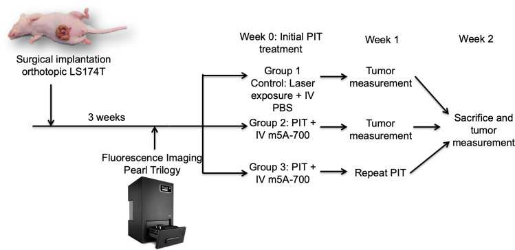
Mice were randomly divided into three groups; Group 1 control mice (n = 3) were irradiated with the NIR laser without pre-administration of the antibody-fluorophore conjugate; group 2 (n = 6) received PIT only one time; and group 3 (n = 6) received repeated PIT treatment one week after initial treatment. Fig 1 illustrates the experimental protocol. After three weeks of tumor growth, Group 2 and 3 mice were administered 50 μg of m5A-700 reconstituted in PBS via tail vein injection. After 24 hours, mice were anesthetized and tumors were surgically exposed through a midline incision. Tumor size was measured with a caliper. Imaging was obtained on the Pearl Trilogy Small Animal Imaging System prior to treatment. The laser beam was centered on the tumor and treatment was delivered at 150 mW/cm2 over 30 minutes for a total delivery of 270 J/cm2. Distance from the laser source and the tumor was standardized at 15 cm for each mouse. After treatment, repeat imaging was obtained on the Pearl Trilogy. Group 3 received repeated PIT treatment one week later. Tumors were surgically exposed and measured with calipers weekly for three weeks. Images were analyzed on the Image Studio Small Animal Imaging Analysis Version 5.2 (LI-COR, Lincoln, NE).

Fig 1. Experimental protocol.

Fig 1

Surgical implantation of an LS174T tumor fragment (1 mm3) onto the cecum was initially performed. Tumors grew 3 weeks and then were imaged with the Pearl Trilogy Small Animal Imaging System. At week 0, initial PIT treatment was performed. At week 1, group 3 received repeat PIT and tumors were measured with calipers. At week 2, mice were sacrificed and tumor size measured.

Statistical analysis

Statistical analysis was performed using SPSS Statistics version 24 (IBM, Armonk, NY). Percent cell death was determined for each well for in vitro PIT and mean percent cell death per group was calculated. Imaging analysis was performed using Image Studio Software Small Animal Imaging Analysis (LI-COR, Lincoln, NE). The skin was set as the background and an area of interest around the tumor fluorescence drawn with a minimum of 250 pixels and at least 2.5 standard deviations from the background signal. Maximum tumor signal before and after PIT was determined from the software analysis. Mean tumor fluorescence signal was calculated before and after PIT and means were compared with the univariate student’s-t test. Significance was determined with p-value cutoff of 0.05, with 2-tailed analysis. Tumor volume was calculated for each time period using the equation ((width x width x length)/2). Mean and average tumor volume was determined for each group at each tumor measurement time point. The univariate student’s-t test was used to compare means between groups at each time point. Significance was determined with p-value cutoff of 0.05, with 2-tailed analysis.

Results

In vitro PIT

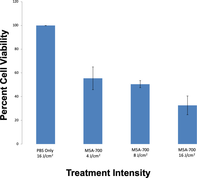
After PIT was delivered to cells in vitro, cell survival analysis was performed. Dose response in vitro PIT demonstrated 44.6%, 49.5% and 61.3% cell death with laser intensities of 4 J/cm2, 8 J/cm2 and 16 J/cm2 respectively (p < 0.001, Fig 2).

Fig 2. In vitro PIT cell viability with increasing exposure to laser intensities.

Fig 2

LS174T cells (2,000) were seeded into each well. Treatment groups were incubated with media containing m5A-700. Lower levels of cell viability were demonstrated in a dose-dependent fashion, with the lowest percentage of cell viability in wells that received 16 J/cm2 of laser exposure (p < 0.001).

Orthotopic PIT

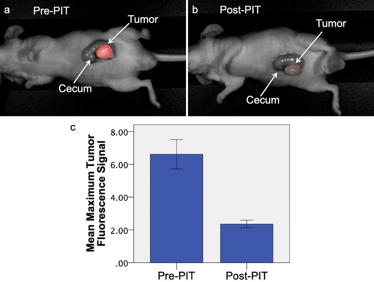
NIR fluorescence signal of the tumor on pre-treatment imaging was compared to signal on post-treatment imaging (n = 12). Prior to treatment, the mean maximum tumor fluorescence was 6.62 (SD ± 3.09). After treatment with PIT, the mean maximum NIR tumor fluorescence decreased to 2.36 (SD ± 0.81), which is indicative of effective PIT treatment (Fig 3). Mean tumor fluorescence before PIT was significantly decreased compared to mean tumor fluorescence after PIT (p < 0.001).

Fig 3. Comparison of pre-treatment and post-treatment fluorescence imaging in orthotopic LS174T mouse models.

Fig 3

The mouse received 25 μg m5A-700 24 hours before treatment. Prior to PIT treatment (a), tumor margins had a distinct NIR fluorescence signal (maximum tumor fluorescence 4.54). After PIT treatment (b), maximum fluorescence signal decreased to 2.82. (c) Mean maximum fluorescence tumor signal before and after PIT (n = 12). There is a significant difference between mean tumor fluorescence signal before and after treatment with PIT (p < 0.001). Error bars represent standard error of the mean.

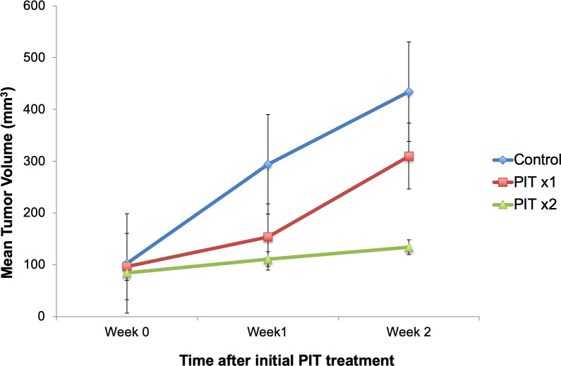
Tumor volumes were measured at weekly intervals with calipers. Fig 4 demonstrates average tumor volume for each group. Control mice demonstrated persistent tumor growth over time. One week after initial PIT treatment, average tumor volume was lower in mice treated with PIT compared to control mice (131.5 mm3 versus 294.1 mm3, respectively, p = 0.063). Mice that received only one PIT treatment did not have a statistically significant difference in tumor volume at week 2 compared to control mice (p = 0.481) Mice that were retreated with PIT one week later had significantly lower tumor volume at week 2 than control mice (p = 0.015).

Fig 4. Tumor volume over time.

Fig 4

PIT was delivered to both treatment groups at week 0. PITx2 group received repeated PIT at week 1. There was a significant difference between PITx2 tumor volume and control tumor volume at week 2 (p < 0.05).

Toxicity and adverse effects

Mice were monitored after treatment for adverse effects. During the study period, no mice were observed to have any adverse events. After mice were euthanized, laparotomy was performed to assess intra-abdominal organs. No local toxicity from treatment was detected on any intra-abdominal organs.

Discussion

In the present study, we initially demonstrated that PIT induces colon cancer cytotoxicity in vitro. This was achieved in a dose-dependent response, which demonstrated higher percentage of cell death with higher laser intensities. Orthotopic colon cancer mouse models were used to analyze the effects of repeated exposure to PIT therapy. Orthotopic mouse models that received PIT treatment only once had an increased rate of tumor growth one week after treatment with PIT. The increased rate of growth after cessation of PIT treatment compared to the control group may be explained by an increase in local growth factors after treatment. Future studies are needed to test this hypothesis. Mice that were treated with a repeated exposure to PIT one week after initial treatment demonstrated persistently inhibited tumor growth. Tumor volume was significantly different between mice that received multiple PIT treatments and control mice two weeks after treatment.

Ogata et al. assessed repeated exposure to PIT in subcutaneous breast and epithelioid cancer mouse models within 24 hours after initial treatment to determine if there was a greater response to PIT with repeated exposure [12]. The results of Ogata et al demonstrated that PIT causes enhanced permeability of tumor vasculature and increasing the delivery of drug in the first few hours after PIT treatment. Repeated exposure to PIT within the first three hours enhanced cancer cell death [12]. While this is a promising approach for superficial cancer types, this may be difficult to perform clinically for intra-abdominal tumors. Therefore, repeated exposure to PIT seven to ten days after initial treatment may be easier to clinically translate and perform on intra-abdominal tumors to promote enhanced suppression of tumor growth. In clinical applications, the easiest disease process to administer repeated treatments of PIT would be rectal cancer, as this is similar to superficial diseases.

The present study is the first application of PIT in an orthotopic mouse model of colon cancer, which makes the results translatable to the clinic than subcutaneous tumor models. For intra-abdominal colorectal cancers in clinical settings, PIT would have to be administered at the time of a surgical procedure. For colon cancer, this would be most applicable in the setting of non-resectable tumors to decrease tumor burden. In addition to primary therapy, PIT would likely be effective as a secondary treatment after resection. Although the rate of radial margin positivity, defined as the presence of colon cancer cells at the resected margins, is low after colectomy for colon cancer, the presence of radial margin positivity significantly reduces survival and leads to a higher risk of recurrence [13]. Delivering PIT to the colectomy margins at the time of surgery may decrease the rate of positive radial margins and lead to improved recurrence rates and survival. PIT is delivered using a small catheter that transmits the laser light. Given the small caliber of this catheter, PIT could be administered to patients undergoing a minimally-invasive resection of colorectal cancer.

Another area that PIT may be the most clinically useful is in the treatment of rectal cancer, as mentioned above. It has been previously reported that the rate of recurrence of colorectal cancers is the highest in tumors of rectal origin [14]. This may be explained by the difficulty of obtaining negative margins in the small area afforded by the pelvis. Therefore, it may also prove useful to use PIT as a directed treatment to the resection area in the pelvis after surgical treatment of rectal cancers to reduce or eliminate residual cancer cells. Future mouse model studies with rectal cancer cell lines can provide information on the efficacy of PIT for rectal tumors.

A potential limitation to the use of PIT for colorectal cancer is the ability to administer treatment since colorectal tumors are largely intraluminal. PIT may be most effective for tumor debulking when the tumor has grown out of the serosa or for locally-advanced or metastatic disease. One area that currently has limited treatment guidelines is peritoneal metastases in colorectal cancer. The current guidelines for colorectal peritoneal metastases are unclear; therefore, PIT provides promising treatment for peritoneal metastases that are not amenable to surgical resection. Preclinical studies have demonstrated efficacy in using anti-HER2 monoclonal antibody trastuzumab conjugated to IR700 in the treatment of ovarian cancer peritoneal metastases in mouse models and demonstrated effective cell killing of peritoneal implants [15]. In addition to peritoneal metastases, PIT has been proven to reduce metastatic disease of breast origin to the lungs in preclinical models [16]. These results provide further promise for the potential clinical uses of PIT since the lung is the second most common site of metastases in colorectal cancer [17]. In addition, PIT has the potential to be useful during endoscopic resections of polyps. Prior studies have demonstrated that 51.3% of patients undergoing polypectomy alone had positive resection margins [18]. Performing PIT to the resection bed after polypectomy can be advantageous to treat residual disease and decrease the rate of recurrence and need for further operation.

The mechanism of PIT has been extensively described in prior studies. Kobayashi et al described the leading mechanism as physical changes to the cell membrane that occur after exposure of a tumor with surface-bound monoclonal antibody conjugated to IRDye700DX to a laser light source [1]. After exposure, a “photoinduced ligand reaction” occurs which induces irreversible physical changes to the cell membrane [1]. These changes in the cell membrane allow for rapid entry of water, cellular swelling and eventual bursting of the cell [1, 3]. Activation of the surrounding immune cells and immunogenic cell death may play a role in the tumor suppressive effects of PIT [1, 2]. In the present study, we utilized athymic nude mice that lack T cells, which are not representative of the complex conditions of the immune system in patients. However, even in the absence of T cells, PIT was shown to be effective, suggesting that T cells are not the major factor in immunogenic cell death. Prior to translation into clinical studies, studies will be performed in orthotopic syngeneic models, humanized or genetically engineered mice, all with an intact immune system. Further limitations to the study include the use of a single colon cancer cell line and a short study period, which was due to rapid tumor growth. Future studies with additional cell lines and patient-derived tumor mouse models will increase the translatability and reliability of this technology. This study serves as a proof-of-principle to demonstrate the effectiveness of PIT in arresting orthotopic colorectal cancer tumor growth, the experimental time was short and detailed histology was not performed. Future studies will include detailed histology to identify characteristics of residual tumor and a longer experimental time prior to translation to clinical studies.

In conclusion, PIT arrested tumor growth in orthotopic colon cancer nude-mouse models. Repeated PIT treatment arrests colon cancer growth for a longer period of time. PIT may be a useful therapy as an adjunct to surgical resection or as primary therapy to suppress tumor progression.

Data Availability

Data is available on the public repository Figshare. https://doi.org/10.6084/m9.figshare.12056157

Funding Statement

The research was supported by VA Merit Review grant numbers 1 I01 BX003856-01A1 and 1 I01 BX004494-01 (MB), and NIH/NCI grant number T32CA121938 (HH). The funder provided support in the form of salaries for authors [HH], but did not have any additional role in the study design, data collection and analysis, decision to publish, or preparation of the manuscript. Dr. Hoffman is affiliated with the commercial company AntiCancer Inc. This commercial affiliation did not provide salaries for any authors and did not have any additional role in the study design, data collection and analysis, decision to publish, or preparation of the manuscript. The specific roles of these authors are articulated in the ‘author contributions’ section.

References

  • 1.Kobayashi H, Choyke PL. Near-Infrared Photoimmunotherapy of Cancer. Acc Chem Res. 2019; 52(8): 2332–2339. 10.1021/acs.accounts.9b00273 [DOI] [PMC free article] [PubMed] [Google Scholar]
  • 2.Ogawa M, Tomita Y, Nakamura Y, Lee MJ, Lee S, Tomita S, et al. Immunogenic cancer cell death selectively induced by near infrared photoimmunotherapy initiates host tumor immunity. Oncotarget. 2017; 8(6):10425–10436. 10.18632/oncotarget.14425 [DOI] [PMC free article] [PubMed] [Google Scholar]
  • 3.Kobayashi H, Griffiths GL, Choyke PL. Near-Infrared Photoimmunotherapy: Photoactivatable Antibody-Drug Conjugates (ADCs). Bioconj Chem. 2019. E pub ahead of print. 10.1021/acs.bioconjchem.9b00546 [DOI] [PMC free article] [PubMed] [Google Scholar]
  • 4.Maawy AA, Hiroshima Y, Zhang Y, Heim R, Making L, Garcia-Guzman M, et al. Near Intra-Red Photoimmunotherapy with Anti-CEA-IR700 Results in Extensive Tumor Lysis and a Significant Decrease in Tumor Burden in Orthotopic Mouse Models of Pancreatic Cancer. PLoS ONE. 2015; 10(3):e0121989 10.1371/journal.pone.0121989 [DOI] [PMC free article] [PubMed] [Google Scholar]
  • 5.Maawy AA, Hiroshima Y, Zhang Y, Garcia-Guzman M, Luiken GA, Kobayashi H, et al. Photoimmunotherapy lowers recurrence after pancreatic cancer surgery in orthotopic nude mouse models. J Surg Res. 2015; 197: 5–11. 10.1016/j.jss.2015.02.037 [DOI] [PMC free article] [PubMed] [Google Scholar]
  • 6.Hiroshima Y, Maawy AA, Zhang Y, Garcia-Guzman M, Heim R, Makings L, et al. Photoimmunotherapy Inhibits Tumor Recurrence After Surgical Resection in Pancreatic Cancer Patient-Derived Orthotopic Xenograft (PDOX) Nude Mouse Model. Ann Surg Onc. 2015; 22:S1469–S1474. 10.1245/s10434-015-4553-9 [DOI] [PMC free article] [PubMed] [Google Scholar]
  • 7.Hammarstrom S. The carcinoembryonic antigen (CEA) family: structures, suggested functions and expression in normal and malignant tissues. Semin Cancer Biol. 1999; 9:67–81. 10.1006/scbi.1998.0119 [DOI] [PubMed] [Google Scholar]
  • 8.Tiernan JP, Perry SL, Verghese ET, West NP, Yeluri S, Jayne G, et al. Carcinoembryonic antigen is the preferred biomarker for in vivo colorectal cancer targeting. Br J Cancer. 2013;108:662–7. 10.1038/bjc.2012.605 [DOI] [PMC free article] [PubMed] [Google Scholar]
  • 9.Yazaki PJ, Sherman MA, Shively JE, Ikle D, Williams LE, Wong J, et al. Humanization of the anti-CEA T84.66 antibody based on crystal structure data. Protein engineering, design & selection. PEDS 2004;17:481–9. [DOI] [PubMed] [Google Scholar]
  • 10.DeLong JC, Murakami T, Yazaki PJ, et al. Near-infrared-conjugated humanized anti-carcinoembryonic antigen antibody targets colon cancer in an orthotopic nude-mouse model. J Surg Res. 2017;218:139–143. 10.1016/j.jss.2017.05.069 [DOI] [PMC free article] [PubMed] [Google Scholar]
  • 11.Hoffman RM. Patient-Derived Mouse Models of Cancer. Humana Press, Springer International Publishing AG; 2017. 10.1007/978-3-319-57424-0 [DOI] [Google Scholar]
  • 12.Ogata F, Nagaya T, Nakamura Y, Sato K, Okuyama S, Maruoka Y, et al. Near-infrared photoimmunotherapy: a comparison of light dosing schedules. Oncotarget. 2017; 8(21): 35069–35075. 10.18632/oncotarget.17047 [DOI] [PMC free article] [PubMed] [Google Scholar]
  • 13.Amri R, Bordeianou LG, Sylla P, Berger DL. Association of Radial Margin Positivity with Colon Cancer. JAMA Surg. 2015; 150(9): 890–8. oi: 10.1001/jamasurg.2015.1525 [DOI] [PubMed] [Google Scholar]
  • 14.Zare-Bandamiri M, Fararouei M, Zohourinia S, Daneshi N, Dianatinasab M. Risk Factors Predicting Colorectal Cancer Recurrence Following Initial Treatment: A 5-year Cohort Study. Asian Pac J Cancer Prev. 2017; 18(9): 2465–2470. 10.22034/APJCP.2017.18.9.2465 [DOI] [PMC free article] [PubMed] [Google Scholar]
  • 15.Sato K, Hanaoka H, Watanabe R, Nakajima T, Choyke PL, Kobayashi H. Near infrared photoimmunotherapy in the treatment of disseminated peritoneal ovarian cancer. Mol Cancer Ther. 2015; 14(1): 141–50. 10.1158/1535-7163.MCT-14-0658 [DOI] [PMC free article] [PubMed] [Google Scholar]
  • 16.Sato K, Nagaya T, Nakamura Y, Harada T, Choyke PL, Kobayashi H. Near infrared photoimmunotherapy prevents lung cancer metastases in a murine model. Oncotarget. 2015; 6(23): 19757–58. 10.18632/oncotarget.3850 [DOI] [PMC free article] [PubMed] [Google Scholar]
  • 17.Villenueve PJ, Sundaresan RS. Surgical Management of Colorectal Lung Metastasis. Clin Colon Rectal Surg. 2009; 22(4): 233–241. 10.1055/s-0029-1242463 [DOI] [PMC free article] [PubMed] [Google Scholar]
  • 18.Jang EJ, Kim DD, Cho CH. Value and Interpretation of Resection Margin after a Colonoscopic Polypectomy for Malignant Polyps. J Korean Soc Coloproctol. 2011; 27(4): 194–201. 10.3393/jksc.2011.27.4.194 [DOI] [PMC free article] [PubMed] [Google Scholar]

Decision Letter 0

Irina V Lebedeva

19 Mar 2020

PONE-D-20-02950

Near-Infrared Photoimmunotherapy is Effective Treatment for Colorectal Cancer in Orthotopic Nude-Mouse Models

PLOS ONE

Dear Dr Bouvet,

Thank you for submitting your manuscript to PLOS ONE. After careful consideration, we feel that it has merit but does not fully meet PLOS ONE’s publication criteria as it currently stands. Therefore, we invite you to submit a revised version of the manuscript that addresses the points raised during the review process.

Quantification of fluorescent intensity and tumor size need to be provided.  Also histology images should be provided.

What is the rationale for the cell line used? Have experiments been done with other cell lines? What is the rationale for treatment time?

The limitations should be discussed for the mouse model and for technology application.

Are there any considerations for the mechanism(s) involved?

We would appreciate receiving your revised manuscript by May 03 2020 11:59PM. When you are ready to submit your revision, log on to https://www.editorialmanager.com/pone/ and select the 'Submissions Needing Revision' folder to locate your manuscript file.

If you would like to make changes to your financial disclosure, please include your updated statement in your cover letter.

To enhance the reproducibility of your results, we recommend that if applicable you deposit your laboratory protocols in protocols.io, where a protocol can be assigned its own identifier (DOI) such that it can be cited independently in the future. For instructions see: http://journals.plos.org/plosone/s/submission-guidelines#loc-laboratory-protocols

Please include the following items when submitting your revised manuscript:

  • A rebuttal letter that responds to each point raised by the academic editor and reviewer(s). This letter should be uploaded as separate file and labeled 'Response to Reviewers'.

  • A marked-up copy of your manuscript that highlights changes made to the original version. This file should be uploaded as separate file and labeled 'Revised Manuscript with Track Changes'.

  • An unmarked version of your revised paper without tracked changes. This file should be uploaded as separate file and labeled 'Manuscript'.

Please note while forming your response, if your article is accepted, you may have the opportunity to make the peer review history publicly available. The record will include editor decision letters (with reviews) and your responses to reviewer comments. If eligible, we will contact you to opt in or out.

We look forward to receiving your revised manuscript.

Kind regards,

Irina V. Lebedeva, Ph.D.

Academic Editor

PLOS ONE

Journal Requirements:

1. When submitting your revision, we need you to address these additional requirements.


Please ensure that your manuscript meets PLOS ONE's style requirements, including those for file naming. The PLOS ONE style templates can be found at

http://www.journals.plos.org/plosone/s/file?id=wjVg/PLOSOne_formatting_sample_main_body.pdf and http://www.journals.plos.org/plosone/s/file?id=ba62/PLOSOne_formatting_sample_title_authors_affiliations.pdf

2. PLOS requires an ORCID iD for the corresponding author in Editorial Manager on papers submitted after December 6th, 2016. Please ensure that you have an ORCID iD and that it is validated in Editorial Manager. To do this, go to ‘Update my Information’ (in the upper left-hand corner of the main menu), and click on the Fetch/Validate link next to the ORCID field. This will take you to the ORCID site and allow you to create a new iD or authenticate a pre-existing iD in Editorial Manager. Please see the following video for instructions on linking an ORCID iD to your Editorial Manager account: https://www.youtube.com/watch?v=_xcclfuvtxQ

3. We note that you have a patent relating to material pertinent to this article. Please provide an amended statement of Competing Interests to declare this patent (with details including name and number), along with any other relevant declarations relating to employment, consultancy, patents, products in development or modified products etc. Please confirm that this does not alter your adherence to all PLOS ONE policies on sharing data and materials, as detailed online in our guide for authors http://journals.plos.org/plosone/s/competing-interests by including the following statement: "This does not alter our adherence to  PLOS ONE policies on sharing data and materials.” If there are restrictions on sharing of data and/or materials, please state these. Please note that we cannot proceed with consideration of your article until this information has been declared.

* This information should be included in your cover letter; we will change the online submission form on your behalf.

4. Thank you for stating the following in the Financial Disclosure section:

"This study was funded by VA Merit Review grant numbers 1 I01 BX003856-01A1 and 1 I01 BX004494-01 (MB), NIH/NCI T32CA121938 (HH).  "

We note that one or more of the authors are employed by a commercial company: 'AntiCancer Inc'.

1.     Please provide an amended Funding Statement declaring this commercial affiliation, as well as a statement regarding the Role of Funders in your study. If the funding organization did not play a role in the study design, data collection and analysis, decision to publish, or preparation of the manuscript and only provided financial support in the form of authors' salaries and/or research materials, please review your statements relating to the author contributions, and ensure you have specifically and accurately indicated the role(s) that these authors had in your study. You can update author roles in the Author Contributions section of the online submission form.

Please also include the following statement within your amended Funding Statement.

“The funder provided support in the form of salaries for authors [insert relevant initials], but did not have any additional role in the study design, data collection and analysis, decision to publish, or preparation of the manuscript. The specific roles of these authors are articulated in the ‘author contributions’ section.”

If your commercial affiliation did play a role in your study, please state and explain this role within your updated Funding Statement.

2. Please also provide an updated Competing Interests Statement declaring this commercial affiliation along with any other relevant declarations relating to employment, consultancy, patents, products in development, or marketed products, etc.  

Within your Competing Interests Statement, please confirm that this commercial affiliation does not alter your adherence to all PLOS ONE policies on sharing data and materials by including the following statement: "This does not alter our adherence to  PLOS ONE policies on sharing data and materials.” (as detailed online in our guide for authors http://journals.plos.org/plosone/s/competing-interests) . If this adherence statement is not accurate and  there are restrictions on sharing of data and/or materials, please state these. Please note that we cannot proceed with consideration of your article until this information has been declared.

* Please include both an updated Funding Statement and Competing Interests Statement in your cover letter. We will change the online submission form on your behalf.

Please know it is PLOS ONE policy for corresponding authors to declare, on behalf of all authors, all potential competing interests for the purposes of transparency. PLOS defines a competing interest as anything that interferes with, or could reasonably be perceived as interfering with, the full and objective presentation, peer review, editorial decision-making, or publication of research or non-research articles submitted to one of the journals. Competing interests can be financial or non-financial, professional, or personal. Competing interests can arise in relationship to an organization or another person. Please follow this link to our website for more details on competing interests: http://journals.plos.org/plosone/s/competing-interests

5. In the Methods, please provide the formula by which tumour volume was calculated.

[Note: HTML markup is below. Please do not edit.]

Reviewers' comments:

Reviewer's Responses to Questions

Comments to the Author

1. Is the manuscript technically sound, and do the data support the conclusions?

The manuscript must describe a technically sound piece of scientific research with data that supports the conclusions. Experiments must have been conducted rigorously, with appropriate controls, replication, and sample sizes. The conclusions must be drawn appropriately based on the data presented.

Reviewer #1: Partly

Reviewer #2: Yes

**********

2. Has the statistical analysis been performed appropriately and rigorously?

Reviewer #1: No

Reviewer #2: Yes

**********

3. Have the authors made all data underlying the findings in their manuscript fully available?

The PLOS Data policy requires authors to make all data underlying the findings described in their manuscript fully available without restriction, with rare exception (please refer to the Data Availability Statement in the manuscript PDF file). The data should be provided as part of the manuscript or its supporting information, or deposited to a public repository. For example, in addition to summary statistics, the data points behind means, medians and variance measures should be available. If there are restrictions on publicly sharing data—e.g. participant privacy or use of data from a third party—those must be specified.

Reviewer #1: No

Reviewer #2: Yes

**********

4. Is the manuscript presented in an intelligible fashion and written in standard English?

PLOS ONE does not copyedit accepted manuscripts, so the language in submitted articles must be clear, correct, and unambiguous. Any typographical or grammatical errors should be corrected at revision, so please note any specific errors here.

Reviewer #1: Yes

Reviewer #2: Yes

**********

5. Review Comments to the Author

Please use the space provided to explain your answers to the questions above. You may also include additional comments for the author, including concerns about dual publication, research ethics, or publication ethics. (Please upload your review as an attachment if it exceeds 20,000 characters)

Reviewer #1: In this manuscript, Hollandsworth et al. describe the application of photoimmunotherapy (PIT) in the treatment of a human colon cancer cell line in vitro and in an orthotopic cecal transplantation model. The authors demonstrate a dose-dependent response to PIT in a cancer cell line. They then show that a single dose of PIT effectively treats the cecal transplantation model. However, a key weakness is that this manuscript fails to provide any mechanism whatsoever to support the conclusion that this technology may be useful for clinical application.

Comments:

1. The authors state that PIT suppressed tumor growth in subcutaneous colon cancer mouse model but this conclusion is demonstrated only in a single mouse image in Figure 3. Quantification of fluorescence intensity and tumor size measurements need to be provided to support this claim. Figure 4 should also be accompanied with quantification and images of tumors.

2. What is the purpose of performing subcutaneous transplant studies if the authors also perform cecal transplants, which are presumably better?

3. Finally, they show inhibition of tumor growth in orthotopic nude mouse models with repeated exposure to PIT one week after initial treatment. Overall the results are promising as this is the first study of PIT in an orthotopic mouse model of colon. However, the conclusions drawn are largely hypothetical as the tumors were grown in athymic nude mice without a competent immune system. Although these mice have dendritic cells, they lack T cells. Thus, this model may have little relevance to humans. Studies in a mouse cell line or organoid transplant model or genetically engineered model would be more relevant.

4. The authors also failed to discuss why the tumors rapidly increased in size following PIT cessation in comparison to control tumors.

5. No experiments were performed to study the possible mechanism of action of PIT in colon cancer. Thus, these experiments add little value to the literature on PIT.

6. Why did the authors select this particular cell line, and why were studies not repeated in additional cell lines or organoid lines?

7. As the authors themselves note, the application of this technology is questionable since the tumor will need to be exposed to the light source. One possible application not discussed by the authors is ablation of positive margins of adenomas during colonoscopy.

8. Why was the treatment only provided for 1-2 weeks? This time scale is extremely short.

9. Histology of the tumors should be provided.

Reviewer #2: The figure legends and results discussion appear to be out of order for several figures. This appears to be an issue of uploading but does not impact the content. The study is technically proficient and highlights an important potential approach to margin positive disease and potentially for peritoneal metastasis.

**********

6. PLOS authors have the option to publish the peer review history of their article (what does this mean?). If published, this will include your full peer review and any attached files.

If you choose “no”, your identity will remain anonymous but your review may still be made public.

Do you want your identity to be public for this peer review? For information about this choice, including consent withdrawal, please see our Privacy Policy.

Reviewer #1: No

Reviewer #2: Yes: Tony R Reid MD, Ph.D.

[NOTE: If reviewer comments were submitted as an attachment file, they will be attached to this email and accessible via the submission site. Please log into your account, locate the manuscript record, and check for the action link "View Attachments". If this link does not appear, there are no attachment files to be viewed.]

While revising your submission, please upload your figure files to the Preflight Analysis and Conversion Engine (PACE) digital diagnostic tool, https://pacev2.apexcovantage.com/. PACE helps ensure that figures meet PLOS requirements. To use PACE, you must first register as a user. Registration is free. Then, login and navigate to the UPLOAD tab, where you will find detailed instructions on how to use the tool. If you encounter any issues or have any questions when using PACE, please email us at figures@plos.org. Please note that Supporting Information files do not need this step.

Attachment

Submitted filename: Review PIT for colorectal cancer.docx

PLoS One. 2020 Jun 18;15(6):e0234643. doi: 10.1371/journal.pone.0234643.r002

Author response to Decision Letter 0


4 Apr 2020

Thank you for your email and reviewers’ comments regarding our manuscript entitled “Near-Infrared Photoimmunotherapy is Effective Treatment for Colorectal Cancer in Orthotopic Nude-Mouse Models.” Thank you also for your interest and the thoughtful comments provided by the Reviewers. For our revision submission, we are providing a revised manuscript that addresses all Reviewer comments and suggestions.

Below, please find the Reviewer’s comments and how each was addressed in the revised manuscript. The Reviewer comments are numbered and our responses to each comment are below in bold.

We thank you again for the opportunity to submit this revised manuscript and look forward to your comments.

Reviewers' comments:

Reviewer 1:

1. The authors state that PIT suppressed tumor growth in subcutaneous colon cancer mouse model but this conclusion is demonstrated only in a single mouse image in Figure 3. Quantification of fluorescence intensity and tumor size measurements need to be provided to support this claim. Figure 4 should also be accompanied with quantification and images of tumors.

Figure 4 legend (now labelled Figure 3) and the Results now include quantification of fluorescence intensity in the orthotopic models.

Please see answer to question 2 below.

2. What is the purpose of performing subcutaneous transplant studies if the authors also perform cecal transplants, which are presumably better?

Data on subcutaneous models was removed from the revised manuscript.

3. Finally, they show inhibition of tumor growth in orthotopic nude mouse models with repeated exposure to PIT one week after initial treatment. Overall the results are promising as this is the first study of PIT in an orthotopic mouse model of colon. However, the conclusions drawn are largely hypothetical as the tumors were grown in athymic nude mice without a competent immune system. Although these mice have dendritic cells, they lack T cells. Thus, this model may have little relevance to humans. Studies in a mouse cell line or organoid transplant model or genetically engineered model would be more relevant.

A section in the discussion has been included to discuss the limitation highlighted above: “In the present study, we utilized athymic nude mice that lack T cells, which are not representative of the complex conditions of the immune system in patients. However, even in the absence of T cells, PIT was shown to be effective, suggesting that T cells are not the major factor in immunogenic cell death. Prior to translation into clinical studies, we will perform PIT on orthotopic syngeneic models, humanized or genetically engineered mice, all with an intact immune system.”

4. The authors also failed to discuss why the tumors rapidly increased in size following PIT cessation in comparison to control tumors.

This is an important question. We can only speculate that the first PIT caused the production of growth factors, such is sometimes the case in surgery, where residual tumors grow faster. Future experiments are needed to answer this question. This is stated in the revised manuscript.

5. No experiments were performed to study the possible mechanism of action of PIT in colon cancer. Thus, these experiments add little value to the literature on PIT.

The present studies are a proof-of-principle that PIT is effective on orthotopic models of colon cancer, as previous studies were only on subcutaneous colon cancer models, which are artificial. This is stated in the revised manuscript. However, general mechanisms of PIT are discussed in the revised manuscript.

6. Why did the authors select this particular cell line, and why were studies not repeated in additional cell lines or organoid lines?

We have done previous studies showing human colon cancer LS174T cell line is targeted well by anti-CEA antibodies (1) and would be appropriate for an orthotopic PIT study and proof-of-principle. This is stated in the revised manuscript.

7. As the authors themselves note, the application of this technology is questionable since the tumor will need to be exposed to the light source. One possible application not discussed by the authors is ablation of positive margins of adenomas during colonoscopy.

PIT can be an intra-operative procedure; therefore, illuminating an intra-abdominal tumor is feasible. This is discussed in the revised version. The potential application for polyp margins during endoscopy is also now included in the Discussion.

8. Why was the treatment only provided for 1-2 weeks? This time scale is extremely short.

The limitation of the time scale is included in the Discussion. This also includes the reason for short time period, which was tumor growth in the control group that exceeded the allowed tumor burden. It was discussed that survival and recurrence studies can increase clinical applicability of PIT in colorectal cancer.

9. Histology of the tumors should be provided.

As the present study was a proof-of-principle study that PIT could arrest an orthotopic tumor, histology studies were not done. Detailed histology studies will be done in the future. This is discussed in the revised edition.

As part of this submission, we have included a revised version of the main manuscript file that shows changes made by highlighting, as well as a clean version of the revised manuscript. Again, we thank the editors and reviewers for their efforts in improving our manuscript. We would be happy to answer any further questions and/or concerns.

Attachment

Submitted filename: Response to Reviewers.docx

Decision Letter 1

Irina V Lebedeva

5 May 2020

PONE-D-20-02950R1

Near-Infrared Photoimmunotherapy is Effective Treatment for Colorectal Cancer in Orthotopic Nude-Mouse Models

PLOS ONE

Dear Dr Bouvet,

Thank you for submitting your manuscript to PLOS ONE. After careful consideration, we feel that it has merit but does not fully meet PLOS ONE’s publication criteria as it currently stands. Therefore, we invite you to submit a revised version of the manuscript that addresses the points raised during the review process.

We would appreciate receiving your revised manuscript by Jun 19 2020 11:59PM. When you are ready to submit your revision, log on to https://www.editorialmanager.com/pone/ and select the 'Submissions Needing Revision' folder to locate your manuscript file.

If you would like to make changes to your financial disclosure, please include your updated statement in your cover letter.

To enhance the reproducibility of your results, we recommend that if applicable you deposit your laboratory protocols in protocols.io, where a protocol can be assigned its own identifier (DOI) such that it can be cited independently in the future. For instructions see: http://journals.plos.org/plosone/s/submission-guidelines#loc-laboratory-protocols

Please include the following items when submitting your revised manuscript:

  • A rebuttal letter that responds to each point raised by the academic editor and reviewer(s). This letter should be uploaded as separate file and labeled 'Response to Reviewers'.

  • A marked-up copy of your manuscript that highlights changes made to the original version. This file should be uploaded as separate file and labeled 'Revised Manuscript with Track Changes'.

  • An unmarked version of your revised paper without tracked changes. This file should be uploaded as separate file and labeled 'Manuscript'.

Please note while forming your response, if your article is accepted, you may have the opportunity to make the peer review history publicly available. The record will include editor decision letters (with reviews) and your responses to reviewer comments. If eligible, we will contact you to opt in or out.

We look forward to receiving your revised manuscript.

Kind regards,

Irina V. Lebedeva, Ph.D.

Academic Editor

PLOS ONE

[Note: HTML markup is below. Please do not edit.]

Reviewers' comments:

Reviewer's Responses to Questions

Comments to the Author

1. If the authors have adequately addressed your comments raised in a previous round of review and you feel that this manuscript is now acceptable for publication, you may indicate that here to bypass the “Comments to the Author” section, enter your conflict of interest statement in the “Confidential to Editor” section, and submit your "Accept" recommendation.

Reviewer #1: All comments have been addressed

Reviewer #2: All comments have been addressed

**********

2. Is the manuscript technically sound, and do the data support the conclusions?

The manuscript must describe a technically sound piece of scientific research with data that supports the conclusions. Experiments must have been conducted rigorously, with appropriate controls, replication, and sample sizes. The conclusions must be drawn appropriately based on the data presented.

Reviewer #1: Yes

Reviewer #2: Yes

**********

3. Has the statistical analysis been performed appropriately and rigorously?

Reviewer #1: Yes

Reviewer #2: Yes

**********

4. Have the authors made all data underlying the findings in their manuscript fully available?

The PLOS Data policy requires authors to make all data underlying the findings described in their manuscript fully available without restriction, with rare exception (please refer to the Data Availability Statement in the manuscript PDF file). The data should be provided as part of the manuscript or its supporting information, or deposited to a public repository. For example, in addition to summary statistics, the data points behind means, medians and variance measures should be available. If there are restrictions on publicly sharing data—e.g. participant privacy or use of data from a third party—those must be specified.

Reviewer #1: Yes

Reviewer #2: Yes

**********

5. Is the manuscript presented in an intelligible fashion and written in standard English?

PLOS ONE does not copyedit accepted manuscripts, so the language in submitted articles must be clear, correct, and unambiguous. Any typographical or grammatical errors should be corrected at revision, so please note any specific errors here.

Reviewer #1: Yes

Reviewer #2: Yes

**********

6. Review Comments to the Author

Please use the space provided to explain your answers to the questions above. You may also include additional comments for the author, including concerns about dual publication, research ethics, or publication ethics. (Please upload your review as an attachment if it exceeds 20,000 characters)

Reviewer #1: The author's has appropriately limited the scope of their conclusions and added appropriate discussion. Below please see our comments about specific points with minor revision requests.

Reviewers' Response to comments:

1. The authors state that PIT suppressed tumor growth in subcutaneous colon cancer

mouse model but this conclusion is demonstrated only in a single mouse image in

Figure 3. Quantification of fluorescence intensity and tumor size measurements need

to be provided to support this claim. Figure 4 should also be accompanied with

quantification and images of tumors.

Author’s Response: Figure 4 legend (now labelled Figure 3) and the Results now include quantification of fluorescence intensity in the orthotopic models.

Please see answer to question 2 below.

Reviewer’s Response:

How many fluorescence measurements were calculated? It would strengthen the claim if this data was shared as a graph in updated Figure 3.

2. What is the purpose of performing subcutaneous transplant studies if the authors

also perform cecal transplants, which are presumably better?

Author’s Response: Data on subcutaneous models was removed from the revised manuscript.

Reviewer’s Response:

Author’s can include other relevant experiments as supplemental figures.

3. Finally, they show inhibition of tumor growth in orthotopic nude mouse models with

repeated exposure to PIT one week after initial treatment. Overall the results are

promising as this is the first study of PIT in an orthotopic mouse model of colon.

However, the conclusions drawn are largely hypothetical as the tumors were grown in

athymic nude mice without a competent immune system. Although these mice have

dendritic cells, they lack T cells. Thus, this model may have little relevance to humans.

Studies in a mouse cell line or organoid transplant model or genetically engineered

model would be more relevant.

Author’s Response: A section in the discussion has been included to discuss the limitation highlighted above: “In the present study, we utilized athymic nude mice that lack T cells, which are not representative of the complex conditions of the immune system in patients. However, even in the absence of T cells, PIT was shown to be effective, suggesting that T cells are not the major factor in immunogenic cell death. Prior to translation into clinical studies, we will perform PIT on orthotopic syngeneic models, humanized or genetically engineered mice, all with an intact immune system.”

Reviewer’s Response:

This is acceptable.

4. The authors also failed to discuss why the tumors rapidly increased in size following

PIT cessation in comparison to control tumors.

Author’s Response: This is an important question. We can only speculate that the first PIT caused the production of growth factors, such is sometimes the case in surgery, where residual tumors grow faster. Future experiments are needed to answer this question. This is

stated in the revised manuscript.

Reviewer’s Response:

Histology of the tumors on future studies can also help identify characteristics of the residual tumor.

5. No experiments were performed to study the possible mechanism of action of PIT in

colon cancer. Thus, these experiments add little value to the literature on PIT.

Author’s Response: The present studies are a proof-of-principle that PIT is effective on orthotopic models of colon cancer, as previous studies were only on subcutaneous colon cancer models, which are artificial. This is stated in the revised manuscript. However, general mechanisms of PIT are discussed in the revised manuscript.

Reviewer’s Response:

The new information provided will help readers understand the possible future directions.

6. Why did the authors select this particular cell line, and why were studies not

repeated in additional cell lines or organoid lines?

Author’s Response:

We have done previous studies showing human colon cancer LS174T cell line is

targeted well by anti-CEA antibodies (1) and would be appropriate for an orthotopic PIT

study and proof-of-principle. This is stated in the revised manuscript.

7. As the authors themselves note, the application of this technology is questionable

since the tumor will need to be exposed to the light source. One possible application

not discussed by the authors is ablation of positive margins of adenomas during

colonoscopy.

Author’s Response:

PIT can be an intra-operative procedure; therefore, illuminating an intra-abdominal

tumor is feasible. This is discussed in the revised version. The potential application for

polyp margins during endoscopy is also now included in the Discussion.

8. Why was the treatment only provided for 1-2 weeks? This time scale is extremely

short.

Author’s Response:

The limitation of the time scale is included in the Discussion. This also includes the

reason for short time period, which was tumor growth in the control group that

exceeded the allowed tumor burden. It was discussed that survival and recurrence

studies can increase clinical applicability of PIT in colorectal cancer.

9. Histology of the tumors should be provided.

Author’s Response:

As the present study was a proof-of-principle study that PIT could arrest an orthotopic

tumor, histology studies were not done. Detailed histology studies will be done in the

future. This is discussed in the revised edition.

As part of this submission, we have included a revised version of the main manuscript

file that shows changes made by highlighting, as well as a clean version of the revised

manuscript. Again, we thank the editors and reviewers for their efforts in improving our

manuscript. We would be happy to answer any further questions and/or concerns.

Reviewer #2: The study demonstrates dose dependent tumor cell killing with PIT and the potential application of this technology to surgical settings. The non-toxic aspect of PIT in the absence of the conjugated antibody offers the potential to treat otherwise surgically inoperable disease as well as debulk disease. The paper offers an important potential path to managing complex intra-abdominal metastatic disease.

**********

7. PLOS authors have the option to publish the peer review history of their article (what does this mean?). If published, this will include your full peer review and any attached files.

If you choose “no”, your identity will remain anonymous but your review may still be made public.

Do you want your identity to be public for this peer review? For information about this choice, including consent withdrawal, please see our Privacy Policy.

Reviewer #1: No

Reviewer #2: Yes: Tony Reid MD, Ph.D.

[NOTE: If reviewer comments were submitted as an attachment file, they will be attached to this email and accessible via the submission site. Please log into your account, locate the manuscript record, and check for the action link "View Attachments". If this link does not appear, there are no attachment files to be viewed.]

While revising your submission, please upload your figure files to the Preflight Analysis and Conversion Engine (PACE) digital diagnostic tool, https://pacev2.apexcovantage.com/. PACE helps ensure that figures meet PLOS requirements. To use PACE, you must first register as a user. Registration is free. Then, login and navigate to the UPLOAD tab, where you will find detailed instructions on how to use the tool. If you encounter any issues or have any questions when using PACE, please email us at figures@plos.org. Please note that Supporting Information files do not need this step.

PLoS One. 2020 Jun 18;15(6):e0234643. doi: 10.1371/journal.pone.0234643.r004

Author response to Decision Letter 1


8 May 2020

May 7, 2020

To: Drs. Joreg Heber and Irina V. Lebedeva, Editor-in Chief and Academic Editor, PLOS One

Manuscript – “Near-Infrared Photoimmunotherapy is Effective Treatment for Colorectal Cancer in Orthotopic Nude-Mouse Models”

Drs. Heber and Lebedeva,

Thank you for your email and reviewers’ comments regarding our manuscript entitled “Near-Infrared Photoimmunotherapy is Effective Treatment for Colorectal Cancer in Orthotopic Nude-Mouse Models.” Thank you also for your interest and the thoughtful comments provided by the Reviewers. For our revision submission, we are providing a revised manuscript that addresses all Reviewer comments and suggestions.

Below, please find the Reviewer’s comments and how each was addressed in the revised manuscript. The most recent Reviewer comments are italicized and our responses to each comment are below in bold.

We thank you again for the opportunity to submit this revised manuscript and look forward to your comments.

Reviewers' Response to comments:

1. The authors state that PIT suppressed tumor growth in subcutaneous colon cancer

mouse model but this conclusion is demonstrated only in a single mouse image in

Figure 3. Quantification of fluorescence intensity and tumor size measurements need

to be provided to support this claim. Figure 4 should also be accompanied with

quantification and images of tumors.

Author’s Response: Figure 4 legend (now labelled Figure 3) and the Results now include quantification of fluorescence intensity in the orthotopic models.

Please see answer to question 2 below.

Reviewer’s Response:

How many fluorescence measurements were calculated? It would strengthen the claim if this data was shared as a graph in updated Figure 3.

Figure 3 has been updated to include a graph depicting the difference in mean maximum tumor fluorescence signal before and after PIT. The figure legend provides description of how many fluorescence measurements were calculated and statistical significance. Materials and methods have been revised to include image analysis and statistical analysis of tumor fluorescence.

2. What is the purpose of performing subcutaneous transplant studies if the authors

also perform cecal transplants, which are presumably better?

Author’s Response: Data on subcutaneous models was removed from the revised manuscript.

Reviewer’s Response:

Author’s can include other relevant experiments as supplemental figures.

All relevant experiments are presented in the main manuscript.

3. Finally, they show inhibition of tumor growth in orthotopic nude mouse models with

repeated exposure to PIT one week after initial treatment. Overall the results are

promising as this is the first study of PIT in an orthotopic mouse model of colon.

However, the conclusions drawn are largely hypothetical as the tumors were grown in

athymic nude mice without a competent immune system. Although these mice have

dendritic cells, they lack T cells. Thus, this model may have little relevance to humans.

Studies in a mouse cell line or organoid transplant model or genetically engineered

model would be more relevant.

Author’s Response: A section in the discussion has been included to discuss the limitation highlighted above: “In the present study, we utilized athymic nude mice that lack T cells, which are not representative of the complex conditions of the immune system in patients. However, even in the absence of T cells, PIT was shown to be effective, suggesting that T cells are not the major factor in immunogenic cell death. Prior to translation into clinical studies, we will perform PIT on orthotopic syngeneic models, humanized or genetically engineered mice, all with an intact immune system.”

Reviewer’s Response:

This is acceptable.

4. The authors also failed to discuss why the tumors rapidly increased in size following

PIT cessation in comparison to control tumors.

Author’s Response: This is an important question. We can only speculate that the first PIT caused the production of growth factors, such is sometimes the case in surgery, where residual tumors grow faster. Future experiments are needed to answer this question. This is

stated in the revised manuscript.

Reviewer’s Response:

Histology of the tumors on future studies can also help identify characteristics of the residual tumor.

This point was added to the revised discussion.

5. No experiments were performed to study the possible mechanism of action of PIT in

colon cancer. Thus, these experiments add little value to the literature on PIT.

Author’s Response: The present studies are a proof-of-principle that PIT is effective on orthotopic models of colon cancer, as previous studies were only on subcutaneous colon cancer models, which are artificial. This is stated in the revised manuscript. However, general mechanisms of PIT are discussed in the revised manuscript.

Reviewer’s Response:

The new information provided will help readers understand the possible future directions.

6. Why did the authors select this particular cell line, and why were studies not

repeated in additional cell lines or organoid lines?

Author’s Response:

We have done previous studies showing human colon cancer LS174T cell line is

targeted well by anti-CEA antibodies (1) and would be appropriate for an orthotopic PIT

study and proof-of-principle. This is stated in the revised manuscript.

7. As the authors themselves note, the application of this technology is questionable

since the tumor will need to be exposed to the light source. One possible application

not discussed by the authors is ablation of positive margins of adenomas during

colonoscopy.

Author’s Response:

PIT can be an intra-operative procedure; therefore, illuminating an intra-abdominal

tumor is feasible. This is discussed in the revised version. The potential application for

polyp margins during endoscopy is also now included in the Discussion.

8. Why was the treatment only provided for 1-2 weeks? This time scale is extremely

short.

Author’s Response:

The limitation of the time scale is included in the Discussion. This also includes the

reason for short time period, which was tumor growth in the control group that

exceeded the allowed tumor burden. It was discussed that survival and recurrence

studies can increase clinical applicability of PIT in colorectal cancer.

9. Histology of the tumors should be provided.

Author’s Response:

As the present study was a proof-of-principle study that PIT could arrest an orthotopic

tumor, histology studies were not done. Detailed histology studies will be done in the

future. This is discussed in the revised edition.

As part of this submission, we have included a revised version of the main manuscript

file that shows changes made by highlighting, as well as a clean version of the revised

manuscript. Again, we thank the editors and reviewers for their efforts in improving our

manuscript. We would be happy to answer any further questions and/or concerns.

As part of this submission, we have included a revised version of the main manuscript file that shows changes made by highlighting, as well as a clean version of the revised manuscript. Again, we thank the editors and reviewers for their efforts in improving our manuscript. We would be happy to answer any further questions and/or concerns.

Sincerely,

Hannah Hollandsworth, MD

Attachment

Submitted filename: Response to Reviewers_2.docx

Decision Letter 2

Irina V Lebedeva

1 Jun 2020

Near-Infrared Photoimmunotherapy is Effective Treatment for Colorectal Cancer in Orthotopic Nude-Mouse Models

PONE-D-20-02950R2

Dear Dr. Bouvet,

We are pleased to inform you that your manuscript has been judged scientifically suitable for publication and will be formally accepted for publication once it complies with all outstanding technical requirements.

Within one week, you will receive an e-mail containing information on the amendments required prior to publication. When all required modifications have been addressed, you will receive a formal acceptance letter and your manuscript will proceed to our production department and be scheduled for publication.

Shortly after the formal acceptance letter is sent, an invoice for payment will follow. To ensure an efficient production and billing process, please log into Editorial Manager at https://www.editorialmanager.com/pone/, click the "Update My Information" link at the top of the page, and update your user information. If you have any billing related questions, please contact our Author Billing department directly at authorbilling@plos.org.

If your institution or institutions have a press office, please notify them about your upcoming paper to enable them to help maximize its impact. If they will be preparing press materials for this manuscript, you must inform our press team as soon as possible and no later than 48 hours after receiving the formal acceptance. Your manuscript will remain under strict press embargo until 2 pm Eastern Time on the date of publication. For more information, please contact onepress@plos.org.

With kind regards,

Irina V. Lebedeva, Ph.D.

Academic Editor

PLOS ONE

Additional Editor Comments (optional):

Reviewers' comments:

Reviewer's Responses to Questions

Comments to the Author

1. If the authors have adequately addressed your comments raised in a previous round of review and you feel that this manuscript is now acceptable for publication, you may indicate that here to bypass the “Comments to the Author” section, enter your conflict of interest statement in the “Confidential to Editor” section, and submit your "Accept" recommendation.

Reviewer #1: All comments have been addressed

**********

2. Is the manuscript technically sound, and do the data support the conclusions?

The manuscript must describe a technically sound piece of scientific research with data that supports the conclusions. Experiments must have been conducted rigorously, with appropriate controls, replication, and sample sizes. The conclusions must be drawn appropriately based on the data presented.

Reviewer #1: Yes

**********

3. Has the statistical analysis been performed appropriately and rigorously?

Reviewer #1: Yes

**********

4. Have the authors made all data underlying the findings in their manuscript fully available?

The PLOS Data policy requires authors to make all data underlying the findings described in their manuscript fully available without restriction, with rare exception (please refer to the Data Availability Statement in the manuscript PDF file). The data should be provided as part of the manuscript or its supporting information, or deposited to a public repository. For example, in addition to summary statistics, the data points behind means, medians and variance measures should be available. If there are restrictions on publicly sharing data—e.g. participant privacy or use of data from a third party—those must be specified.

Reviewer #1: Yes

**********

5. Is the manuscript presented in an intelligible fashion and written in standard English?

PLOS ONE does not copyedit accepted manuscripts, so the language in submitted articles must be clear, correct, and unambiguous. Any typographical or grammatical errors should be corrected at revision, so please note any specific errors here.

Reviewer #1: Yes

**********

6. Review Comments to the Author

Please use the space provided to explain your answers to the questions above. You may also include additional comments for the author, including concerns about dual publication, research ethics, or publication ethics. (Please upload your review as an attachment if it exceeds 20,000 characters)

Reviewer #1: All review questions / comments have been addressed. I recommend proceeding with acceptance and publication.

**********

7. PLOS authors have the option to publish the peer review history of their article (what does this mean?). If published, this will include your full peer review and any attached files.

If you choose “no”, your identity will remain anonymous but your review may still be made public.

Do you want your identity to be public for this peer review? For information about this choice, including consent withdrawal, please see our Privacy Policy.

Reviewer #1: Yes: Jatin Roper

Acceptance letter

Irina V Lebedeva

3 Jun 2020

PONE-D-20-02950R2

Near-Infrared Photoimmunotherapy is Effective Treatment for Colorectal Cancer in Orthotopic Nude-Mouse Models

Dear Dr. Bouvet:

I'm pleased to inform you that your manuscript has been deemed suitable for publication in PLOS ONE. Congratulations! Your manuscript is now with our production department.

If your institution or institutions have a press office, please let them know about your upcoming paper now to help maximize its impact. If they'll be preparing press materials, please inform our press team within the next 48 hours. Your manuscript will remain under strict press embargo until 2 pm Eastern Time on the date of publication. For more information please contact onepress@plos.org.

If we can help with anything else, please email us at plosone@plos.org.

Thank you for submitting your work to PLOS ONE and supporting open access.

Kind regards,

PLOS ONE Editorial Office Staff

on behalf of

Dr. Irina V. Lebedeva

Academic Editor

PLOS ONE

Associated Data

    This section collects any data citations, data availability statements, or supplementary materials included in this article.

    Supplementary Materials

    Attachment

    Submitted filename: Review PIT for colorectal cancer.docx

    Attachment

    Submitted filename: Response to Reviewers.docx

    Attachment

    Submitted filename: Response to Reviewers_2.docx

    Data Availability Statement

    Data is available on the public repository Figshare. https://doi.org/10.6084/m9.figshare.12056157


    Articles from PLoS ONE are provided here courtesy of PLOS

    RESOURCES