

Abstract
Unlocking and penetration of the screws and displacement or breakage of the plates are some commonly reported complications associated with the cervical implants. It is imperative to provide immediate surgical intervention along with a complete workup. Timely diagnosis and management can reduce further complications and morbidities.
Keywords: anterior spine fusion, dysphagia, esophageal perforation, hypopharynx
Unlocking and penetration of the screws and displacement or breakage of the plates are some commonly reported complications associated with the cervical implants. It is imperative to provide immediate surgical intervention along with a complete workup. Timely diagnosis and management can reduce further complications and morbidities.
1. INTRODUCTION
Following cervical trauma, an anterior cervical fusion is one of the most widely performed surgical procedures which includes insertion of anterior cervical plates.1 However, unlocking and penetration of the screws and displacement or breakage of the plates are some commonly reported complications.2 These can also lead to disk degeneration and esophageal perforations, demanding revision surgeries.3 The loosening of the plate is likely to be the result of osteoporosis and false joint formation (pseudarthrosis of the spine). Advancements have led to the betterment in the design and techniques of these implants such as the use of meshes and cages.4, 5 These complications can present a variety of symptoms including pain in neck and arms and dysphagia, in rare cases.6, 7
2. CASE PRESENTATION
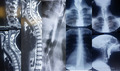
The patient was a 54‐year‐old man with a 2.5‐year‐old history of cervical disk surgery. He was reported to our center with dysphagia and odynophagia, repeated coughs, and the weight loss of 30 kg. MRI and CT scan findings indicated the displacement of the cervical plate (Figure 1). He was admitted to the surgical ward where blood tests, cardiac consultation, and radiography were performed. After the induction of anesthesia, we noticed the presence of an external object in the cervical‐esophagus region for which the operation was halted. Through stereoscopy, normal recurrent laryngeal nerves were reported while the examination of the esophagus did not report the plate in the esophagus. In performing B‐Swallow (barium swallow), normal esophagus was seen from piriform sinus. Spirometry was normal before surgery: FEV: 115%, FVC: 113%. Paraclinical findings included the following: Laboratory: WBC: 9.6, Hb: 13.5, CV: 0.8, BUN: 14, and chest X‐ray: normal. Initially, under the sedation, the examination of oral and pharyngeal cavities was performed, where, from pharyngoscopy, the plate was seen in the pharynx. Central venous catheter was placed (number 7.5), whereas the plate was displaced from the posterior to the anterior throat (Figure 2). In the supine position, the right side of the head was exploited for the surgical procedure where the omohyoid muscle was excised, followed by exploratory procedure and the release of adhesion. The left carotid plate was pulled out from the thyroid bridge, meticulously preventing any damage to the parathyroid and CRCN nerves. Subsequently, an incision in the neck was made to examine the upper mediastinal region. The cervical and anatomical elements of the site and nerve LRLN were removed from the plate site, and the laryngeal pharynx region was vertically opened. Subsequently, NG and rectal tubes were placed through laryngeal pharynx space to prevent stenosis at the time of restoration. The corrugated drain was placed in a SIMPLE surgical procedure, and the patient was transferred to the ICU. Left recurrent nerve rupture was normalized after the surgery. The oral feeding was resumed with water, and the patient was discharged with the good general condition. Postoperative laboratory findings: BUN: 15, Hb: 13.8, WBC: 17.7, and CT scan: normal.
Figure 1.

CT scan findings indicated the displacement of the cervical plate
Figure 2.

Displacement of the plate from the posterior to the anterior throat
3. DISCUSSION
Anterior cervical plate implants are extensively used for the fusion and stabilization of the spine, in response to trauma, infection, or other pathologies. Nonetheless, several perioperative and postoperative complications are reported such as excessive bleeding, tissue swelling, hematoma, hypopharyngeal and esophageal raptures/perforations, and infections. These events can also be led by the loosening of screws or plates that can occur years after the procedure.4 Kothari and Almouradi7 reported a case of a 53‐year‐old woman with adenocarcinoma of upper gastrointestinal tract where anterior spine fusion plate had displaced into the hypopharynx region, above the esophageal sphincter. They reported the erosion of the plate owing to her carcinogenic condition and other morbidities, which was clinically corrected. Similarly, dysphagia was also reported in 4.1% of the anterior cervical fusion patients which was the consequence of implant displacement and esophageal rupture and was commonest at the mid‐cervical level.8
Torretta et al9 presented a similar case of a 53‐year‐old man with dysphagia, weight loss, and dyspnea, 4 years following anterior cervical fusion. Radiographic findings revealed the migration of the cage anterior to the 4th cervical vertebra, till the posterior of hypopharyngeal and laryngeal walls. After the infection control by the means of antibiotics, surgical intervention was considered, which aided the patient to return to a normal and healthy life.
Our report presents a similar case where 54‐year‐old man presented dysphagia, and extreme weight loss was found to have a displacement of the cervical plate in the inner surface of the hypopharynx region. Nonetheless, timely surgical intervention helped to restore normal feeding and healthy life in the patient.
4. CONCLUSION
Displacement of the part of the cervical fusion system has been reported in a few cases, with discrepancies in the clinical findings. Long‐term postoperative follow‐up and considering other medical conditions can help clinicians to provide safer and better therapeutic and preventive measures.
CONFLICT OF INTEREST
The authors report no conflict of interest.
AUTHOR CONTRIBUTIONS
RA: conceived and designed the study, acquired or analysed the data, and interpreted the data. ZA: drafted the article or revised it critically for important intellectual content. SM and ZM: involved in final approval of the version published, agreed to be accountable for the article, and ensured that all questions regarding the accuracy or integrity of the article are investigated and resolved.
Alizadeh R, Aghsaeifard Z, Marzbanrad Z, Marzban‐Rad S. An unusual displacement of the cervical plate to the inner surface of the hypopharynx. Clin Case Rep. 2020;8:999–1001. 10.1002/ccr3.2790
REFERENCES
- 1. Tasiou A, Giannis T, Brotis AG, et al. Anterior cervical spine surgery‐associated complications in a retrospective case‐control study. J Spine Surg. 2017;3(3):444. [DOI] [PMC free article] [PubMed] [Google Scholar]
- 2. Finn MA, MacDonald JD. C2–C3 anterior cervical fusion: technical report. Clin Spine Surg. 2016;29(10):E536‐E541. [DOI] [PubMed] [Google Scholar]
- 3. Spinelli J, Neal CJ, Rosner MK. Performance of cervical arthroplasty at a pseudarthrosed level of a multilevel anterior cervical discectomy and fusion: case report. Mil Med. 2016;181(6):e621‐e624. [DOI] [PubMed] [Google Scholar]
- 4. Salis G, Pittore B, Balata G, Bozzo C. A rare case of hypopharyngeal screw migration after spine stabilization with plating. Case Rep Otolaryngol. 2013;2013:1‐4. [DOI] [PMC free article] [PubMed] [Google Scholar]
- 5. Ning X, Wen Y, Xiao‐Jian Y, et al. Anterior cervical locking plate‐related complications; prevention and treatment recommendations. Int Orthop. 2008;32(5):649‐655. [DOI] [PMC free article] [PubMed] [Google Scholar]
- 6. Park S, Lee DH, Ha JK, et al. How does screw migration or fracture after anterior cervical plate fixation affect the radiographic and clinical outcomes? Clin Spine Surg. 2019;32(9):398‐402. [DOI] [PubMed] [Google Scholar]
- 7. Kothari S, Almouradi T. Dysphagia caused by cervical plate erosion through the hypopharynx. ACG Case Rep J. 2018;5(12):e104‐e204. [DOI] [PMC free article] [PubMed] [Google Scholar]
- 8. Carucci LR, Turner MA, Yeatman CF. Dysphagia secondary to anterior cervical fusion: radiologic evaluation and findings in 74 patients. AJR Am J Roentgenol. 2015;204(4):768‐775. [DOI] [PubMed] [Google Scholar]
- 9. Torretta S, Brevi A, Guastella C, Rampini P, Locatelli M, Pignataro L. An unusual hypopharyngeal foreign body causing dyspnea after vertebroplasty. Ear Nose Throat J. 2019;98(6):321‐323. [DOI] [PubMed] [Google Scholar]