Abstract
Cohesin, a multisubunit protein complex, is required for holding sister chromatids together during mitosis and meiosis. The recruitment of cohesin by the sister chromatid cohesion 2/4 (SCC2/4) complex has been extensively studied in Saccharomyces cerevisiae mitosis, but its role in mitosis and meiosis remains poorly understood in multicellular organisms, because complete loss-of-function of either gene causes embryonic lethality. Here, we identified a weak allele of Atscc2 (Atscc2-5) that has only minor defects in vegetative development but exhibits a significant reduction in fertility. Cytological analyses of Atscc2-5 reveal multiple meiotic phenotypes including defects in chromosomal axis formation, meiosis-specific cohesin loading, homolog pairing and synapsis, and AtSPO11-1-dependent double strand break repair. Surprisingly, even though AtSCC2 interacts with AtSCC4 in vitro and in vivo, meiosis-specific knockdown of AtSCC4 expression does not cause any meiotic defect, suggesting that the SCC2-SCC4 complex has divergent roles in mitosis and meiosis. SCC2 homologs from land plants have a unique plant homeodomain (PHD) motif not found in other species. We show that the AtSCC2 PHD domain can bind to the N terminus of histones and is required for meiosis but not mitosis. Taken together, our results provide evidence that unlike SCC2 in other organisms, SCC2 requires a functional PHD domain during meiosis in land plants.
Author summary
Cohesin is required to hold sister chromatids together during mitosis and meiosis. The recruitment of cohesin, mediated by the sister chromatid cohesion 2/4 (SCC2/4) complex, has been extensively studied in yeast mitosis. Because complete loss-of-function of either gene causes embryonic lethality in multicellular organisms, its role in mitosis and meiosis remains poorly understood. Here, we show that Arabidopsis SCC2 functions in meiosis in an AtSCC4-independent manner. We also demonstrate that SCC2 in land plants has a PHD domain not found in animal or fungal homologs and is critical for meiotic function but not mitosis.
Introduction
The faithful transmission of chromosomes to daughter cells is an essential feature of the cell cycle in most eukaryotes. Improper chromosome segregation during mitosis or meiosis leads to aneuploidy, which in turn can cause defects in growth, development and reproduction [1, 2]. During meiosis, to ensure the proper segregation of chromosomes, the cohesin complex holds sister chromatids together from the end of S phase until anaphase II [3].
Cohesin is a member of the ancient and conserved SMC (Structural Maintenance of Chromosomes) protein family [3]. In S. cerevisiae, the core cohesin complex consists of Smc1, Smc3, Scc3 and an α-kleisin (Scc1 and Rec8 in mitosis and meiosis respectively) [4]. Smc1 interacts with Smc3 via their hinge domains, and their ATPase head domains are linked by the kleisin subunit [5]. Scc3 is able to interact with the kleisin subunit [6]. These four subunits form a ring-shaped structure [7]. Arabidopsis has single copies of AtSMC1/AtTTN8, AtSMC3/AtTTN7 and AtSCC3 [8–11]. However, in Arabidopsis, there are four kleisin proteins: SYN1/DIF1/REC8, SYN2/RAD21.1, SYN3/RAD21.2, and SYN4/RAD21.3 [10, 12–14]. Among them, AtSYN3 is required for male and female meiosis and may have functions other than as a subunit of cohesin, including a role in regulating nucleolar structure [10, 15]. AtSYN1, the orthologue of yeast Rec8, is a meiosis-specific cohesin subunit and is essential for chromosome condensation, sister chromatid cohesion, double strand break (DSB) repair and mono-orientation of meiotic chromosome [9, 16, 17]. Other accessory proteins such as AtSWITCH1 (AtSWI1) and AtWAPL1/AtWAPL2, also help mediate cohesin association or disassociation with chromatin [18–21].
Cohesin is recruited onto chromosomes by the conserved heterodimeric SCC2-SCC4 complex in most model organisms [21–25]. The SCC2 C terminus contains several HEAT repeats that are required for forming a hook-like structure, which is critical for loading cohesin onto DNA [26]. The N-terminal end of SCC2 can interact with SCC4 to form a globular head domain [26]. Arabidopsis SCC4 is a small, 726 amino acid protein with a predicted tetratricopeptide repeat (TPR) 12 domain [27]. In S. cerevisiae mitosis, Scc4 may stabilize Scc2 in vivo and facilitates cohesin loading at centromeres [26, 28, 29]. In addition, in vitro experiments with the C-terminal end of human Scc2 showed it can interact specifically with the HsSmc1-HsSmc3 heterodimer, but HsScc4 does not bind to cohesin [24]. Loading assays using Schizosaccharomyces pombe in vitro reconstituted cohesin complexes indicated that Mis4Scc2 is sufficient for cohesin loading onto DNA, in the absence of Ssl3Scc4 [30]. Recent biochemical and genetic analyses in S. cerevisiae also support the idea that Scc2 is sufficient for stimulating cohesin’s ATPase activity in the absence of Scc4 [31]. Furthermore, the mechanisms that recruit SCC4 to specific chromatin sites have been reported in several species. In S. cerevisiae, Scc4 can be directly recruited to centromeres by the phosphorylated kinetochore protein Ctf19 [29]. In Xenopus, a complex of Scc4 and the N terminus of Scc2 is sufficient to bind chromatin, through interacting with pre-replication complex (pre-RC), but cannot recruit cohesin [32, 33]. The human SCC2-SCC4 complex also interacts with the MCM2-7 complex [34]. In Zea mays (maize), SCC4/Dek15 interacts with several chromatin remodeling proteins [35]. Together, these results suggest that recruitment of SCC2/4 onto chromatin likely depends on SCC4-interacting factors such as Ctf19 and the MCM2-7 complex, while SCC2 is important for loading cohesin.
Arabidopsis has single copy of SCC2 and homozygous full loss-of-function alleles are embryonic lethal [36]. Plants with RNAi-induced knock-down of AtSCC2 exhibit reduced fertility and meiotic defects in pairing of homologous chromosomes, chromosomal axis formation and sister chromatid cohesion [36]. Interestingly, unlike other taxa, SCC2 orthologs from plants contain a PHD domain [36]. However, the function of the SCC2 PHD domain in mitosis and meiosis has not been investigated. Recently, SCC4 was also identified in Arabidopsis and maize. Similar to AtSCC2, complete loss of function of SCC4 is also embryonic lethal in both Arabidopsis and maize, consistent with their roles in mitotic cell division [27, 35, 37]. Nonetheless, the gene expression pattern of AtSCC2 does not fully overlap with AtSCC4 [27]. In addition, they have non-overlapping roles in process such as endosperm development [27, 36].
We recovered a mutant in a screen for male sterility and identified the locus as AtSCC2. The non-lethal Atscc2 mutant (designated Atscc2-5) provided an opportunity to study its meiotic function. Consistent with previous AtSCC2 RNAi knock-down phenotypes, Atscc2-5 has meiotic defects in chromosomal axis formation, pairing of homologous chromosomes, synapsis and recombination. Our analyses demonstrate that AtSCC2 acts in the same pathway as AtSYN1 and AtWAPL1/2, and participates in AtSPO11-1-depedent DSB repair. We also provide evidence that the N-terminal end of AtSCC2 interacts with AtSCC4 both in vitro and in vivo, and meiosis-specific knockdown of AtSCC4 does not cause any meiotic defects, suggesting that the role of the SCC2-SCC4 complex is divergent in mitosis and meiosis. Using in vitro binding assays we also show that the plant-specific AtSCC2 PHD domain binds to the N terminus of H2A, H3 and H4. Further in vivo functional analyses demonstrate that the PHD domain of AtSCC2 is required for meiotic function, but not for vegetative growth, which adds to our mechanistic understanding of cohesins in plants.
Results
Identification of a hypomorphic Atscc2 allele
To better understand the genetic mechanisms controlling meiosis, we screened an EMS mutagenized population and identified a sterile plant (line88). After backcrossing line88 with wild type (WT) Col-0 for three generations, the stable line88 plants have a slight vegetative growth defect at four and eight-week-old stages compared to WT (S1A and S1B Fig). However, line88 plants are almost completely sterile and produce very few seeds (0.7 ± 0.8 seeds per silique, n = 21, p-value = 4.9E-48, two-tailed Student’s t test) compared to WT (55.5 ± 2.5 seeds per silique, n = 21) (S1C and S1D Fig). WT stamens are plump, and their mature stigmas are covered with pollen (S1E Fig), while line88 plants lack pollen on their stigmas suggesting a defect in male gamete production (S1F Fig). Alexander staining for pollen viability showed a significant reduction in the number of viable pollen grains per anther in line88 (average = 20.3 ± 12.4, n = 19, p-value = 2.1E-31, two-tailed Student’s t test), compared to WT (average = 581.1 ± 58.3, n = 19) (S1H and S1G Fig). Consistent with these observations, toluidine blue staining of tetrad-stage microspores showed that 56.1% (n = 57) of male meioses in line88 plants result in polyads with variable sized microspores (S1J Fig), while WT plants produce only tetrads with four similarly sized microspores (S1I Fig, n = 48). These phenotypes suggest a defect in meiotic chromosome segregation in line88.
Heterozygous F1 plants (line88 as the female parent and WT Lansberg erecta (Ler) as the male parent) have normal fertility, indicating that the mutation is recessive. We allowed the F1 plants to self-fertilize and used bulked-segregant analysis of 97 sterile F2 plants to map the mutant locus to a region on the upper arm of chromosome 5 (S2A and S2B Fig). The region includes four candidate genes, including AT5G15540 (AtSCC2) which contains a mutation compared to the WT reference sequence. The single nucleotide polymorphism is a G to A transition in the C terminus of AtSCC2 (5,049,797 bp) that does not result in an amino acid change, but is predicted to disrupt the splice site at the 3' end of exon 22 (S2C Fig). Genotyping the alleles in the F2 progeny shows that the ratio of homozygous WT (n = 409) and heterozygotes (n = 816) is consistent with a 1:2 ratio (p(χ2) = 0.99, chi-square test), whereas the ratio of mutant (n = 278) and WT phenotypes (n = 1225) deviates significantly from a 1:3 ratio (S1 Table) (χ2 = 33.6 > χ20.05 = 3.8, chi-square test). These segregation ratios are consistent with a single recessive allele with an incompletely penetrant embryonic lethal phenotype.
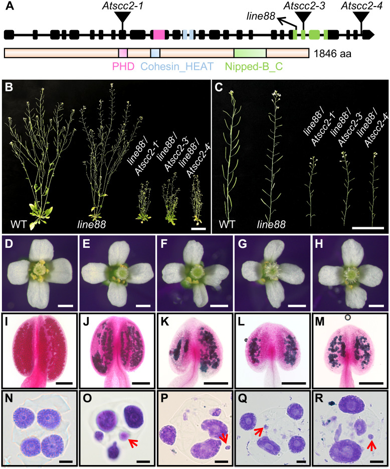
Previous studies showed that complete loss-of-function of AtSCC2 leads to embryonic lethality [36]. To confirm that the line88 phenotype is caused by the mutation we mapped to AtSCC2, we crossed line88 with heterozygotes of three T-DNA alleles of AtSCC2 (Atscc2-1, Atscc2-3 and Atscc2-4) (Fig 1A and 1B). These three T-DNA alleles are embryonic lethal as homozygotes as shown by the 1:2 segregation ratio of homozygous WT plants and heterozygotes in the F2 progeny of Atscc2-1+/- (67/110, p(χ2) = 0.23, chi-square test), Atscc2-3+/- (33/63, p(χ2) = 0.91, chi-square test) and Atscc2-4+/- (33/58, p(χ2) = 0.63, chi-square test) plants (S1 Table), and the complete lack of homozygous mutant F2 (S1 Table). Consistently, the ratio of compound heterozygous F1 plants of two independent alleles (line88-/Atscc2-1- and line88-/Atscc2-3-) with their corresponding Atscc2-5+/- heterozygous F1 plants is 1:1 (χ2 ≤ χ0.052 = 3.84, chi-square test) (S2 Table). The ratio of F1 compound heterozygous of line88-/Atscc2-4- with Atscc2-5+/- heterozygous F1 plants is not consistent with 1:1 (χ2 = 4.02 > χ0.052 = 3.84), probably due to low population numbers or an incompletely penetrant embryonic lethal phenotype. All of the compound heterozygous F1 progeny had severe vegetative growth defects compared with the homozygous line88 plants and WT (Fig 1B), supporting the essential role of AtSCC2 in mitosis. These compound heterozygous plants also have short siliques and no pollen on their stigmas (Fig 1C and 1F–1H) compared with WT (Fig 1C and 1D), similar to line88 (Fig 1C and 1E). Pollen viability in the compound heterozygous plants is reduced (Fig 1J–1M), compared with WT (Fig 1I). The compound heterozygotes also have abnormal tetrad-stage microspores with polyads of uneven size (Fig 1P–1R), similar to line88 (Fig 1O) and unlike WT (Fig 1N). These results demonstrate that line88 is a mutant allele of AtSCC2 which we will henceforth call Atscc2-5.
Fig 1. Identification of Atscc2-5.
(A) A diagram of AtSCC2 gene and AtSCC2 protein structure. Mutant alleles are marked above the gene structure (inverted triangles and diagonal arrow). (B) WT, line88 and three compound heterozygous mutants of line88 with individual Atscc2 alleles. Bar = 3 cm. (C) Primary stems of WT, line88 and three compound heterozygous plants. Bar = 3 cm. (D-H) Open flowers of WT, line88 and three compound heterozygous plants. Bar = 0.5 mm. (I-M) Alexander staining of WT, line88 and three compound heterozygous plant anthers. Bar = 100 μm. (N-R) Toluidine blue staining of tetrad-stage microspores of WT, line88 and three compound heterozygous plants. Red arrows indicate the abnormal micronucleus in tetrad stage. Bar = 5 μm.
We also used trans-complementation with the WT AtSCC2 coding sequence fused to a FLAG-tag and expressed by the AtACTIN7 promoter to confirm that the Atscc2-5 phenotypes are caused by a mutation in the AtSCC2 locus. The AtACT7::AtSCC2-FLAG transgene is able to rescue the fertility, pollen viability and meiotic defects of Atscc2-5-/- plants (S3A–S3G Fig). Western blotting with anti-FLAG antibody confirmed that the AtSCC2-FLAG fusion is expressed at the expected size in the transgenic plants (211.6 kD; S3E Fig). These results validate the point mutation in AtSCC2 as the causative lesion for the mutant phenotypes we observe in Atscc2-5.
The Atscc2-5 point mutation at the border of the exon 22 (S2C and S4A Figs) is predicted to disrupt a splice site. To test this hypothesis, we amplified the AtSCC2 transcript using primers spanning the point mutation and found that Atscc2-5 expresses two transcripts: one WT AtSCC2 transcript and one 47 bp deletion version (S4B Fig). Quantitative real-time reverse transcription PCR (qRT-PCR) demonstrated that the full length AtSCC2 transcript is significantly lower (~5% in leaves and ~2% in meiocytes) in Atscc2-5 compared to WT (S4C Fig). This is also supported by RNA-seq data from WT and Atscc2-5 meiocytes (S5A Fig). We speculate that the reduced level of full length AtSCC2 transcript in Atscc2-5 may be caused by a premature termination codon inducing nonsense-mediated mRNA decay (NMD) [38]. Disruption of the splice site appears to trigger the use of an upstream cryptic splice site, resulting in a 47 bp deletion in the mRNA which creates a premature stop codon and may yield a truncated AtSCC2 protein (1–1400 amino acids) in Atscc2-5 (S5B Fig). We speculate that the residual intact AtSCC2 transcripts in Atscc2-5 are sufficient to rescue embryonic lethality, but still cause aberrant meiotic phenotypes.
The Atscc2-5 mutant shows multiple meiotic defects
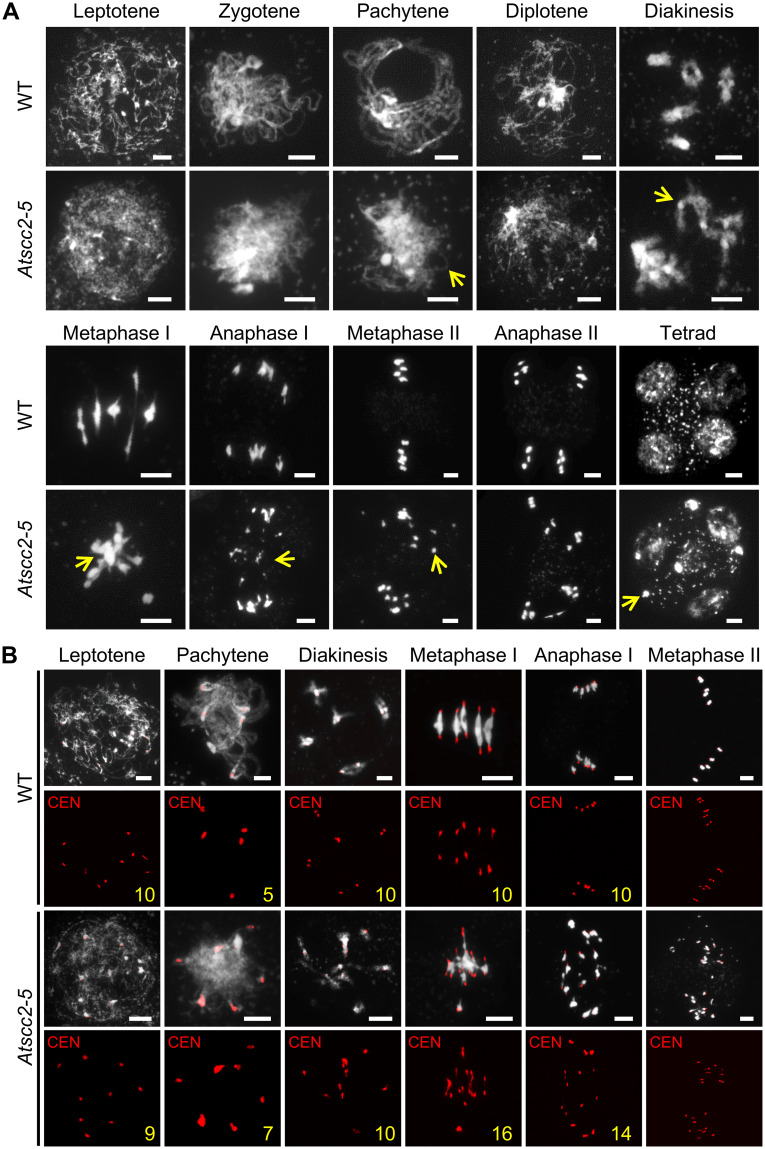
We stained chromosome spreads from WT and mutant pollen mother cells (PMCs) with 4’, 6-diamidino-2-phenylindole (DAPI) to investigate the meiotic defects in Atscc2-5 (Fig 2A). At leptotene, the Atscc2-5 chromosomes are very rough and appear less condensed compared to WT which appear as distinct thin threads. At zygotene and pachytene, WT chromosomes continue to condense, homologs align, and synapsis results in thick thread-like structures. In Atscc2-5 meiocytes, chromosome at similar stages remain relatively thin and defuse, indicating a defect in synapsis. Following desynapsis at diplotene, WT homologs remain associated through chiasmata at crossover sites and form five highly condensed bivalents at diakinesis. In contrast, condensation of Atscc2-5 diplotene chromosomes appears normal, but entangled chromosomes or multivalents are observed at diakinesis. At metaphase I, Atscc2-5 chromosomes are associated in a multivalent mass while WT meiocytes have five bivalents aligned on the equatorial plate. At anaphase, the WT homologs segregate to opposite poles, while the Atscc2-5 meiocytes display improper chromosome segregation and chromosome fragmentation, which is consistent with previous observations in AtSCC2-RNAi plants [36]. In WT, the segregation of sister chromatids during meiosis II results in the formation of four nuclei. Atscc2-5, by comparison, produces polyads containing several micronuclei.
Fig 2. Chromosome morphology of Atscc2-5 and WT male meiocytes.
(A) Chromosome spreads of WT and Atscc2-5 male meiocytes stained by DAPI. Yellow arrows indicate the asynaptic chromosomes, univalent, abnormal chromosomal entanglements or fragments in Atscc2-5. Bar = 5 μm. (B) Fluorescence in situ hybridization (FISH) of WT and Atscc2-5 chromosomes using a centromere probe. Yellow numbers indicate the number of centromeres in the meiocytes. Bar = 5 μm.
To further examine the Atscc2-5 chromosome segregation defect we used fluorescence in situ hybridization (FISH) with a 180 bp centromeric repeat probe. We did not observe any obvious difference in the number of centromere signals between WT and Atscc2-5 at leptotene (Fig 2B). This result confirms that duplicated sister chromatids are associated with each other at centromeric regions in mutant and WT meiocytes, which suggests that centromeric cohesin loading is initially sufficient. At pachytene, synapsis of WT homologs creates five pairs of centromere signals, while Atscc2-5 meiocytes have more than five signals, indicating a defect in homolog paring at centromeres. Similarly, at diakinesis WT has five pairs of signals, whereas most signals in Atscc2-5 meiocytes are unpaired. At metaphase I and anaphase I, WT centromere signals segregate to opposite poles, while Atscc2-5 meiocytes have more than 10 centromere signals (16 shown in Fig 2B), including some that appear to lag, indicating precocious sister chromatid separation (PSCS) [39]. At metaphase II, WT has 10 paired centromere signals aligned on the equatorial plate, but Atscc2-5 has a mixture of poorly organized, paired and unpaired signals which is also consistent with PSCS. These results suggest that the maintenance of centromere cohesin is compromised beginning in pachytene and continues through the end of meiosis.
We also analyzed chromosome morphology and dynamics of the three compound heterozygous mutant plants, and found their meiotic phenotypes are similar to the Atscc2-5 single mutant (S6 Fig). These results demonstrate that the meiotic defects in Atscc2-5 plants are due to a reduction of full-length AtSCC2, rather than the expression of a truncated protein.
AtSCC2 is required for loading meiosis-specific cohesin and genetically acts in the same pathway as AtSYN1 and AtWAPL1/2
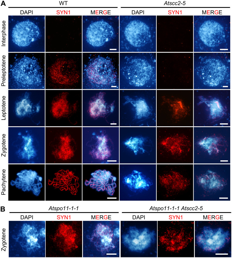
Because SCC2 is widely reported to be responsible for loading cohesin [5], including in Arabidopsis [36], we used immunofluorescence staining of AtSYN1 to investigate the localization of meiosis-specific cohesin. In WT, AtSYN1 signals appear in preleptotene as diffuse foci, and extend the length of the chromosomes at leptotene (Fig 3A). Beginning at diplotene, AtWAPL1 and AtWAPL2 disassociate cohesins from chromosome arms [19, 20]. However, in Atscc2-5, AtSYN1 signals are barely observable at preleptotene and start to be discontinuous from leptotene onward (Fig 3A), suggesting a possible defect in the initial establishment of cohesion at centromeres and chromosome arms. It was previously reported that AtSYN1 signals in Atspo11-1-1 and Atscc3-1 single mutant meiocytes are similar to wild type, while AtSYN1 signals are disrupted in Atspo11-1-1 Atscc3-1 double mutant, suggesting that the association of AtSYN1 with chromosomes needs both AtSCC3 and AtSPO11-1 [9]. Unlike the enhanced AtSYN1 defects in Atspo11-1-1 Atscc3-1 [9], Atspo11-1-1 Atscc2-5 double mutant exhibits similar AtSYN1 defects compared to Atscc2-5 at zygotene (Fig 3B), indicating that AtSPO11-1 does not play a synergetic role with AtSCC2 in the process of AtSYN1 localization, which is consistent with the previous findings that loading of SYN1/REC8 is SPO11-1 independent in rice [40].
Fig 3. AtSCC2 is required to load meiosis-specific cohesin subunit AtSYN1.
(A) The distribution of AtSYN1 from interphase to pachytene in WT and Atscc2-5. Bar = 5 μm. (B) The distribution of AtSYN1 in Atspo11-1-1 and Atspo11-1-1 Atscc2-5 zygotene chromosomes. Bar = 5 μm.
Since AtSYN1 is a meiosis-specific cohesin subunit and AtWAPL1/AtWAPL2 are responsible for cohesin removal in late prophase, particularly on chromosome arms, we analyzed the Atsyn1 Atscc2-5 double and Atwapl1-1 Atwapl2 Atscc2-5 triple mutants. The chromosome phenotypes of the Atsyn1 Atscc2-5 double mutant (S7D Fig) are similar to Atscc2-5 and Atsyn1 single mutant at all stages examined (S7B and S7C Fig), including diffuse chromosomes, aberrant pachytene morphology, entangled multivalents, PSCS, and chromosome fragmentation (S7A Fig). Because AtWAPL1 and AtWAPL2 function to remove cohesin and AtSCC2 is thought to function in loading cohesin, we hypothesized that AtWAPL1/AtWAPL2 mutations might partially rescue the Atscc2-5 meiotic defect. Unexpectedly, the Atwapl1 Atwapl2 Atscc2-5 triple mutant is sterile and has atypical pachytene chromosomes, chromosome entanglements and chromosome fragmentation (S7F Fig), similar to the Atscc2-5 single mutant (S7B Fig), implying that AtSCC2, AtWAPL1 and AtWAPL2 are epistatic to one another. It is possible that the PSCS observed in Atscc2-5 is due to insufficient AtSYN1-mediated centromere cohesion in addition to the defects in chromosome arm cohesion.
Formation of DSBs appears normal, but their repair is affected in Atscc2-5
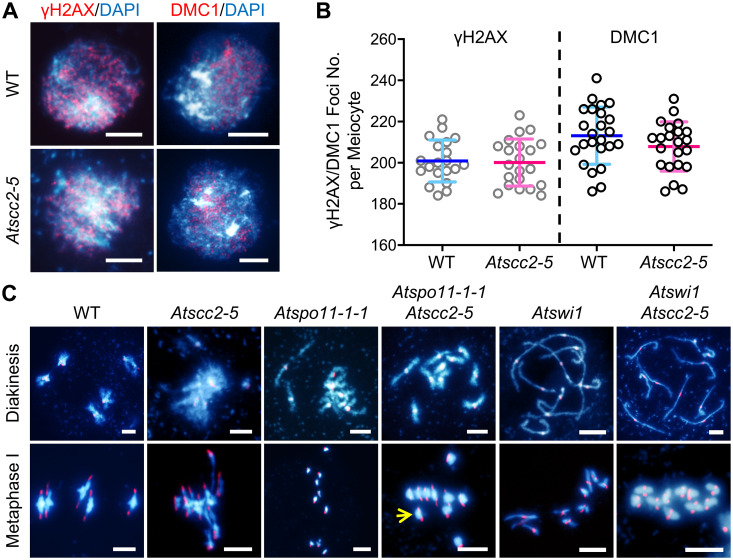
The observation of chromosome fragmentation in Atscc2-5 suggests that AtSCC2 may participate in meiotic DSB repair. We examined whether the lack of AtSCC2 or chromosome-bound cohesin impacts the formation of meiotic DSBs, by using immunofluorescence staining of two DSB markers, γH2AX, a phosphorylated variant histone [41], and AtDMC1, a recombinase [42], in WT and Atscc2-5 zygotene meiocytes (Fig 4A). We did not observe any significant difference (both p-value > 0.05, two tailed Student’s t test) in the number of AtDMC1 or γH2AX foci between WT (n = 21 cells for AtDMC1; n = 22 cells for γH2AX) and Atscc2-5 (n = 24 for AtDMC1; n = 22 cells for γH2AX; Fig 4B), suggesting that AtSCC2 is not required for DSB formation. This is consistent with the previous report in Caenorhabditis elegans [43].
Fig 4. AtSCC2 is dispensable for DSB formation but is indispensable for AtSPO11-1-dependent DSB repair.
(A) Localization of γH2AX and DMC1 in WT and Atscc2-5 zygotene male meiocytes. Bar = 5 μm. (B) Plots of the γH2AX and DMC1 foci numbers in WT and Atscc2-5 zygotene male meiocytes (two-tailed Student’s t test). (C) Fluorescence in situ hybridization of WT, Atscc2-5, Atspo11-1-1, Atspo11-1-1 Atscc2-5, Atswi1, and Atswi1 Atscc2-5 double mutant chromosomes using centromere probes. Yellow arrows indicate the separated centromeres of sister-chromatids at metaphase I. Bar = 5 μm.
To test whether the DSB repair defects are SPO11-1-dependent, we introduced the Atspo11-1-1 mutation [44] into the Atscc2-5 mutant background. AtSPO11-1 is required for generating meiotic DSBs, and the Atspo11-1-1 mutant has univalents at diakinesis which segregate randomly at metaphase/anaphase I (Fig 4C). The Atspo11-1-1 Atscc2-5 double mutant has 10 unfragmented univalents at diakinesis and no multivalents at metaphase I, suggesting that AtSCC2 participates in AtSPO11-1-dependent DSB repair. In addition, the metaphase I Atspo11-1-1 Atscc2-5 univalents exhibit PSCS, possibly due to the defective AtSYN1 localization in Atscc2-5, providing additional evidence that centromeric cohesin between sister chromatids is compromised. AtSWI1 is required for the establishment of sister chromatid cohesin and the initiation of meiotic recombination [18, 45]. Atswi1 mutants have univalents which segregate randomly at metaphase I and have noticeable PSCS. In the Atswi1 Atscc2-5 double mutants, sister chromatids are mono-oriented and there is no chromosome fragmentation, which resembles the meiotic defects in Atspo11-1-1 Atscc2-5 double mutants. These results provide additional evidence that AtSCC2 participates in meiotic recombination likely through loading cohesin.
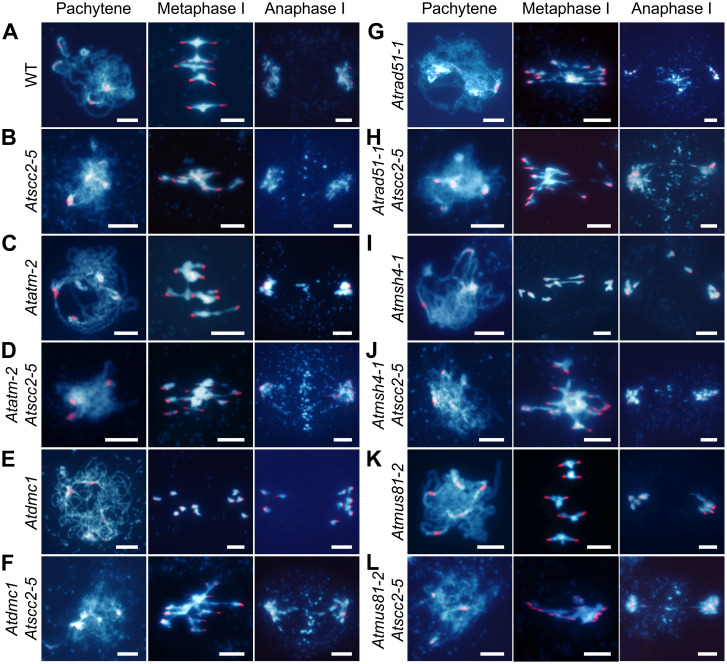
To investigate whether AtSCC2 has a role during other stages of meiotic recombination, we generated double mutants of Atscc2-5 with Atatm-2 (DSB response), Atdmc1 (strand invasion), Atrad51-1 (strand invasion), Atmsh4-1 (CO resolution), and Atmus81-2 (CO resolution) (Fig 5). Compared with Atscc2-5 and Atatm-2 single mutants, chromosome fragmentation is aggravated in Atatm-2 Atscc2-5 at anaphase I (Fig 5D), suggesting that AtSCC2 acts synergistically with AtATM in mediating meiotic recombination. Atdmc1 meiocytes have 10 univalents, but no chromosome fragmentation or entanglements, presumably because AtRAD51 is able to repair DSBs using sister chromatids as a template [42]. The Atdmc1 Atscc2-5 double mutant has chromosome entanglements at metaphase I and chromosome fragments at anaphase I (Fig 5F), similar to the Atscc2-5 single mutant (Fig 5B), indicating that AtSCC2 and AtDMC1 are epistatic to one another during meiotic recombination. The Atrad51-1 Atscc2-5 double mutant has similar severe chromosome entanglement and fragmentation phenotypes (Fig 5H) compared to the Atrad51-1 single mutant (Fig 5G), indicating that AtRAD51 and AtSCC2 are epistatic to one another. Taken together, these results support the idea that AtSCC2 is required for efficient DSB repair and acts together with AtRAD51 in a manner that is distinct from the action of the meiosis-specific recombinase AtDMC1. Alternatively, because Atscc2-5 still expresses very low levels of wild type AtSCC2 transcript, these results could indicate that AtRAD51 is more sensitive than AtDMC1 to AtSCC2 levels.
Fig 5. Genetic analyses of AtSCC2 in meiotic recombination mutants.
DAPI stained chromosome spreads at pachytene, metaphase I and anaphase I in (A) WT, (B) Atscc2-5, (C) Atatm-2, (D) Atatm-2 Atscc2-5, (E) Atdmc1, (F) Atdmc1 Atscc2-5, (G) Atrad51-1, (H) Atrad51-1 Atscc2-5, (I) Atmsh4-1, (J) Atmsh4-1 Atscc2-5, (K) Atmus81-2, (L) Atmus81-2 Atscc2-5. Bar = 5 μm.
Arabidopsis has two classes of CO: Type I COs are sensitive to a regulatory phenomenon called CO interference and are associated with the ZMM class of proteins, including AtMSH4; and Type II COs are insensitive to interference and are AtMUS81-dependent [46, 47]. To investigate whether AtSCC2 is involved with one or the other, or both pathways, we compared Atmsh4-1 Atscc2-5 and Atmus81-2 Atscc2-5 double mutants with the corresponding single mutants. Neither Atmsh4-1 nor Atmus81-2 has chromosome entanglement or fragmentation phenotypes (Fig 5I and 5K). In contrast, the Atmsh4-1 Atscc2-5 and Atmus81-2 Atscc2-5 double mutants have similar chromosome entanglement and fragmentation phenotypes compared to Atscc2-5 (Fig 5J and 5L), implying that AtSCC2 likely functions upstream of AtMSH4 and AtMUS81. Taken together, these data suggest that during meiotic recombination, AtSCC2 is not required for DSB formation, but is critical during steps prior to CO resolution in a manner that impinges on AtRAD51 to a greater extent than AtDMC1.
AtSCC2 is required for axial element and synaptonemal complex (SC) formation
As described above, Atscc2-5 zygotene chromosomes appear less condensed compared to WT (Fig 2A). It has been reported that reduced expression of AtSCC2 affects axial element formation [36]. We used immunofluorescence staining of AtASY1, which plays important roles in the coordination of axis/SC morphogenesis [48], and AtZYP1, a component of the SC transverse elements [49] to examine axial element formation in Atscc2-5. Punctate AtASY1 signals are associated with chromosomes at leptotene, and then appear to be linear on chromosomes at zygotene in WT (S8A Fig). From pachytene to diakinesis, as homologous chromosomes condense, synapsis and desynapse, AtASY1 signals gradually diminish and only remain prominent in highly condensed heterochromatic regions. In Atscc2-5 leptotene meiocytes, punctate AtASY1 signals are similar to WT, but are less concentrated, suggesting that the initial assembly of the axis is not severely compromised in Atscc2-5. At zygotene, AtASY1 appears as a mixture of discontinuous and linear signals, due to lack of homolog alignment and synapsis, their removal at pachytene is delayed, suggesting a defect in axis assembly completion in Atscc2-5. By diakinesis, AtASY1 signals in Atscc2-5 are weaker relative to WT. These observations indicate that axis formation can initiate in Atscc2-5, but proceeds inefficiently and is aberrantly disassembled which further supports a defect in synapsis. The AtASY1 localization at zygotene in the Atspo11-1 Atscc2-5 double mutant shows additive defects relative to that of the Atscc2-5 single mutant (S8B Fig), suggesting that AtSCC2 has a synergistic role with AtSPO11-1 in assembly of ASY1 during meiosis, consistent with recent results reported in maize [50].
Because Atscc2-5 plants have aberrant pachytene chromosomes and AtASY1 assembly, we speculated that their SC transverse elements may be also defective. The AtZYP1 signals in pachytene meiocytes are greatly diminished in Atscc2-5, compared to the linear AtZYP1 distribution on WT chromosomes (S8C Fig). Taken together, our results provide strong evidence that AtSCC2 is required for axial element assembly and SC formation.
AtSCC2 interacts with AtSCC4 in vivo, but AtSCC4 is dispensable for male meiosis
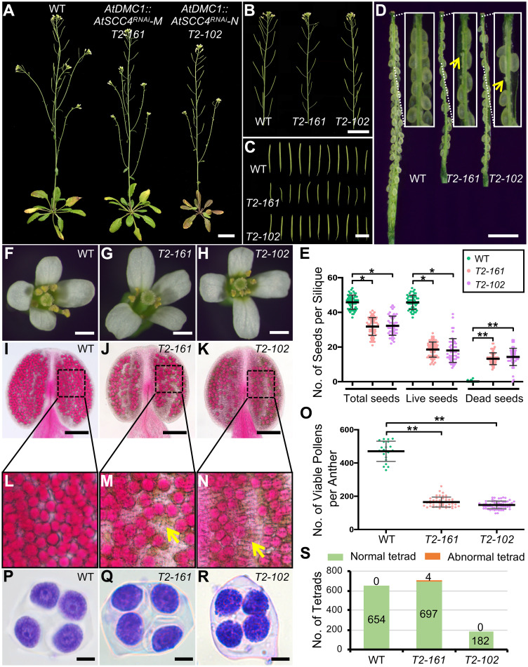
SCC2 has been reported to interact with SCC4 in several organisms, including humans, maize and Arabidopsis [25, 27, 35]. Our yeast two-hybrid assay also confirmed their physical interaction (S9A and S9B Fig). The interaction was further validated by bimolecular fluorescence complementation (BiFC) in tobacco cells (S9C Fig). A recent study demonstrated that the N terminus of AtSCC2 (1–824 aa) interacts with AtSCC4 [27]. To further refine the specific interacting regions, we divided the N-terminus into AtSCC2N1 (1–254 aa) and AtSCC2N2 (255–427 aa) (S9A Fig) and found that only AtSCC2N1 interacts with AtSCC4 (S9B Fig). Because AtSCC4 is essential in mitosis [27], we used a tissue-specific RNAi strategy to test its role in meiosis by targeting two AtSCC4 regions expressed from the meiosis-specific AtDMC1 promoter (AtDMC1::AtSCC4RNAi-M and AtDMC1::AtSCC4RNAi-N; S9D and S9E Fig), and obtained 87 positive transformants, including 37 AtDMC1::AtSCC4RNAi-M and 50 AtDMC1::AtSCC4RNAi-N plants. Five AtDMC1::AtSCC4RNAi-M plants and ten AtDMC1::AtSCC4RNAi-N plants had relatively normal vegetative growth, but reduced fertility. We selected 8 sterile lines for subsequent study. The pollen viability and number of seeds in all 8 lines were significantly reduced compared to WT (n = 10, S10A and S10B Fig). qRT-PCR revealed that AtSCC4 expression levels in the meiocytes of the RNAi lines are significantly reduced compared to WT (p-value < 0.01, two-tailed Student’s t test, S10C Fig). We further analyzed two representative RNAi lines (T2-102 and T2-161), which have reduced fertility, short siliques, few seeds and less viable pollen relative to WT (Fig 6A–6O), but their tetrad-stage microspores have no obvious differences compared to WT (Fig 6P–6S). Analysis of male meiotic chromosome spreads confirmed that stages in the RNAi plants were similar to WT (S11 Fig). This result suggests that AtSCC4 does not play a prominent role in male meiosis, but it is formally possible that the residual gene product, after knocking down gene expression by 90%, is sufficient for wild type function. The RNAi plants appeared to produce sufficient pollen to allow pollination, so we hypothesized that female fertility may be impaired in AtSCC4RNAi plants. To test this hypothesis, we reciprocally crossed the WT and AtSCC4RNAi plants. WT pistils pollenated with T2-161 or T2-102 pollen produced indistinguishable seeds per silique respectively compared with WT (S3 Table and S12A Fig). As female parents the transgenic plants produced only 15.9 and 14.6 normal seeds, respectively (S3 Table and S12A Fig). However, no obvious female meiotic defects were observed in WT (n = 159), T2-102 (n = 86) or T2-161 (n = 54) (S12B Fig). A previous study showed that AtSCC4 is required for embryo development [27]. Consistently, 11.8% (n = 17) T2-161 and 28.6% (n = 14) T2-102 embryos exhibited asymmetric cell division at globular stage compared with WT (n = 15) (S12C Fig). Taken together, these results suggest that AtSCC4 is dispensable for male and female meiosis. It is possible that the Arabidopsis SCC2-SCC4 complex is only required for loading cohesin during mitosis, while AtSCC4 does not play a role in meiosis.
Fig 6. AtDMC1::AtSCC4RNAi transgenic plants have reduced fertility.
(A) WT, AtDMC1::AtSCC4RNAi-M T2-161 and AtDMC1::AtSCC4RNAi-N T2-102 (M and N refer the fragments used for the construction of RNAi plasmids) plants. Bar = 3 cm. (B) Primary stems of WT, T2-161 and T2-102. Bar = 3 cm. (C) Siliques of WT, T2-161 and T2-102. Bar = 1 cm. (D) Stripped siliques of WT, T2-161 and T2-102. Yellow arrows indicate undeveloped embryos. Bar = 1 mm. (E) Plots of total seeds, live seeds and dead seeds in WT, T2-161 and T2-102 (* P < 0.05 or ** P < 0.01, the significance of reduced seed number in AtSCC4RNAi transgenic plants versus WT, by two-tailed Student’s t test; each dot represents the number of seeds in one silique). (F-H) Open flowers of WT, T2-161 and T2-102. Bar = 1 mm. (I-K) Alexander staining of WT, T2-161 and T2-102 anthers. Bar = 100 μm. (L-N) Zoom-in of WT, T2-161 and T2-102 pollens. (O) Plots of viable pollen in WT, T2-161 and T2-102 (** P < 0.01, the significance of reduced pollen number in AtSCC4RNAi transgenic plants versus WT, by two-tailed Student’s t test; each dot represents the number of pollen in one anther). (P-Q) Toluidine blue dye staining of WT, T2-161 and T2-102 tetrad-stage microspores. Bar = 100 μm. (S) Plots of normal and abnormal tetrad-stage microspores in WT, T2-161 and T2-102.
The AtSCC2 PHD domain binds to histones in vitro
Among SCC2 homologs, only those of plants contain a plant homeodomain (PHD) with a C4-H-C3 amino acid motif (S13A and S13B Fig). Phylogenetic analysis demonstrates that the SCC2 PHD domain is highly conserved in land plants, but absent in the green algae Chlamydomonas reinhardtii, Volvox carteri and Micromonas (S13B Fig), suggesting that the SCC2 PHD domain may have played an important role during the adaptation of plants to terrestrial environments.
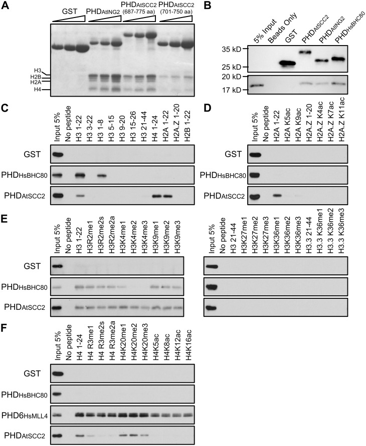
Some PHD domains can bind to unmodified H3K4 or methylated H3K4 in animals and plants [51, 52]. To investigate the potential histone binding specificity of the AtSCC2 PHD domain, we aligned several plant SCC2 PHD sequences and compared them to PHD domains with known histone binding targets (S13C Fig). PHD domains that recognize methylated H3K4 possess three aromatic amino acids (Y-Y-W), but these are absent in the AtSCC2 PHD domain, suggesting that AtSCC2 may not bind methylated H3K4. Based on the alignments, the AtSCC2 PHD domain is more similar to the human BHC80 PHD domain which can bind unmodified H3K4 [53]. To test whether the AtSCC2-PHD has a similar binding affinity, we used an in vitro pull-down assay of two different length AtSCC2-PHD constructs (701–750 aa and 687–775 aa) and a known AtING2 PHD domain as positive control [52] with calf thymus histones. The result showed that the AtSCC2-PHD is able to bind to histones, similar to the AtING2-PHD positive control, but the longer AtSCC2-PHD has a stronger binding affinity than the shorter one (Fig 7A). The pull-down assay using H3 from calf thymus showed AtSCC2-PHD can bind to H3 (Fig 7B). We further tested AtSCC2-PHD with different length unmodified histone peptides. As expected, the HsBHC80-PHD positive control was able to bind to N terminal unmethylated H3 peptides. In contrast, AtSCC2-PHD was able to bind either H3 1–22, H2A 1–22 or H4 1–24 peptides, but not H2A variant H2A.Z (Fig 7C). Binding assays with modified histone H3, H4 and H2A tails showed that methylation does not affect the binding affinity, but acetylation inhibits binding (Fig 7D–7F). These results suggest that the AtSCC2 PHD domain may recognize intact histone octamers.
Fig 7. AtSCC2 PHD can bind to histones.
(A) Pull-down assay of GST, GST-AtING2PHD, GST-AtSCC2PHD (687–775 aa), GST-AtSCC2PHD (701–750 aa) with calf thymus histones. (B) Pull-down assay of GST, GST-HsBHC80PHD, GST-AtSCC2PHD (687–775 aa) with H3. (C) Pull-down assay of GST, GST-HsBHC80PHD, GST-AtSCC2PHD (687–775 aa) with different histone peptides in N terminal length. (D) Pull-down assay of GST, GST-HsBHC80PHD, GST-AtSCC2PHD (687–775 aa) with unmodified and modified H2A peptides. (E) Pull-down assay of GST, GST-HsBHC80PHD, GST-AtSCC2PHD (687–775 aa) with unmodified and modified H3 peptides. (F) Pull-down assay of GST, GST-HsBHC80PHD, GST-AtSCC2PHD (687–775 aa) with unmodified and modified H4 peptides.
The AtSCC2 PHD domain is required for meiosis but not mitosis in vivo
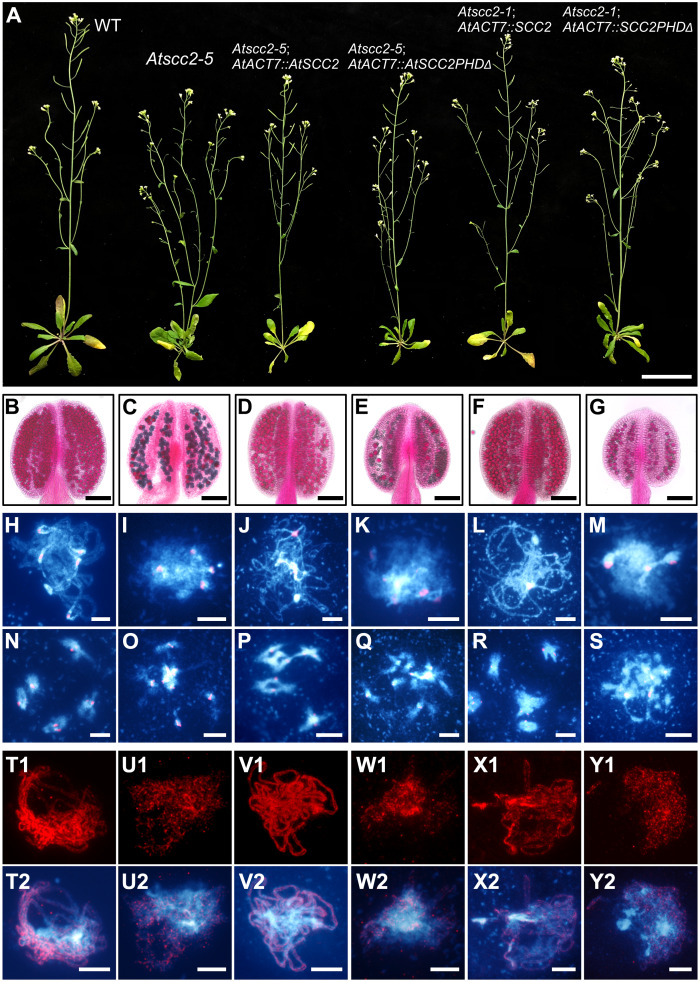
To test the function of the AtSCC2 PHD domain in vivo, we transformed Atscc2-5+/- (Atscc2-5-/- is a weak allele) and Atscc2-1+/- (Atscc2-1-/- is a null allele) mutant plants with constructs encoding full-length AtSCC2 and AtSCC2-PHDΔ (PHD domain deletion in 702–745 aa) expressed from the ubiquitous AtACT7 promoter (Fig 8). qRT-PCR confirmed that the AtACT7::AtSCC2 and AtACT7::AtSCC2PHDΔ transgenes are expressed in four representative lines, compared to WT and Atscc2-5 mutant controls (S14 Fig). Neither AtSCC2 nor AtSCC2-PHDΔ in Atscc2-5 mutant background (called Atscc2-5; AtACT7::AtSCC2 and Atscc2-5; AtACT7::AtSCC2PHDΔ) affect vegetative growth of the transgenic plants compared to WT or Atscc2-5 controls (Fig 8A). However, AtACT7::AtSCC2 is able to rescue (Fig 8D, 8J, 8P and 8V1) the fertility and aberrant meiotic phenotypes of Atscc2-5 (Fig 8C, 8I, 8O and 8U1). In contrast, AtACT7::AtSCC2PHDΔ is not able to rescue the meiotic phenotypes (Fig 8E, 8K, 8Q and 8W1), suggesting that the PHD domain is required for the meiotic functions of AtSCC2.
Fig 8. The AtSCC2 PHD domain is indispensable for male meiosis but not mitosis.
(A) WT, Atscc2-5, Atscc2-5; AtACT7::AtSCC2, Atscc2-5; AtACT7::AtSCC2PHDΔ, Atscc2-1; AtACT7::AtSCC2 and Atscc2-1; AtACT7::AtSCC2PHDΔ plants. Bar = 3 cm. (B-G) Alexander staining of the corresponding anthers. Bar = 100 μm. (H-S) DAPI stained chromosome spreads and FISH with centromere probes at pachytene and diakinesis from the plants shown in panel A. Bar = 5 μm. (T1-Y2) Distribution of AtSYN1 signal at pachytene in meiocytes from the corresponding plants. Bar = 5 μm.
Because the Atscc2-1 null allele is embryonic lethal, we examined whether the AtSCC2 PHD domain is also essential for mitosis, and found that Atscc2-1; AtACT7::AtSCC2 transgenic plants have normal vegetative growth, fertility and meiotic phenotypes (Fig 8A, 8F, 8L, 8R and 8X1), similar to WT. In contrast, Atscc2-1; AtACT7::AtSCC2PHDΔ transgenic plants also have normal vegetative growth, but have reduced fertility, increased pollen inviability, and aberrant meiotic phenotypes (Fig 8G, 8M, 8S and 8Y1), similar to Atscc2-5. These results provide in vivo evidence that the AtSCC2 PHD domain is required for meiosis and fertility, but not for vegetative development.
To further develop a mechanistic understanding of the AtSCC2-PHD domain in meiosis, we modeled the structure of full length AtSCC2 using the SWISS-MODEL web server (https://www.swissmodel.expasy.org) [54]. The crystal structure of Chaetomium thermophilum SCC2 (PDB code: 5T8V) was chosen as the template with the highest rank to build the predicted model. C. thermophilum and Arabidopsis SCC2 share 19.5% sequence identity. The results showed that AtSCC2 full length protein can fold into a hook-like structure (S15A Fig) that is thought to be important for cohesin loading function both in vitro and in vivo [26, 55, 56]. The structure also presents the PHD domain on the surface (S15A Fig), providing the possibility that it is available to bind to histones. We also modeled the uncorrected spliced transcript of Atscc2-5, which produces a protein with a severely attenuated hook-like structure which we speculate is incapable of mediating cohesin loading (S15B Fig).
Discussion
Cohesin is a large protein complex that participates in multiple biological processes including DNA replication, DNA repair, chromosome segregation and gene expression [5]. The cohesin loader SCC2-SCC4 complex has been well studied in yeast [5]. However, because SCC2 is essential in multicellular organisms, including plants [36], its function in both mitosis and meiosis remains unclear. Previous analyses that knocked down AtSCC2 expression revealed some meiotic functions [36], but the underlying molecular mechanism is still unclear. In this study, we generated a hypomorphic allele of AtSCC2 to comprehensively analyze the role of AtSCC2 in meiosis. We found that AtSCC2 is required for AtSPO11-1- and AtRAD51-dependent meiotic DNA repair and works synergistically with AtATM. We also showed that meiotic AtSCC2-mediated cohesin loading may not require AtSCC4, and that AtSCC4 is not required for meiosis. Finally, we showed that AtSCC2 is indispensable for loading AtSYN1 during meiosis, likely via its PHD domain, and that the PHD domain is required for meiosis but not mitosis.
A proposed model for AtSCC2 meiotic function
Cohesin loading requires the SCC2-SCC4 complex in several organisms including plants. Previous studies showed that AtSCC4 interacts with AtSCC2, and its localization on chromosomes is AtSCC2-independent [27], probably depending instead on chromosome remodeling proteins [35]. Furthermore, AtSCC4 is required for loading the mitotic cohesin subunit AtSYN4 into chromosomes [27]. Based on previous findings and our data, we proposed a model to show how AtSCC2 functions in meiosis (S15C Fig). In wild type, at preleptotene, meiosis-specific cohesin is gradually loaded onto chromosomes, which requires AtSCC2. Because AtSCC4 is dispensable for meiosis, we hypothesize that AtSCC2 uses its PHD domain binding histones to determine the SYN1 loading sites. After leptotene, meiosis-specific cohesin is fully localized on chromosomes. In the Atscc2-5 mutant, the truncated AtSCC2 retains its N-terminal PHD domain but lacks the C-terminal hook (S15B Fig) rendering it incapable of cohesin loading. Atscc2-5 still produces a residual amount of full-length protein which is efficient for mitotic cell divisions, perhaps due to the action of AtSCC4. This is similar to the findings in yeast where a 70% reduction in mitotic cohesin levels impedes DNA repair but is still sufficient to support chromosome segregation [57]. However, in Atscc2-5, at similar stages, the presence of only residual AtSCC2 significantly reduces the efficiency for AtSYN1 loading (S15C Fig), leading to multiple meiotic defects.
The importance of the SCC2 PHD domain in land plant
PHD fingers are structurally conserved modules found in a variety of proteins including those that modulate gene expression [58]. They are comprised of 50–80 amino acids that typically form a two-stranded, anti-parallel β-sheet, a C terminal α-helix and a Cys4–His–Cys3 motif which coordinates two zinc cations [51]. Many PHD fingers are able to recognize the N terminal tail of histone H3, including unmethylated and methylated H3K4, H3R2 and acetylated H3K14 [51]. In Arabidopsis, only two PHD finger containing proteins, AtMMD1/AtDUET and AtSCC2, are known to be required for meiosis [36, 59, 60]. The PHD finger of AtMMD1/AtDUET is unique and highly conserved in plants and can bind to methylated H3K4, thereby regulating AtTDM1 and AtCAP-D3 gene expression [59, 60]. As described above, the SCC2 PHD finger is also conserved in land plants, but not in algae, animals or fungi (S13 Fig). Land plants evolved from an ancestral charophycean alga about 450 million years ago and dominate the terrestrial environment [61]. We speculate that SCC2 may have acquired its PHD finger during the evolutionary transition of plants from aquatic to terrestrial environments. Unlike Arabidopsis and Oryza sativa, our understanding of algal meiosis is still limited [62], making difficult to interpret the conservation and divergence of PHD domain between algae and higher plants. One possibility is that, algal meiosis is mechanistically more similar to the processes in ancestral species in which meiosis first evolved and may not need the replacement of mitosis-specific cohesin by a meiosis-specific one [63]. We also cannot exclude the possibility that SCC2-PHD domain functions diverge among plant taxa. It is notable that SCC2 has acquired other lineage-specific domains over evolutionary time, especially on its N terminus. Recently, human SCC2 was found to participate in some biological process via its N terminal, independently of SCC4. Human HP1 recruits SCC2 by interacting with its N terminus (996–1009 aa, HP1-interacting motif) in a SCC4-independent manner during DNA damage repair [64], while this motif is not existed in Arabidopsis SCC2.
In our study, we found the AtSCC2 PHD domain, independently of AtSCC4, directly binds to calf histones and N terminal region of H2A, H3 and H4 (Fig 7). Furthermore, in vivo evidence supports the idea that the AtSCC2 PHD domain is important for fertility, meiosis and cohesin loading. The mechanisms we identified, involving the SCC2 PHD domain in meiosis and/or cohesin loading, are likely conserved at least in plants.
Conservation and divergence of SCC2 in cohesin loading across species
Arabidopsis SCC2 is required for loading the cohesin subunits AtSYN1 (in our study) and AtSCC3 onto meiotic chromosomes [36]. In yeast and animal mitosis, SCC2 always works together with SCC4 and SCC4 seems to determine the location of cohesin binding along chromosomes. It has been reported that the recruitment of SCC2-SCC4 onto chromosomes may depend on either the pre-replication complex during S phase or chromosome remodeling complex [32, 34, 35, 65, 66]. In Xenopus, the recruitment of SCC2 onto chromosomes depends on MCM2-7 [32, 65]. Similar mechanisms may also exist in humans [34]. In budding yeast, RSC (remodels the structure of chromatin) can facilitate the loading of cohesin onto chromosome arms [66]. Recently, maize SCC4/Dek15 was found to be able to interact with several chromosome remodeling proteins, providing additional potential for SCC4-dependent SCC2 recruitment [35]. Compared to mitosis, our understanding of meiotic SCC2 recruitment is much less complete. Here, we provide several lines of evidence that Arabidopsis SCC2 has a unique PHD domain that is required for meiosis, while AtSCC4 is dispensable for meiosis, supporting a distinct role of SCC2-SCC4 in plant meiosis compared with other organisms.
Materials and methods
Plant material and genotyping
The Atscc2-5 mutant was isolated from an EMS-mutagenized mutant library of FTL interval “I3” (CFP and DsRed transgenes insertions on chromosome 3 in Col-3 background) [67]. Wild type (Col-0), Atspo11-1-1 [44], Atscc2-1 (SALK_151009) [36], Atscc2-3 (SALK_052585) [36], Atscc2-4 (SALK_079431), Atatm-2 (SALK_006953) [68], Atdmc1, Atrad51-1 (GABI_134A01) [69], Atmsh4-1 (SALK_136296) [46], Atmsu81-2 (SALK_107515) [70], Atwapl1-1 atwapl2 [19], Atsyn1 (SALK_137095) [71] and Atswi1 (SAIL_654_C06) [20] used in this study were genotyped using PCR primers as described in S4 Table.
Growth conditions
Plants were cultivated in a growth chamber under a 16-hour day/8-hour night photoperiod, at 20°C with 70% humidity. For in vitro culture, seeds were sterilized with 70% ethanol and plated on 1/2 Murashige and Skoog medium (MS medium). After incubation for 48 hours at 4°C in the dark, plants were then transferred to soil and cultivated in a growth chamber.
Mutagenesis
EMS mutagenesis was performed as described previously [72]. Briefly, 120 mg of seeds were incubated with gentle agitation at room temperature for 16 hours in 45 mL ddH2O with 0.27% ethylmethane sulfonate (EMS). Mutagenized seeds were rinsed twice with 45 mL water for 4 hours followed by 9 additional 45 mL rapid rinses. Rinsed seeds were suspended in 45 mL of 0.05% agarose and incubated at 4°C for 3 days. The cold treated seeds were transferred to 100 mL of fresh 0.05% agarose solution and planted on soil.
Cloning of AtSCC2 by whole genome sequencing
Atscc2-5 was crossed with Ler to acquire mapping populations. Genomic DNA was extracted from 97 sterile F2 progeny and mixed. The bulked DNA was sequenced on an Illumina HiSeq 3000 platform, providing 48 million 150-bp paired-end reads (7.2 Gb, ~ 60X coverage). We downloaded the 2X100 bp paired-end whole genome resequencing datasets of Col (SRX202246, 9.6 Gb, ~ 80X coverage) and Ler (SRX202247, 8.4 Gb, ~ 70X coverage) from the NCBI SRA database [73]. The raw reads of Col, Ler and the F2 bulk were trimmed to remove potential adapter and low-quality sequences using Trimmomatic 0.36 [74] with the parameter “LEADING:3 TRAILING:3 SLIDINGWINDOW:4:15 MINLEN:50”. The filtered short reads were mapped onto the TAIR10 Arabidopsis thaliana (Col) reference genome [75] by BWA [76]. To obtain Single-nucleotide polymorphism (SNP) markers between Col and Ler, we collected SNPs from both the 1001 Genomes project website (http://1001genomes.org/projects/MPISchneeberger2011/index.html) and the mapping results of Col and Ler reads by using inGAP [77]. inGAP-sv [78] was employed to detect larger-scale structural variants. The SNPs were examined using the methods described by Qi et al. [79] to avoid artificial variants from false mapping of non-allelic reads. To identify candidate regions that capture causal mutations, we used a sliding window analysis to estimate allelic ratios, with a window size of 100 kb and a sliding step of 50 kb. Novel SNPs that exhibit G-A or C-T nonsynonymous substitutions in the “valley” regions were considered as candidate causal mutations for subsequent analysis.
qRT-PCR for transcript expression analysis
Total RNAs were extracted from meiocytes or core inflorescences using Trizol reagent (Invitrogen, USA). cDNA synthesis was performed using PrimeScript RT with gDNA Eraser (Takara, Japan) following the manufacturer’s instructions. qRT-PCR was performed using iTaq Universal SYBR Green supermix (Bio-Rad, USA) and the gene expression level was calculated employing the 2-ΔΔCt method [80]. AtTIP41-like gene was chosen as the reference gene as previously reported [81]. Each qRT-PCR experiment had three biological replicates and the statistical significance (p-values) of differences in gene expression levels between samples was analyzed using a two tailed Student’s t test. The qRT-PCR primers used are listed in S3 Table.
Plasmid construction and plant transformation
For complementation plasmid construction, the full-length CDS of AtSCC2 or two separated fragment CDS of AtSCC2 lacking PHD domain (AtSCC2ΔPHD) was cloned into modified plasmid pCAMBIA1306 (AtACT7::3*FLAG) by One-step Cloning Kit (Novoprotein, China). To generate transgenic AtSCC4-RNAi plants, two regions of AtSCC4 CDS were amplified using PCR with the primers including NcoI/XbaI and SpeI/SalI restriction sites, respectively. The amplification products were ligated into the pMeioDMC1-intron vector using SpeI/NcoI for the sense fragment and SalI/XbaI for antisense fragment. The constructs were then individually transformed into Agrobacterium tumefaciens GV3101 (Weidi, China) and bacterial cultures were used for dip transformation as previously reported [82]. Positive T1 plants were screened on 1/2 MS medium containing 25 mg/L hygromycin.
Morphological analysis of plants
Whole plants, stems and siliques were photographed using a Canon digital camera SX20 IS (Canon, Japan). Images of dissected seedpods were taken using a Zeiss Stereo Discovery microscope (Zeiss, Germany). Pollen viability was analyzed via modified Alexander red staining at 65°C for 40 min [83]. Tetrad-stage microspores were stained with toluidine blue dye as previously described [84]. Images of tetrad and mature pollen were collected using a Zeiss Axio Scope A1 microscope (Zeiss, Germany). Excel 2018 (Microsoft, USA) was used to calculate the significances (p-values) of seed numbers and pollen numbers between WT and AtSCC4RNAi transgenic plants using a two tailed Student’s t test.
Embryo morphogenesis observation
Siliques were fixed in Carnoy’s fixative for more than one hour at room temperature. Wash the fixed siliques three times with ddH2O. Seeds were taken out of the siliques, incubated on a sample glass in chloral hydrate solution (4 g chloral hydrate, 1 mL glycerol, 2 mL water) for 3–5 min and covered with a cover slip. The embryos were observed with DIC optics using the AxioScope A2 microscope.
Cytological analysis
Chromosome spreading, fluorescence in situ hybridization (FISH), and immunofluorescence staining were all conducted following the procedures as described previously [84]. Rabbit-sourced polyclonal AtASY1 antibody and rat-sourced AtZYP1 antibody were used at a 1:200 dilution in blocking buffer as previously described [85]. The rabbit-sourced polyclonal AtDMC1 antibody was used at a 1:500 dilution as previously described [86]. The rabbit-sourced polyclonal AtSYN1 antibody was newly generated (Shanghai Ango Biotechnology CO, China) and used at a 1:200 dilution. The secondary antibodies Alexa Fluor 488 Goat Anti-Rat IgG (H+L) (A-21208) and Alexa Fluor 555 Goat Anti-Rabbit IgG (H+L) (A-21428) (Invitrogen, USA) were used at 1:500 and 1:1000-fold dilutions, respectively. All cytological images were taken using a Zeiss Axio Scope A1 microscope (Zeiss, Germany). Statistical analysis of the significance (p-values) of the differences in the number of AtDMC1 and AtγH2AX foci between WT and Atscc2-5 was done using a two tailed Student’s t test.
Western blot
In brief, total proteins were extracted using a protein extraction buffer (20 mM Tris-HCl pH 8.0, 150 mM NaCl, 1 mM EDTA, 10% glycerol, 1 mM PMSF) mixed with inflorescences ground to a fine powder in liquid nitrogen. The supernatant was for SDS-PAGE electrophoresis after 3 h incubation at 4°C and 30 min centrifugation at 12,000 rpm. Proteins were transferred to nitrocellulose (NC) membranes (Abm, China) and incubated in monoclonal anti-FLAG antibody (GNI, Japan) at a 1:1000 dilution. HRP-conjugated anti-mouse antibody (1:5000, GNI, Japan) was used as the secondary antibody. Protein–antibody conjugates were revealed using Clarity Western ECL Substrate (Bio-Rad, USA) according to the manufacturer’s protocol.
Histone peptide binding assay
AtSCC2 cDNA regions encoding different length PHD finger (residues 687–775 and 701–750) were cloned into the pGEX 4T-1 vector using BamHI/SalI restriction sites. Constructs were transformed to E. coli Rosetta (DE3). GST-fusion proteins were induced by 0.02 mM/L IPTG and purified using GST•Bind Resin (Merck, Germany). Biotinylated histone peptides were synthesized at Beijing Scilight Biotechnology Ltd. Co. or purchased from Millipore. Briefly, 1 μg of peptides were incubated with 2 μg of GST-fusion protein in 300 μL binding buffer (50 mM Tris-HCl, pH 7.5, 150 mM NaCl, 0.05% (v/v) NP-40, 1 mM phenylmethylsulphonyl fluoride (PMSF)) for two hours at 4°C. 5 μL Streptavidin magnetic beads (Pierce) were added, followed by another 1 h of incubation at 4°C. After washing three times with binding buffer, the beads were boiled and subjected to SDS-PAGE and WB.
Phylogenetic tree construction
The construction of the green plant phylogenetic analysis was mainly based on two previous studies [87, 88].
Accesion numbers
Arabidopsis Genome Initiative (AGI) gene identifiers used in this study are as follows: AtSCC2 (AT5G15540), AtSCC4 (AT5G51340), AtSPO11-1 (AT3G13170), AtASY1 (AT1G67370), AtZYP1 (AT1G22260), AtRAD51 (AT5G20850), AtDMC1 (AT3G22880), AtSYN1 (AT5G05490), AtATM (AT3G48190), AtMSH4 (AT4G17380), AtMSU81 (AT4G30870), AtSWI1 (AT5G51330), AtWAPL1 (AT1G11060), AtWAPL2 (AT1G61030) and AtTIP41-like (AT4G34270).
Supporting information
(A) The rosette leaves of four-week old WT and line88. Bar = 3 cm. (B) Comparison of an eight-week old WT plant and a line88 mutant plant. Bar = 3 cm. (C) Comparison of the WT and line88 stems. The yellow arrow indicates a short silique in line88. Bar = 3 cm. (D) The first 18 siliques of WT and line88. Bar = 1 cm. (E and F) The open flowers of WT and line88. Bar = 1 mm. (G and H) WT and line88 pollens stained with Alexander dye. Bar = 100 μm. (I and J) WT and line88 tetrads stained with Toluidine blue dye. Red arrows indicate the micronuclei. Bar = 5 μm.
(TIF)
(A) Genotypic ratios are evaluated on sliding windows of 100-kb with step of 50-kb. The candidate genomic region is marked by a pink bar. (B) Distribution of genotypic ratio of SNPs in 4,403,200–5,324,800 bp on chromosome 5. (C) Mapping details of resequencing reads from line88 along with causal mutation (red triangle) on AT5G15540 gene.
(TIF)
(A) Comparison of a WT plant, Atscc2-5 and Atscc2-5; AtACT7::AtSCC2 transgenic plant. Bar = 3 cm. (B) Comparison of the stems of WT, Atscc2-5 and Atscc2-5; AtACT7::AtSCC2 transgenic plant. Bar = 3 cm. (C) The first 6 siliques of WT, Atscc2-5 and Atscc2-5; AtACT7::AtSCC2 transgenic plant. Bar = 1 cm. (D) The stripped siliques of WT, Atscc2-5 and Atscc2-5; AtACT7::AtSCC2 transgenic plant. Bar = 1 mm. (E) Western blotting by Flag antibody in WT, Atscc2-5 homozygote, Atscc2-5; AtACT7::AtSCC2 transgenic plant and Atscc2-5 heterozygote plant. (F) Pollens of WT and Atscc2-5; AtACT7::AtSCC2 transgenic plant stained by Alexander dye. Bar = 100 μm. (G) Chromosome spreads of pachytene and metaphase I in WT and Atscc2-5; AtACT7::AtSCC2 transgenic plant meiocytes, hybridized with centromere probe and stained by DAPI. Bar = 5 μm.
(TIF)
(A) The schematic diagram of normal and spliced AtSCC2 transcript structure. (B) Nucleic acid electrophoresis of PCR products amplified by P7 primer in WT and Atscc2-5. Yellow arrows indicate the full length AtSCC2 transcripts in Atscc2-5 leaf and meiocyte, respectively. (C) Expression level of AtSCC2 in leaves and meiocytes of WT and Atscc2-5 mutant. Values are means ± SD of three independent experiments (* P < 0.05, ** P < 0.01, the significance of AtSCC2 gene expression in WT leaf Vs Atscc2-5 leaf, WT meiocyte Vs Atscc2-5 meiocyte, Atscc2-5 meiocyte Vs Atscc2-5 leaf, by two-tailed Student’s t test).
(TIF)
(A) RNA-seq data show the read distribution of the 3’ terminal AtSCC2 mRNA in WT and Atscc2-5. Orange arrow indicates the incorrectly spliced AtSCC2 transcriptional reads in Atscc2-5 meiocytes. (B) The AtSCC2 coding amino acid sequences in WT and Atscc2-5.
(TIF)
Chromosome spreads of WT, line88, line88-/Atscc2-1-, line88-/Atscc2-3- and line88-/Atscc2-4- compound heterozygous mutant male meiocytes, hybridized with centromere probe and stained by DAPI from zygotene to tetrad stage. Yellow arrows indicate chromosomal fragments. Yellow digitals indicate the number of centromeres at metaphase I. Bar = 5 μm.
(TIF)
DAPI stained chromosome and FISH with a centromere probe at zygotene, pachytene, metaphase I, telophase I, prophase II and metaphase II in (A) WT, (B) Atscc2-5, (C) Atsyn1, (D) Atsyn1 Atscc2-5, (E) Atwapl1-1 Atwapl2, (F) Atwapl1-1 Atwapl2 Atscc2-5. Bar = 5 μm.
(TIF)
(A) The distribution of AtASY1 from leptotene to diakinesis in WT and Atscc2-5. Bar = 5 μm. (B) The distribution of AtASY1 in Atspo11-1-1 and Atspo11-1-1 Atscc2-5 zygotene chromosomes. Bar = 5 μm. (C) The AtZYP1 signals in WT and Atscc2-5 pachytene chromosomes. Bar = 5 μm.
(TIF)
(A) The truncated AtSCC2 N terminal protein was used in yeast two-hybrid assay. (B) Yeast two-hybrid of the AtSCC2 N terminus with AtSCC4. (C) Validation of the AtSCC2-AtSCC4 interaction by Bimolecular Fluorescence Complementation (BiFC). (D) The schematic diagram of AtSCC4 protein and its transcript. (E) The two AtDMC1::AtSCC4RNAi-M and AtDMC1::AtSCC4RNAi-N plasmids used for transgenic plants.
(TIF)
(A) Alexander staining anthers of WT and 8 AtDMC1::AtSCC4RNAi transgenic plant. Bar = 100 μm. (B) Plots of live seeds per silique in WT and 8 AtDMC1::AtSCC4RNAi transgenic plants (* P < 0.05 or ** P < 0.01, the significance of reduced seed number in AtSCC4RNAi transgenic plants versus WT, by two-tailed Student’s t test). (C) The AtSCC4 gene expression level in WT, AtDMC1::AtSCC4RNAi-N T2-102, T2-129, T2-144, T2-147, AtDMC1::AtSCC4RNAi-M T2-161, T2-169, T2-171 and T2-180 male meiocytes. Data were mean ± SD (two times repeated, * P < 0.05 or ** P < 0.01, the significance of AtSCC4 gene expression in transgenic plants was compared with WT by two-tailed Student’s t test).
(TIF)
Chromosome spreads of WT, T2-102, T2-129, T2-144, T2-147, T2-161, T2-169, T2-171 and T2-180 transgenic plant meiocytes at pachytene, metaphase I and metaphase II stage. Chromosomes were hybridized with centromere probe and stained by DAPI. Bar = 5 μm.
(TIF)
(A) Siliques and seeds of the reciprocal cross lines between WT and AtSCC4RNAi (Scale bar = 1 mm). (B) Chromosome spreads of WT, T2-102 and T2-161 in transgenic plant female meiocytes from leptotene to telophase II (Bar = 5 μm). (C) The embryo morphogenesis at globular stage in WT, T2-102 and T2-161 transgenic plants (Bar = 5 μm).
(TIF)
(A) The schematic diagram of SCC2 protein structures in Ashbya gossypii, Saccharomyces cerevisiae, Arabidopsis thaliana and Homo sapiens. (B) Alignment of PHD domains in plants. (C) Amino acid sequence alignment of the AtSCC2 PHD domain with other PHD domains. Stars indicate conserved “cysteine” or “histidine” amino acids.
(TIF)
(Values are means ± SD of three independent experiments. ** P < 0.01, the significance of AtSCC2 gene expression in transgenic plants compared with WT or Atscc2-5 mutant by two-tailed Student’s t test).
(TIF)
(A) The predicted full-length protein structure of AtSCC2. Purple indicates the PHD domain and yellow indicates Nipped_B domain at the C terminus. The AtSCC2 C terminus forms a hook-like structure. (B) The predicted AtSCC2 truncated protein structure in Atscc2-5. Purple indicates the PHD domain, yellow indicates Nipped_B domain and green indicates the extra translated amino acids at C terminus. The AtSCC2-5 C terminus has a severely attenuated hook-like structure. (C) In wild type, at pre-leptotene stage, meiosis-specific cohesins start to be gradually loaded onto duplicated sister chromatids in an AtSCC2-dependent manner. When cells enter into leptotene, meiosis-specific cohesins are fully localized on chromosomes. The AtSYN1 loading may be mediated by AtSCC2 PHD domain binding to histones. In Atscc2-5, the tiny AtSCC2 can still load some cohesin from preleptotene to leptotene. However, the reduced AtSYN1 localization in chromosome and centromere ultimately causes meiotic defects.
(TIF)
In these four alleles of Atscc2 (Atscc2-1, Atscc2-3, Atscc2-4 and Atscc2-5), the ratio of heterozygotes and wild types followed a 2:1 segregation pattern [p (χ2) > 0.23 in each case].
(DOCX)
The ratio of compound heterozygous F1 plants of two independent alleles (Atscc2-5-/Atscc2-1- and Atscc2-5-/Atscc2-3-) with their corresponding Atscc2-5 heterozygous F1 plants is 1:1 (χ2 ≤ χ0.052 = 3.84, chi-square test). The ratio of F1 compound heterozygous of Atscc2-5-/Atscc2-4- with Atscc2-5 heterozygous F1 plants is not consistent with 1:1 (χ2 = 4.02 > χ0.052 = 3.84), probably due to the low population number or an incompletely penetrant embryonic lethal phenotype.
(DOCX)
(DOCX)
(DOCX)
Acknowledgments
We thank Dr. Xiaobing Shi (Center for Epigenetics, Van Andel Institute, Grand Rapids, MI, USA) for great support and helpful discussions on peptide pull-down assays.
Data Availability
All relevant data are within the manuscript and its Supporting Information files.
Funding Statement
This work was supported by the National Science Foundation of China grant (31925005 and 31870293); the US National Science Foundation grant (IOS-1844264) and funds from the State Key Laboratory of Genetic Engineering, Fudan University; University of North Carolina at Chapel Hill. The funders had no role in study design, data collection and analysis, decision to publish, or preparation of the manuscript.
References
- 1.Zamariola L, Tiang CL, De Storme N, Pawlowski W, Geelen D. Chromosome segregation in plant meiosis. Front Plant Sci. 2014;5:279 Epub 2014/07/06. 10.3389/fpls.2014.00279 . [DOI] [PMC free article] [PubMed] [Google Scholar]
- 2.Potapova T, Gorbsky GJ. The consequences of chromosome segregation errors in mitosis and meiosis. Biology (Basel). 2017;6(1). Epub 2017/02/18. 10.3390/biology6010012 . [DOI] [PMC free article] [PubMed] [Google Scholar]
- 3.Makrantoni V, Marston AL. Cohesin and chromosome segregation. Curr Biol. 2018;28(12):R688–R93. Epub 2018/06/20. 10.1016/j.cub.2018.05.019 . [DOI] [PMC free article] [PubMed] [Google Scholar]
- 4.Haering CH, Gruber S. SnapShot: SMC protein complexes part I. Cell. 2016;164(1–2):326–e1. Epub 2016/01/16. 10.1016/j.cell.2015.12.026 . [DOI] [PubMed] [Google Scholar]
- 5.Litwin I, Wysocki R. New insights into cohesin loading. Curr Genet. 2018;64(1):53–61. 10.1007/s00294-017-0723-6 [DOI] [PubMed] [Google Scholar]
- 6.Li Y, Muir KW, Bowler MW, Metz J, Haering CH, Panne D. Structural basis for Scc3-dependent cohesin recruitment to chromatin. Elife. 2018;7 Epub 2018/08/16. 10.7554/eLife.38356 . [DOI] [PMC free article] [PubMed] [Google Scholar]
- 7.Nasmyth K, Haering CH. Cohesin: its roles and mechanisms. Annu Rev Genet. 2009;43:525–58. Epub 2009/11/06. 10.1146/annurev-genet-102108-134233 . [DOI] [PubMed] [Google Scholar]
- 8.Liu Cm CM, McElver J, Tzafrir I, Joosen R, Wittich P, Patton D, et al. Condensin and cohesin knockouts in Arabidopsis exhibit a titan seed phenotype. Plant J. 2002;29(4):405–15. Epub 2002/02/16. 10.1046/j.1365-313x.2002.01224.x . [DOI] [PubMed] [Google Scholar]
- 9.Chelysheva L, Diallo S, Vezon D, Gendrot G, Vrielynck N, Belcram K, et al. AtREC8 and AtSCC3 are essential to the monopolar orientation of the kinetochores during meiosis. J Cell Sci. 2005;118(Pt 20):4621–32. Epub 2005/09/24. 10.1242/jcs.02583 . [DOI] [PubMed] [Google Scholar]
- 10.Yuan L, Yang X, Ellis JL, Fisher NM, Makaroff CA. The Arabidopsis SYN3 cohesin protein is important for early meiotic events. Plant J. 2012;71(1):147–60. Epub 2012/03/03. 10.1111/j.1365-313X.2012.04979.x . [DOI] [PubMed] [Google Scholar]
- 11.Lam WS, Yang XH, Makaroff CA. Characterization of Arabidopsis thaliana SMC1 and SMC3: evidence that AtSMC3 may function beyond chromosome cohesion. J Cell Sci. 2005;118(14):3037–48. 10.1242/jcs.02443 [DOI] [PubMed] [Google Scholar]
- 12.Bai X, Peirson BN, Dong F, Xue C, Makaroff CA. Isolation and characterization of SYN1, a RAD21-like gene essential for meiosis in Arabidopsis. Plant Cell. 1999;11(3):417–30. 10.1105/tpc.11.3.417 . [DOI] [PMC free article] [PubMed] [Google Scholar]
- 13.Dong F, Cai X, Makaroff CA. Cloning and characterization of two Arabidopsis genes that belong to the RAD21/REC8 family of chromosome cohesin proteins. Gene. 2001;271(1):99–108. 10.1016/s0378-1119(01)00499-1 [DOI] [PubMed] [Google Scholar]
- 14.da Costa-Nunes JA, Bhatt AM, O'Shea S, West CE, Bray CM, Grossniklaus U, et al. Characterization of the three Arabidopsis thaliana RAD21 cohesins reveals differential responses to ionizing radiation. J Exp Bot. 2006;57(4):971–83. Epub 2006/02/21. 10.1093/jxb/erj083 . [DOI] [PubMed] [Google Scholar]
- 15.Jiang L, Xia M, Strittmatter LI, Makaroff CA. The Arabidopsis cohesin protein SYN3 localizes to the nucleolus and is essential for gametogenesis. Plant J. 2007;50(6):1020–34. 10.1111/j.1365-313X.2007.03106.x [DOI] [PubMed] [Google Scholar]
- 16.Cai X, Dong F, Edelmann RE, Makaroff CA. The Arabidopsis SYN1 cohesin protein is required for sister chromatid arm cohesion and homologous chromosome pairing. J Cell Sci. 2003;116(Pt 14):2999–3007. Epub 2003/06/05. 10.1242/jcs.00601 . [DOI] [PubMed] [Google Scholar]
- 17.Bhatt AM, Lister C, Page T, Fransz P, Findlay K, Jones GH, et al. The DIF1 gene of Arabidopsis is required for meiotic chromosome segregation and belongs to the REC8/RAD21 cohesin gene family. Plant J. 1999;19(4):463–72. Epub 1999/10/03. 10.1046/j.1365-313x.1999.00548.x . [DOI] [PubMed] [Google Scholar]
- 18.Mercier R, Vezon D, Bullier E, Motamayor JC, Sellier A, Lefevre F, et al. SWITCH1 (SWI1): a novel protein required for the establishment of sister chromatid cohesion and for bivalent formation at meiosis. Genes Dev. 2001;15(14):1859–71. Epub 2001/07/19. 10.1101/gad.203201 . [DOI] [PMC free article] [PubMed] [Google Scholar]
- 19.De K, Sterle L, Krueger L, Yang X, Makaroff CA. Arabidopsis thaliana WAPL is essential for the prophase removal of cohesin during meiosis. PLoS Genet. 2014;10(7):e1004497 Epub 2014/07/18. 10.1371/journal.pgen.1004497 . [DOI] [PMC free article] [PubMed] [Google Scholar]
- 20.Yang C, Hamamura Y, Sofroni K, Böwer F, Stolze SC, Nakagami H, et al. SWITCH 1/DYAD is a WINGS APART-LIKE antagonist that maintains sister chromatid cohesion in meiosis. Nat Commun. 2019;10(1):1755 10.1038/s41467-019-09759-w [DOI] [PMC free article] [PubMed] [Google Scholar]
- 21.Bolanos-Villegas P, De K, Pradillo M, Liu D, Makaroff CA. In favor of establishment: regulation of chromatid cohesion in plants. Front Plant Sci. 2017;8:846 Epub 2017/06/08. 10.3389/fpls.2017.00846 . [DOI] [PMC free article] [PubMed] [Google Scholar]
- 22.Ciosk R, Shirayama M, Shevchenko A, Tanaka T, Toth A, Shevchenko A, et al. Cohesin's binding to chromosomes depends on a separate complex consisting of Scc2 and Scc4 proteins. Mol Cell. 2000;5(2):243–54. Epub 2000/07/06. 10.1016/s1097-2765(00)80420-7 . [DOI] [PubMed] [Google Scholar]
- 23.Kogut I, Wang J, Guacci V, Mistry RK, Megee PC. The Scc2/Scc4 cohesin loader determines the distribution of cohesin on budding yeast chromosomes. Genes Dev. 2009;23(19):2345–57. Epub 2009/10/03. 10.1101/gad.1819409 . [DOI] [PMC free article] [PubMed] [Google Scholar]
- 24.Bermudez VP, Farina A, Higashi TL, Du F, Tappin I, Takahashi TS, et al. In vitro loading of human cohesin on DNA by the human Scc2-Scc4 loader complex. Proc Natl Acad Sci U S A. 2012;109(24):9366–71. Epub 2012/05/26. 10.1073/pnas.1206840109 . [DOI] [PMC free article] [PubMed] [Google Scholar]
- 25.Watrin E, Schleiffer A, Tanaka K, Eisenhaber F, Nasmyth K, Peters JM. Human Scc4 is required for cohesin binding to chromatin, sister-chromatid cohesion, and mitotic progression. Curr Biol. 2006;16(9):863–74. Epub 2006/05/10. 10.1016/j.cub.2006.03.049 . [DOI] [PubMed] [Google Scholar]
- 26.Chao WC, Murayama Y, Munoz S, Costa A, Uhlmann F, Singleton MR. Structural studies reveal the functional modularity of the Scc2-Scc4 cohesin loader. Cell Rep. 2015;12(5):719–25. Epub 2015/07/28. 10.1016/j.celrep.2015.06.071 . [DOI] [PubMed] [Google Scholar]
- 27.Minina EA, Reza SH, Gutierrez-Beltran E, Elander PH, Bozhkov PV, Moschou PN. The Arabidopsis homolog of Scc4/MAU2 is essential for embryogenesis. J Cell Sci. 2017;130(6):1051–63. Epub 2017/02/01. 10.1242/jcs.196865 . [DOI] [PubMed] [Google Scholar]
- 28.Hinshaw SM, Makrantoni V, Kerr A, Marston AL, Harrison SC. Structural evidence for Scc4-dependent localization of cohesin loading. Elife. 2015;4:e06057 Epub 2015/06/04. 10.7554/eLife.06057 . [DOI] [PMC free article] [PubMed] [Google Scholar]
- 29.Hinshaw SM, Makrantoni V, Harrison SC, Marston AL. The kinetochore receptor for the cohesin loading complex. Cell. 2017;171(1):72–84.e13. Epub 2017/09/25. 10.1016/j.cell.2017.08.017 . [DOI] [PMC free article] [PubMed] [Google Scholar]
- 30.Murayama Y, Uhlmann F. Biochemical reconstitution of topological DNA binding by the cohesin ring. Nature. 2014;505(7483):367–71. Epub 2013/12/03. 10.1038/nature12867 . [DOI] [PMC free article] [PubMed] [Google Scholar]
- 31.Petela NJ, Gligoris TG, Metson J, Lee BG, Voulgaris M, Hu B, et al. Scc2 is a potent activator of cohesin's ATPase that promotes loading by binding Scc1 without Pds5. Mol Cell. 2018;70(6):1134-+. 10.1016/j.molcel.2018.05.022 [DOI] [PMC free article] [PubMed] [Google Scholar]
- 32.Takahashi TS, Yiu P, Chou MF, Gygi S, Walter JC. Recruitment of Xenopus Scc2 and cohesin to chromatin requires the pre-replication complex. Nat Cell Biol. 2004;6(10):991–6. Epub 2004/09/28. 10.1038/ncb1177 . [DOI] [PubMed] [Google Scholar]
- 33.Takahashi TS, Basu A, Bermudez V, Hurwitz J, Walter JC. Cdc7-Drf1 kinase links chromosome cohesion to the initiation of DNA replication in Xenopus egg extracts. Genes Dev. 2008;22(14):1894–905. Epub 2008/07/17. 10.1101/gad.1683308 . [DOI] [PMC free article] [PubMed] [Google Scholar]
- 34.Zheng G, Kanchwala M, Xing C, Yu H. MCM2-7-dependent cohesin loading during S phase promotes sister-chromatid cohesion. Elife. 2018;7 Epub 2018/04/04. 10.7554/eLife.33920 . [DOI] [PMC free article] [PubMed] [Google Scholar]
- 35.He Y, Wang J, Qi W, Song R. Maize Dek15 encodes the cohesin-loading complex subunit SCC4 and is essential for chromosome segregation and kernel development. Plant Cell. 2019. Epub 2019/02/02. 10.1105/tpc.18.00921 . [DOI] [PMC free article] [PubMed] [Google Scholar]
- 36.Sebastian J, Ravi M, Andreuzza S, Panoli AP, Marimuthu MP, Siddiqi I. The plant adherin AtSCC2 is required for embryogenesis and sister-chromatid cohesion during meiosis in Arabidopsis. Plant J. 2009;59(1):1–13. Epub 2009/02/21. 10.1111/j.1365-313X.2009.03845.x . [DOI] [PubMed] [Google Scholar]
- 37.Neuffer MG, Sheridan WF. Defective kernel mutants of maize. I. Genetic and lethality studies. Genetics. 1980;95(4):929–44. Epub 1980/08/01. . [DOI] [PMC free article] [PubMed] [Google Scholar]
- 38.Kurosaki T, Popp MW, Maquat LE. Quality and quantity control of gene expression by nonsense-mediated mRNA decay. Nat Rev Mol Cell Biol. 2019;20(7):406–20. Epub 2019/04/18. 10.1038/s41580-019-0126-2 . [DOI] [PMC free article] [PubMed] [Google Scholar]
- 39.Tanaka T, Fuchs J, Loidl J, Nasmyth K. Cohesin ensures bipolar attachment of microtubules to sister centromeres and resists their precocious separation. Nat Cell Biol. 2000;2(8):492–9. Epub 2000/08/10. 10.1038/35019529 . [DOI] [PubMed] [Google Scholar]
- 40.Yu H, Wang M, Tang D, Wang K, Chen F, Gong Z, et al. OsSPO11-1 is essential for both homologous chromosome pairing and crossover formation in rice. Chromosoma. 2010;119(6):625–36. Epub 2010/07/14. 10.1007/s00412-010-0284-7 . [DOI] [PubMed] [Google Scholar]
- 41.Turinetto V, Giachino C. Multiple facets of histone variant H2AX: a DNA double-strand-break marker with several biological functions. Nucleic Acids Res. 2015;43(5):2489–98. Epub 2015/02/26. 10.1093/nar/gkv061 . [DOI] [PMC free article] [PubMed] [Google Scholar]
- 42.Kurzbauer MT, Uanschou C, Chen D, Schlogelhofer P. The recombinases DMC1 and RAD51 are functionally and spatially separated during meiosis in Arabidopsis. Plant Cell. 2012;24(5):2058–70. Epub 2012/05/17. 10.1105/tpc.112.098459 . [DOI] [PMC free article] [PubMed] [Google Scholar]
- 43.Lightfoot J, Testori S, Barroso C, Martinez-Perez E. Loading of meiotic cohesin by SCC-2 is required for early processing of DSBs and for the DNA damage checkpoint. Curr Biol. 2011;21(17):1421–30. Epub 2011/08/23. 10.1016/j.cub.2011.07.007 . [DOI] [PubMed] [Google Scholar]
- 44.Grelon M, Vezon D, Gendrot G, Pelletier G. AtSPO11-1 is necessary for efficient meiotic recombination in plants. EMBO J. 2001;20(3):589–600. 10.1093/emboj/20.3.589 . [DOI] [PMC free article] [PubMed] [Google Scholar]
- 45.Mercier R, Armstrong SJ, Horlow C, Jackson NP, Makaroff CA, Vezon D, et al. The meiotic protein SWI1 is required for axial element formation and recombination initiation in Arabidopsis. Development. 2003;130(14):3309–18. 10.1242/dev.00550 [DOI] [PubMed] [Google Scholar]
- 46.Higgins JD, Armstrong SJ, Franklin FCH, Jones GH. The Arabidopsis MutS homolog AtMSH4 functions at an early step in recombination: evidence for two classes of recombination in Arabidopsis. Genes Dev. 2004;18(20):2557–70. 10.1101/gad.317504 [DOI] [PMC free article] [PubMed] [Google Scholar]
- 47.Berchowitz LE, Francis KE, Bey AL, Copenhaver GP. The role of AtMUS81 in interference-insensitive crossovers in A. thaliana. PLoS Genet. 2007;3(8):e132 10.1371/journal.pgen.0030132 . [DOI] [PMC free article] [PubMed] [Google Scholar]
- 48.Armstrong SJ, Caryl AP, Jones GH, Franklin FC. Asy1, a protein required for meiotic chromosome synapsis, localizes to axis-associated chromatin in Arabidopsis and Brassica. J Cell Sci. 2002;115(Pt 18):3645–55. 10.1242/jcs.00048 . [DOI] [PubMed] [Google Scholar]
- 49.Higgins JD, Sanchez-Moran E, Armstrong SJ, Jones GH, Franklin FCH. The Arabidopsis synaptonemal complex protein ZYP1 is required for chromosome synapsis and normal fidelity of crossing over. Gene Dev. 2005;19(20):2488–500. 10.1101/gad.354705 [DOI] [PMC free article] [PubMed] [Google Scholar]
- 50.Ku J-C, Ronceret A, Golubovskaya I, Lee DH, Wang C, Timofejeva L, et al. Dynamic localization of SPO11-1 and conformational changes of meiotic axial elements during recombination initiation of maize meiosis. PLoS Genet. 2020;16(4):e1007881 10.1371/journal.pgen.1007881 [DOI] [PMC free article] [PubMed] [Google Scholar]
- 51.Sanchez R, Zhou MM. The PHD finger: a versatile epigenome reader. Trends Biochem Sci. 2011;36(7):364–72. 10.1016/j.tibs.2011.03.005 [DOI] [PMC free article] [PubMed] [Google Scholar]
- 52.Lee WY, Lee D, Chung WI, Kwon CS. Arabidopsis ING and Alfin1-like protein families localize to the nucleus and bind to H3K4me3/2 via plant homeodomain fingers. Plant J. 2009;58(3):511–24. Epub 2009/01/22. 10.1111/j.1365-313X.2009.03795.x . [DOI] [PubMed] [Google Scholar]
- 53.Lan F, Collins RE, De Cegli R, Alpatov R, Horton JR, Shi X, et al. Recognition of unmethylated histone H3 lysine 4 links BHC80 to LSD1-mediated gene repression. Nature. 2007;448(7154):718–22. Epub 2007/08/10. 10.1038/nature06034 . [DOI] [PMC free article] [PubMed] [Google Scholar]
- 54.Waterhouse A, Bertoni M, Bienert S, Studer G, Tauriello G, Gumienny R, et al. SWISS-MODEL: homology modelling of protein structures and complexes. Nucleic Acids Res. 2018;46(W1):W296–W303. Epub 2018/05/23. 10.1093/nar/gky427 . [DOI] [PMC free article] [PubMed] [Google Scholar]
- 55.Kikuchi S, Borek DM, Otwinowski Z, Tomchick DR, Yu H. Crystal structure of the cohesin loader Scc2 and insight into cohesinopathy. Proc Natl Acad Sci U S A. 2016;113(44):12444–9. Epub 2016/11/03. 10.1073/pnas.1611333113 . [DOI] [PMC free article] [PubMed] [Google Scholar]
- 56.Chao WC, Murayama Y, Munoz S, Jones AW, Wade BO, Purkiss AG, et al. Structure of the cohesin loader Scc2. Nat Commun. 2017;8:13952 Epub 2017/01/07. 10.1038/ncomms13952 . [DOI] [PMC free article] [PubMed] [Google Scholar]
- 57.Heidinger-Pauli JM, Mert O, Davenport C, Guacci V, Koshland D. Systematic reduction of cohesin differentially affects chromosome segregation, condensation, and DNA Repair. Curr Biol. 2010;20(10):957–63. 10.1016/j.cub.2010.04.018 [DOI] [PMC free article] [PubMed] [Google Scholar]
- 58.Mellor J. It takes a PHD to read the histone code. Cell. 2006;126(1):22–4. 10.1016/j.cell.2006.06.028 [DOI] [PubMed] [Google Scholar]
- 59.Andreuzza S, Nishal B, Singh A, Siddiqi I. The chromatin protein DUET/MMD1 controls expression of the meiotic gene TDM1 during male meiosis in Arabidopsis. PLoS Genet. 2015;11(9). [DOI] [PMC free article] [PubMed] [Google Scholar]
- 60.Wang J, Niu BX, Huang JY, Wang HK, Yang XH, Dong AW, et al. The PHD finger protein MMD1/DUET ensures the progression of male meiotic chromosome condensation and directly regulates the expression of the condensin gene CAP-D3. Plant Cell. 2016;28(8):1894–909. 10.1105/tpc.16.00040 [DOI] [PMC free article] [PubMed] [Google Scholar]
- 61.Delwiche CF, Cooper ED. The evolutionary origin of a terrestrial flora. Curr Biol. 2015;25(19):R899–910. Epub 2015/10/07. 10.1016/j.cub.2015.08.029 . [DOI] [PubMed] [Google Scholar]
- 62.Loidl J. Conservation and variability of meiosis across the eukaryotes. Annu Rev Genet. 2016;50:293–316. Epub 2016/10/01. 10.1146/annurev-genet-120215-035100 . [DOI] [PubMed] [Google Scholar]
- 63.Niklas KJ, Cobb ED, Kutschera U. Did meiosis evolve before sex and the evolution of eukaryotic life cycles? Bioessays. 2014;36(11):1091–101. 10.1002/bies.201400045 [DOI] [PubMed] [Google Scholar]
- 64.Gao D, Zhu B, Cao X, Zhang M, Wang X. Roles of NIPBL in maintenance of genome stability. Semin Cell Dev Biol. 2019;90:181–6. Epub 2018/08/11. 10.1016/j.semcdb.2018.08.005 . [DOI] [PubMed] [Google Scholar]
- 65.Gillespie PJ, Hirano T. Scc2 couples replication licensing to sister chromatid cohesion in Xenopus egg extracts. Curr Biol. 2004;14(17):1598–603. Epub 2004/09/03. 10.1016/j.cub.2004.07.053 . [DOI] [PubMed] [Google Scholar]
- 66.Huang J, Hsu JM, Laurent BC. The RSC nucleosome-remodeling complex is required for Cohesin's association with chromosome arms. Mol Cell. 2004;13(5):739–50. 10.1016/s1097-2765(04)00103-0 [DOI] [PubMed] [Google Scholar]
- 67.Francis KE, Lam SY, Harrison BD, Bey AL, Berchowitz LE, Copenhaver GP. Pollen tetrad-based visual assay for meiotic recombination in Arabidopsis. Proc Natl Acad Sci U S A. 2007;104(10):3913–8. Epub 2007/03/16. 10.1073/pnas.0608936104 . [DOI] [PMC free article] [PubMed] [Google Scholar]
- 68.Garcia Valérie B H, Camescasse Delphine, Granier Fabienne, Bouchez David, Tissier Alain. AtATM is essential for meiosis and the somatic response to DNA damage in plants. Plant Cell. 2003;15(1):119–32. 10.1105/tpc.006577 [DOI] [PMC free article] [PubMed] [Google Scholar]
- 69.Li WX, Chen CB, Markmann-Mulisch U, Timofejeva L, Schmelzer E, Ma H, et al. The Arabidopsis AtRAD51 gene is dispensable for vegetative development but required for meiosis. P Natl Acad Sci USA. 2004;101(29):10596–601. 10.1073/pnas.0404110101 [DOI] [PMC free article] [PubMed] [Google Scholar]
- 70.Hartung F, Suer S, Bergmann T, Puchta H. The role of AtMUS81 in DNA repair and its genetic interaction with the helicase AtRecQ4A. Nucleic Acids Res. 2006;34(16):4438–48. Epub 2006/09/02. 10.1093/nar/gkl576 . [DOI] [PMC free article] [PubMed] [Google Scholar]
- 71.Schubert V, Weissleder A, Ali H, Fuchs J, Lermontova I, Meister A, et al. Cohesin gene defects may impair sister chromatid alignment and genome stability in Arabidopsis thaliana. Chromosoma. 2009;118(5):591–605. 10.1007/s00412-009-0220-x [DOI] [PubMed] [Google Scholar]
- 72.Sun Y, Ambrose JH, Haughey BS, Webster TD, Pierrie SN, Munoz DF, et al. Deep genome-wide measurement of meiotic gene conversion using tetrad analysis in Arabidopsis thaliana. PLoS Genet. 2012;8(10):e1002968 Epub 2012/10/12. 10.1371/journal.pgen.1002968 . [DOI] [PMC free article] [PubMed] [Google Scholar]
- 73.Yang S, Yuan Y, Wang L, Li J, Wang W, Liu H, et al. Great majority of recombination events in Arabidopsis are gene conversion events. Proc Natl Acad Sci U S A. 2012;109(51):20992–7. Epub 2012/12/06. 10.1073/pnas.1211827110 . [DOI] [PMC free article] [PubMed] [Google Scholar]
- 74.Bolger AM, Lohse M, Usadel B. Trimmomatic: a flexible trimmer for Illumina sequence data. Bioinformatics. 2014;30(15):2114–20. Epub 2014/04/04. 10.1093/bioinformatics/btu170 . [DOI] [PMC free article] [PubMed] [Google Scholar]
- 75.Lamesch P, Berardini TZ, Li D, Swarbreck D, Wilks C, Sasidharan R, et al. The Arabidopsis Information Resource (TAIR): improved gene annotation and new tools. Nucleic Acids Res. 2012;40(Database issue):D1202–10. Epub 2011/12/06. 10.1093/nar/gkr1090 . [DOI] [PMC free article] [PubMed] [Google Scholar]
- 76.Li H, Durbin R. Fast and accurate short read alignment with Burrows-Wheeler transform. Bioinformatics. 2009;25(14):1754–60. Epub 2009/05/20. 10.1093/bioinformatics/btp324 . [DOI] [PMC free article] [PubMed] [Google Scholar]
- 77.Qi J, Zhao F, Buboltz A, Schuster SC. inGAP: an integrated next-generation genome analysis pipeline. Bioinformatics. 2010;26(1):127–9. Epub 2009/11/03. 10.1093/bioinformatics/btp615 . [DOI] [PMC free article] [PubMed] [Google Scholar]
- 78.Qi J, Zhao F. inGAP-sv: a novel scheme to identify and visualize structural variation from paired end mapping data. Nucleic Acids Res. 2011;39(Web Server issue):W567–75. Epub 2011/07/08. 10.1093/nar/gkr506 . [DOI] [PMC free article] [PubMed] [Google Scholar]
- 79.Qi J, Chen Y, Copenhaver GP, Ma H. Detection of genomic variations and DNA polymorphisms and impact on analysis of meiotic recombination and genetic mapping. Proc Natl Acad Sci U S A. 2014;111(27):10007–12. Epub 2014/06/25. 10.1073/pnas.1321897111 . [DOI] [PMC free article] [PubMed] [Google Scholar]
- 80.Livak KJ, Schmittgen TD. Analysis of relative gene expression data using real-time quantitative PCR and the 2(-Delta Delta C(T)) Method. Methods. 2001;25(4):402–8. Epub 2002/02/16. 10.1006/meth.2001.1262 . [DOI] [PubMed] [Google Scholar]
- 81.Czechowski T, Stitt M, Altmann T, Udvardi MK, Scheible WR. Genome-wide identification and testing of superior reference genes for transcript normalization in Arabidopsis. Plant Physiol. 2005;139(1):5–17. Epub 2005/09/17. 10.1104/pp.105.063743 . [DOI] [PMC free article] [PubMed] [Google Scholar]
- 82.Clough SJ, Bent AF. Floral dip: a simplified method for Agrobacterium-mediated transformation of Arabidopsis thaliana. Plant J. 1998;16(6):735–43. 10.1046/j.1365-313x.1998.00343.x . [DOI] [PubMed] [Google Scholar]
- 83.Peterson R, Slovin PJ, Chen C. A simplified method for differential staining of aborted and non-aborted pollen grains. Int J Plant Biol. 2010;1. [Google Scholar]
- 84.Wang Y, Cheng Z, Lu P, Timofejeva L, Ma H. Molecular cell biology of male meiotic chromosomes and isolation of male meiocytes in Arabidopsis thaliana In: Riechmann JL, Wellmer F, editors. Flower Development: Methods and Protocols. New York, NY: Springer New York; 2014. p. 217–30. [DOI] [PubMed] [Google Scholar]
- 85.Wang Y, Cheng Z, Huang J, Shi Q, Hong Y, Copenhaver GP, et al. The DNA replication factor RFC1 is required for interference-sensitive meiotic crossovers in Arabidopsis thaliana. PLoS Genet. 2012;8(11):e1003039 Epub 2012/11/13. 10.1371/journal.pgen.1003039 . [DOI] [PMC free article] [PubMed] [Google Scholar]
- 86.Wang C, Huang J, Zhang J, Wang H, Han Y, Copenhaver GP, et al. The largest subunit of DNA polymerase delta is required for normal formation of meiotic type I crossovers. Plant Physiol. 2018. 10.1104/pp.18.00861 . [DOI] [PMC free article] [PubMed] [Google Scholar]
- 87.Ren R, Wang H, Guo C, Zhang N, Zeng L, Chen Y, et al. Widespread whole genome duplications contribute to genome complexity and species diversity in Angiosperms. Mol Plant. 2018;11(3):414–28. Epub 2018/01/11. 10.1016/j.molp.2018.01.002 . [DOI] [PubMed] [Google Scholar]
- 88.Hori K, Maruyama F, Fujisawa T, Togashi T, Yamamoto N, Seo M, et al. Klebsormidium flaccidum genome reveals primary factors for plant terrestrial adaptation. Nat Commun. 2014;5:3978 Epub 2014/05/29. 10.1038/ncomms4978 . [DOI] [PMC free article] [PubMed] [Google Scholar]
Associated Data
This section collects any data citations, data availability statements, or supplementary materials included in this article.
Supplementary Materials
(A) The rosette leaves of four-week old WT and line88. Bar = 3 cm. (B) Comparison of an eight-week old WT plant and a line88 mutant plant. Bar = 3 cm. (C) Comparison of the WT and line88 stems. The yellow arrow indicates a short silique in line88. Bar = 3 cm. (D) The first 18 siliques of WT and line88. Bar = 1 cm. (E and F) The open flowers of WT and line88. Bar = 1 mm. (G and H) WT and line88 pollens stained with Alexander dye. Bar = 100 μm. (I and J) WT and line88 tetrads stained with Toluidine blue dye. Red arrows indicate the micronuclei. Bar = 5 μm.
(TIF)
(A) Genotypic ratios are evaluated on sliding windows of 100-kb with step of 50-kb. The candidate genomic region is marked by a pink bar. (B) Distribution of genotypic ratio of SNPs in 4,403,200–5,324,800 bp on chromosome 5. (C) Mapping details of resequencing reads from line88 along with causal mutation (red triangle) on AT5G15540 gene.
(TIF)
(A) Comparison of a WT plant, Atscc2-5 and Atscc2-5; AtACT7::AtSCC2 transgenic plant. Bar = 3 cm. (B) Comparison of the stems of WT, Atscc2-5 and Atscc2-5; AtACT7::AtSCC2 transgenic plant. Bar = 3 cm. (C) The first 6 siliques of WT, Atscc2-5 and Atscc2-5; AtACT7::AtSCC2 transgenic plant. Bar = 1 cm. (D) The stripped siliques of WT, Atscc2-5 and Atscc2-5; AtACT7::AtSCC2 transgenic plant. Bar = 1 mm. (E) Western blotting by Flag antibody in WT, Atscc2-5 homozygote, Atscc2-5; AtACT7::AtSCC2 transgenic plant and Atscc2-5 heterozygote plant. (F) Pollens of WT and Atscc2-5; AtACT7::AtSCC2 transgenic plant stained by Alexander dye. Bar = 100 μm. (G) Chromosome spreads of pachytene and metaphase I in WT and Atscc2-5; AtACT7::AtSCC2 transgenic plant meiocytes, hybridized with centromere probe and stained by DAPI. Bar = 5 μm.
(TIF)
(A) The schematic diagram of normal and spliced AtSCC2 transcript structure. (B) Nucleic acid electrophoresis of PCR products amplified by P7 primer in WT and Atscc2-5. Yellow arrows indicate the full length AtSCC2 transcripts in Atscc2-5 leaf and meiocyte, respectively. (C) Expression level of AtSCC2 in leaves and meiocytes of WT and Atscc2-5 mutant. Values are means ± SD of three independent experiments (* P < 0.05, ** P < 0.01, the significance of AtSCC2 gene expression in WT leaf Vs Atscc2-5 leaf, WT meiocyte Vs Atscc2-5 meiocyte, Atscc2-5 meiocyte Vs Atscc2-5 leaf, by two-tailed Student’s t test).
(TIF)
(A) RNA-seq data show the read distribution of the 3’ terminal AtSCC2 mRNA in WT and Atscc2-5. Orange arrow indicates the incorrectly spliced AtSCC2 transcriptional reads in Atscc2-5 meiocytes. (B) The AtSCC2 coding amino acid sequences in WT and Atscc2-5.
(TIF)
Chromosome spreads of WT, line88, line88-/Atscc2-1-, line88-/Atscc2-3- and line88-/Atscc2-4- compound heterozygous mutant male meiocytes, hybridized with centromere probe and stained by DAPI from zygotene to tetrad stage. Yellow arrows indicate chromosomal fragments. Yellow digitals indicate the number of centromeres at metaphase I. Bar = 5 μm.
(TIF)
DAPI stained chromosome and FISH with a centromere probe at zygotene, pachytene, metaphase I, telophase I, prophase II and metaphase II in (A) WT, (B) Atscc2-5, (C) Atsyn1, (D) Atsyn1 Atscc2-5, (E) Atwapl1-1 Atwapl2, (F) Atwapl1-1 Atwapl2 Atscc2-5. Bar = 5 μm.
(TIF)
(A) The distribution of AtASY1 from leptotene to diakinesis in WT and Atscc2-5. Bar = 5 μm. (B) The distribution of AtASY1 in Atspo11-1-1 and Atspo11-1-1 Atscc2-5 zygotene chromosomes. Bar = 5 μm. (C) The AtZYP1 signals in WT and Atscc2-5 pachytene chromosomes. Bar = 5 μm.
(TIF)
(A) The truncated AtSCC2 N terminal protein was used in yeast two-hybrid assay. (B) Yeast two-hybrid of the AtSCC2 N terminus with AtSCC4. (C) Validation of the AtSCC2-AtSCC4 interaction by Bimolecular Fluorescence Complementation (BiFC). (D) The schematic diagram of AtSCC4 protein and its transcript. (E) The two AtDMC1::AtSCC4RNAi-M and AtDMC1::AtSCC4RNAi-N plasmids used for transgenic plants.
(TIF)
(A) Alexander staining anthers of WT and 8 AtDMC1::AtSCC4RNAi transgenic plant. Bar = 100 μm. (B) Plots of live seeds per silique in WT and 8 AtDMC1::AtSCC4RNAi transgenic plants (* P < 0.05 or ** P < 0.01, the significance of reduced seed number in AtSCC4RNAi transgenic plants versus WT, by two-tailed Student’s t test). (C) The AtSCC4 gene expression level in WT, AtDMC1::AtSCC4RNAi-N T2-102, T2-129, T2-144, T2-147, AtDMC1::AtSCC4RNAi-M T2-161, T2-169, T2-171 and T2-180 male meiocytes. Data were mean ± SD (two times repeated, * P < 0.05 or ** P < 0.01, the significance of AtSCC4 gene expression in transgenic plants was compared with WT by two-tailed Student’s t test).
(TIF)
Chromosome spreads of WT, T2-102, T2-129, T2-144, T2-147, T2-161, T2-169, T2-171 and T2-180 transgenic plant meiocytes at pachytene, metaphase I and metaphase II stage. Chromosomes were hybridized with centromere probe and stained by DAPI. Bar = 5 μm.
(TIF)
(A) Siliques and seeds of the reciprocal cross lines between WT and AtSCC4RNAi (Scale bar = 1 mm). (B) Chromosome spreads of WT, T2-102 and T2-161 in transgenic plant female meiocytes from leptotene to telophase II (Bar = 5 μm). (C) The embryo morphogenesis at globular stage in WT, T2-102 and T2-161 transgenic plants (Bar = 5 μm).
(TIF)
(A) The schematic diagram of SCC2 protein structures in Ashbya gossypii, Saccharomyces cerevisiae, Arabidopsis thaliana and Homo sapiens. (B) Alignment of PHD domains in plants. (C) Amino acid sequence alignment of the AtSCC2 PHD domain with other PHD domains. Stars indicate conserved “cysteine” or “histidine” amino acids.
(TIF)
(Values are means ± SD of three independent experiments. ** P < 0.01, the significance of AtSCC2 gene expression in transgenic plants compared with WT or Atscc2-5 mutant by two-tailed Student’s t test).
(TIF)
(A) The predicted full-length protein structure of AtSCC2. Purple indicates the PHD domain and yellow indicates Nipped_B domain at the C terminus. The AtSCC2 C terminus forms a hook-like structure. (B) The predicted AtSCC2 truncated protein structure in Atscc2-5. Purple indicates the PHD domain, yellow indicates Nipped_B domain and green indicates the extra translated amino acids at C terminus. The AtSCC2-5 C terminus has a severely attenuated hook-like structure. (C) In wild type, at pre-leptotene stage, meiosis-specific cohesins start to be gradually loaded onto duplicated sister chromatids in an AtSCC2-dependent manner. When cells enter into leptotene, meiosis-specific cohesins are fully localized on chromosomes. The AtSYN1 loading may be mediated by AtSCC2 PHD domain binding to histones. In Atscc2-5, the tiny AtSCC2 can still load some cohesin from preleptotene to leptotene. However, the reduced AtSYN1 localization in chromosome and centromere ultimately causes meiotic defects.
(TIF)
In these four alleles of Atscc2 (Atscc2-1, Atscc2-3, Atscc2-4 and Atscc2-5), the ratio of heterozygotes and wild types followed a 2:1 segregation pattern [p (χ2) > 0.23 in each case].
(DOCX)
The ratio of compound heterozygous F1 plants of two independent alleles (Atscc2-5-/Atscc2-1- and Atscc2-5-/Atscc2-3-) with their corresponding Atscc2-5 heterozygous F1 plants is 1:1 (χ2 ≤ χ0.052 = 3.84, chi-square test). The ratio of F1 compound heterozygous of Atscc2-5-/Atscc2-4- with Atscc2-5 heterozygous F1 plants is not consistent with 1:1 (χ2 = 4.02 > χ0.052 = 3.84), probably due to the low population number or an incompletely penetrant embryonic lethal phenotype.
(DOCX)
(DOCX)
(DOCX)
Data Availability Statement
All relevant data are within the manuscript and its Supporting Information files.


