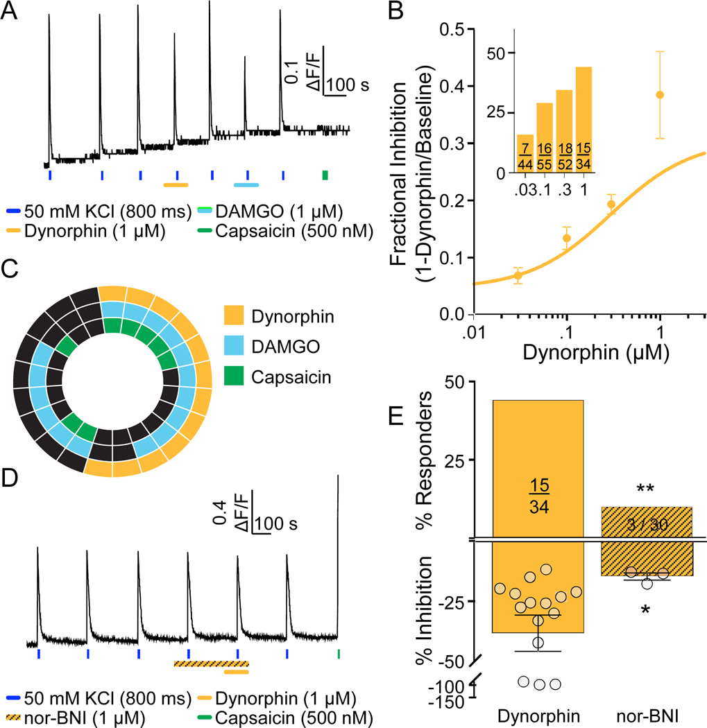
Figure 5. Characterization of functional KOP receptors in human DRG neurons.
A. Example of Ca2+ signaling in a neuron responsive to dynorphin and DAMGO, but not capsaicin. B. Both the magnitude of dynorphin-induced suppression of the evoked Ca2+ transient, and the proportion of neurons responsive to this KOP receptor agonist (inset), were concentration dependent. C. Distribution of neurons responsive to dynorphin, DAMGO, and capsaicin. D. Example of Ca2+ signaling in a neuron in which pre-application of the KOP receptor antagonist, nor-BNI was associated with the absence of a response to dynorphin. This neuron was responsive to capsaicin, however. E. Pooled data for neurons challenged with dynorphin alone or following application of nor-BNI. The proportion of neurons responsive to dynorphin (**p=0.0025, chi-square) and the magnitude of the suppression of the evoked Ca2+ transient (*p=0.027, Mann Whitney t-test) were significantly reduced by nor-BNI.

