Abstract
Cutaneous manifestations are becoming increasingly well‐documented in adults with COVID‐19. There is now also a growing body of literature regarding skin involvement in children, with reports of papulovesicular, petechial and widespread macular and papular lesions, and chilblains (pernio). We describe the case of a 13‐year‐old boy with confirmed COVID‐19 in the United Kingdom who presented with skin findings localized to the plantar aspects of the feet, axillae, and lower limbs. The morphology was predominantly maculopapular but also included petechiae and annular lesions.
Keywords: child, COVID‐19, pediatric, rash, SARS‐CoV‐2; coronavirus
1. INTRODUCTION
Diagnosis of COVID‐19 in children is currently uncommon compared to adults. Children who are symptomatic most commonly display cough, fever, pharyngeal erythema, and gastrointestinal symptoms. 1 In children, cutaneous manifestations include widespread erythematous macular and papular lesions, a skin rash in the context of a multisystem inflammatory state or similar clinical presentation to Kawasaki's disease, and papulovesicular eruptions. 2 , 3 We report a case of confirmed COVID‐19 in a child who presented with skin findings localized to the axillae, lower limbs, and plantar aspects of the feet.
2. CASE REPORT
A 13‐year‐old boy presented in April 2020 with the chief complaint of pain in the soles of his feet. He also had a 24‐hour history of fever, myalgia, and headache. He denied any respiratory symptoms. He had taken acetaminophen infrequently. He was previously fit and well with no comorbidities. His stepfather reported a flu‐like illness and his mother a mild cough.
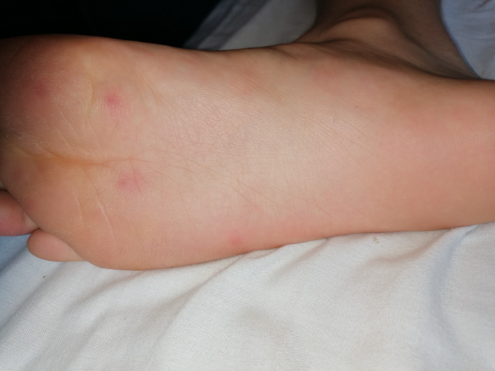
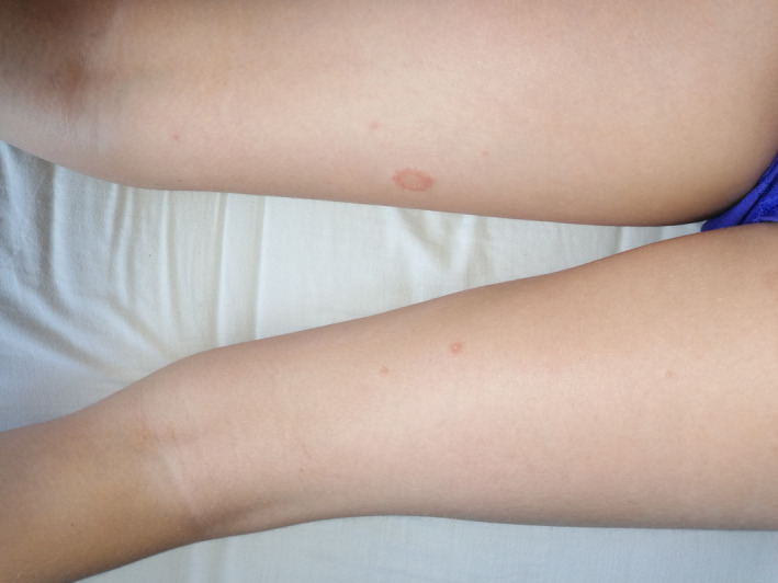
On examination, he was systemically well with normal vital signs. There was an erythematous papular eruption in the axillae with axillary and cervical lymphadenopathy, all nodes measuring <1 cm. On the plantar surface of his feet were multiple tender, erythematous papules each measuring approximately 1 cm (Figure 1). There were no vesicles or pustules. Erythematous macules with associated scattered petechiae, located in close proximity to the macules, were also present on the child's distal lower extremities (Figure 2).
Figure 1.

Feet—Multiple erythematous, tender papules, each measuring approximately 1 cm, were present on the plantar aspects of the feet
Figure 2.

Lower limbs—A macular rash with associated scattered petechiae was present on the lower limbs at presentation. An annular lesion developed following discharge
Coagulation profile and full blood count were within normal range. C‐reactive protein was 10 mg/L. Polymerase chain reaction (PCR) for SARS‐CoV‐2 was positive. PCR was negative for all seasonal respiratory viruses (influenza A, influenza B, parainfluenza, respiratory syncytial virus, rhinovirus, enterovirus, human metapneumovirus, adenovirus, Mycoplasma pneumoniae, enterovirus, and seasonal coronavirus).
The clinical photographs provided were taken by the family 2 days after presentation, at which point annular lesions were evident on the distal lower extremities. He continued to have intermittent mild flu‐like symptoms for 5 days but did not develop any cough or dyspnea. The skin changes fully resolved within 10‐14 days.
3. DISCUSSION
Currently, the most commonly reported symptoms of COVID‐19 in the pediatric population are cough and fever. 1 The adult literature describes erythematous rashes, chilblains, urticaria, vesicular lesions, petechiae, purpura, and papules. 4 There is also reference to localization to the axillae and soles of the feet. 5 A common theme in the adult data is the hypothesis that the skin findings are secondary to a drug reaction in view of the use of experimental treatments such as hydroxychloroquine and lopinavir. 5 Our patient only had exposure to infrequent acetaminophen, supporting the likelihood that the cutaneous manifestations are a direct result of the SARS‐CoV‐2 virus and the host immune response.
At this current time, pediatric health care providers should consider the diagnosis of COVID‐19 in children presenting with not only widespread papulovesicular and macular and papular lesions but also localized lesions with varied morphology including plantar papules, petechiae, and annular lesions.
ACKNOWLEDGMENTS
All authors would like to sincerely thank the patient and his family for taking the clinical photographs and consenting to their use along with the case history.
Klimach and Evans contributed equally as co‐first authors.
REFERENCES
- 1. Lu X, Zhang L, Du H, et al. SARS‐CoV‐2 infection in children. N Engl J Med. 2020;382(17):1663‐1665. [DOI] [PMC free article] [PubMed] [Google Scholar]
- 2. Genovese G, Colonna C, Marzano AV. Varicella‐like exanthem associated with COVID‐19 in an 8‐year‐old girl: a diagnostic clue? Pediatr Dermatol. 2020;37:435‐436. 10.1111/pde.14201 [DOI] [PMC free article] [PubMed] [Google Scholar]
- 3. Jones VG, Mills M, Suarez D, et al. COVID‐19 and kawasaki disease: novel virus and novel case. Hosp Pediatr. 2020;10(6):537‐540. [published online ahead of print, 2020 Apr 7]. [DOI] [PubMed] [Google Scholar]
- 4. Recalcati S. Cutaneous manifestations in COVID‐19: a first perspective. J Eur Acad Dermatol Venereol. 2020;34(5):e212‐e213. 10.1111/jdv.16387 [DOI] [PubMed] [Google Scholar]
- 5. Jimenez‐Cauhe J, Ortega‐Quijano D, Prieto‐Barrios M, Moreno- Arrones OM, Fernandez‐Nieto D. Reply to "COVID‐19 can present with a rash and be mistaken for dengue": petechial rash in a patient with COVID‐19 infection. J Am Acad Dermatol. 2020;S0190‐9622(20)30556‐9. 10.1016/j.jaad.2020.04.016 [published online ahead of print, 2020 Apr 10]. [DOI] [PMC free article] [PubMed] [Google Scholar]


