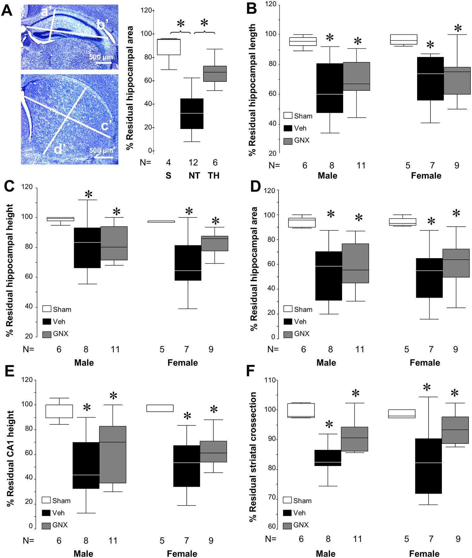
Figure 4:

Regional measurements of brain injury in p40 survivors. [A] Schema of hippocampal (a’, height, b’, length) and striatal measurements (c’, length; d’, height) performed with calibrated ocular filar micrometer at p40 following HI and treatment with vehicle or GNX-4728. Using this analysis schema, significant injury after HI and neuroprotection with therapeutic hypothermia is demonstrated in % residual hippocampal area 8 days following HI at p10 (*P < .05 vs. sham) as expected. Scale bar = 500 μm. [B – F] Measurements with the filar ocular micrometer of the anterior hippocampus, CA1 pyramidal layer thickness and striatum are able to detect greater injury each area in both male and female injured mice exposed to HI than in shams (*P < .05). However, there are no detectable differences between vehicle and GNX-4728 treatment after HI in any of the individual regional measurements or composite measures of residual hippocampal or striatal area.
