Abstract
Purpose
To assess the impact of the Covid-19 pandemic on FDG-PET/CT work volume and to evaluate the occurrence of abnormal imaging findings suspicious or potentially diagnostic for interstitial pneumonia by Covid-19 infection in south Italy.
Methods
We retrospectively reviewed the number and the findings of FDG-PET/CT studies acquired between February and April 2020 during the Covid-19 pandemic at the University of Napoli Federico II. The number and the findings of FDG-PET/CT studies acquired in the corresponding period of 2019 were also assessed for direct comparison.
Results
The number of FDG-PET/CT studies performed during the pandemic (n = 299) and in the corresponding period of 2019 (n = 335) were comparable. The percentage of abnormal FDG-PET/CT findings, suspicious for interstitial pneumonia by Covid-19 infection, was significantly higher during the pandemic (9%) compared with that found in the corresponding period of 2019 (4%) (χ2 5.45, P = 0.02). No significant differences were observed in the distribution of Covid-19 reporting and data system (CO-RADS) classification and in the maximum standardized uptake value between the pandemic (2.6 ± 2.2) and the corresponding period of 2019 (3.2 ± 1.4). Of note, patients with abnormal imaging findings during the pandemic time had clinical data and/or laboratory tests negative for Covid-19 infection.
Conclusion
Despite the restrictive medical measures for the emergency, the number of FDG-PET/CT studies was unchanged during the pandemic compared with the previous year. Our findings also indicate that Covid-19 infection was contained in our series of patients from southern Italy.
Keywords: Covid-19, SARS-CoV-2, FDG-PET/CT, Imaging, Southern Italy
Introduction
The recent Covid-19 emergency in Italy determined the reorganization of the healthcare system with subsequent limitations in diagnostic imaging throughput and potential clinical implications for daily medical practice [1–3]. In particular, the policy measures to limit the spread of infection have determined a decrease in the number of imaging studies requested for clinical indications different from Covid-19 disease. Moreover, hospital adjustments to improve the management of Covid-19 patients are diminishing the volume of conventional medical imaging studies. Of note, outpatient diagnostic imaging may suffer the major impact of these changes, even though the amount of all imaging studies is decreasing. Of course, these changes depend on the site of clinical practice as well as on the evolution of Covid-19 pandemic in different geographic regions. In this emergency context, also 18F-fluorodeoxyglucose (FDG) positron emission tomography (PET)/computed tomography (CT) imaging studies may be reduced despite the fact that cancer patients require timely FDG scan in different phases of their disease. Furthermore, FDG-PET/CT imaging has been reported to be useful to incidentally diagnose Covid-19 infection as demonstrated by initial experiences in Wuhan as well as in northern Italy [4–8]. The aim of this study was to assess the impact of the Covid-19 pandemic on FDG-PET/CT work volume and to evaluate the occurrence of abnormal imaging findings suspicious or potentially diagnostic for interstitial pneumonia by Covid-19 infection in southern Italy.
Methods
Patients
We retrospectively reviewed the number and the findings of FDG-PET/CT imaging studies acquired in 2020 between February 3 and April 30, during the Covid-19 pandemic at the University of Napoli Federico II. The number and the findings of FDG-PET/CT imaging studies acquired in the corresponding period of 2019 (February 4 and April 30) were also assessed for direct comparison.
PET/CT imaging
18F-FDG-PET/CT studies were acquired using a Gemini TF 64 scanner (Philips Healthcare, Best, The Netherlands). All patients fasted for at least 6 h prior to imaging, and blood glucose levels were < 180 mg/dL at the time of tracer injection. PET scans were acquired in 3-D mode starting 60 min after 18F-FDG administration (activity range 200–300 MBq, according to body weight). A low- (70 mAs) and high-dose (230 mAs) CT scans (rotation time 1.5 s, collimation 16 × 0.625) were acquired for attenuation correction of emission data. The sinogram of emission data was reconstructed using the 3-D row action maximum likelihood algorithm, taking into account attenuation, detector efficiency, and scatter and random coincidence corrections. Attenuation correction was performed using CT images. CT and PET images were matched and fused into transaxial, coronal, and sagittal images.
Imaging analysis
CT chest images were evaluated by two experienced radiologists who worked in consensus and reviewed each set-in random order to evaluate the presence and the location of abnormal findings in lung parenchyma according to Covid-19 reporting and data system (CO-RADS) classification [9]. In case of disagreement, a third senior radiologist was consulted to reach a final consensus for CT imaging interpretation. Successively, FDG distribution in the lungs was qualitatively evaluated using PET/CT fusion images and maximum standardized uptake value (SUVmax) was measured on areas of increased FDG uptake corresponding to abnormal CT findings. SUVmax value of the most FDG-avid lung abnormality was recorded. In particular, CO-RADS classification of CT findings [9] represented the level of suspicion of Covid-19 infection graded as not interpretable (CO-RADS 0 = scan technically incomplete or of insufficient quality for artifacts), with no suspicion (CO-RADS 1 = normal CT or non-infectious CT abnormalities), low suspicion (CO-RADS 2 = CT abnormalities consistent with infections other than Covid-19, absence of ground-glass opacities), indeterminate suspicion (CO-RADS 3 = uncertain CT findings for Covid-19 such as small unifocal, perihilar, or homogeneous extensive ground-glass opacities), high suspicion (CO-RADS 4 = unilateral peribronchovascular ground-glass CT opacities without any other typical findings), very high suspicion (CO-RADS 5 = typical bilateral multifocal ground-glass CT opacities with peripheral and/or basal distribution with or without parenchyma consolidations), and as proven very high suspicion (CO-RADS 6 = CO-RADS 5 with positive RT-PCR test for virus-specific nucleic acid).
Statistical analysis
Continuous data are expressed as mean ± standard deviation and categorical data as percentage. Confidence intervals (CI) were calculated using the Poisson distribution. Student’s t test and χ2 test were used to compare the differences in continuous and categorical variables, respectively. Two-tailed P values < 0.05 were considered significant. Statistical analysis was performed with Stata 16 software (StataCorp, College Station, Texas, USA). The correlation between CT findings as CO-RADS and tracer activity at FDG-PET was evaluated calculating Spearman’s correlation coefficient (ρ).
Results
A total of 299 FDG-PET/CT studies in 288 patients (157 men, mean age 60 ± 17 years) were acquired during the pandemic. The majority (94%) of imaging studies was performed for diagnosis/staging (n = 102) or follow-up (n = 178) of cancer patients; in particular, 11 patients were studied twice during follow-up. The remaining 19 (6%) imaging studies were acquired for other non-oncological diseases: vasculitis (n = 4), endocarditis (n = 3), fever of unknown origin (n = 2), histiocytosis x (n = 1), and other inflammatory diseases (n = 9). Similarly, a total of 335 FDG-PET/CT imaging studies in 330 patients (165 men, mean age 61 ± 18 years) were acquired during the corresponding period of 2019. The majority (94%) of imaging studies was performed for diagnosis/staging (n = 114) or follow-up (n = 202) of cancer patients; in particular, 5 patients were studied twice during follow-up. The remaining 19 (6%) imaging studies were acquired for other non-oncological diseases: fever of unknown origin (n = 6), vasculitis (n = 4), endocarditis (n = 2), histiocytosis x (n = 1), mastocytosis (n = 1), Chron’s disease (n = 1), and other inflammatory diseases (n = 4). The number of FDG-PET/CT studies performed during the pandemic (n = 299) and in the corresponding period of 2019 (n = 335) were comparable (P = 0.74).
The percentage of abnormal FDG-PET/CT findings, suspicious for interstitial pneumonia by Covid-19 infection, was significantly higher during the pandemic (9%) compared with that found in the corresponding period of 2019 (4%) (χ2 5.45, P = 0.02).
Abnormal CT findings were observed during the pandemic in 26 patients subjected to the imaging procedure for oncological (n = 23) or non-oncological (n = 3) diseases. The individual clinical characteristics of these patients are reported in Table 1. In particular, CT abnormalities consisted of CO-RADS 1 (n = 1), CO-RADS 2 (n = 6), CO-RADS 3 (n = 15), CO-RADS 4 (n = 3), and CO-RADS 5 (n = 1) grading. Of note, in all these patients, laboratory tests were negative for Covid-19 infection.
Table 1.
Patient characteristics, CO-RADS classification, and extra-thoracic sites with increased FDG uptake during the pandemic
| Sex | Age (year) | Disease | Phase | Symptoms | Treatment | CO-RADS | Extra-thoracic sites with FDG uptake | |
|---|---|---|---|---|---|---|---|---|
| 1 | M | 84 | Colon cancer | Staging | None | None | 2 | Liver |
| 2 | F | 60 | Breast cancer | Follow-up | None | Chemotherapy, surgery | 3 | Local recurrence, axillary LN, thyroid |
| 3 | M | 75 | Lung cancer | Staging | Cough, dyspnea | Immunosuppressive therapy for kidney transplantation | 2 | None |
| 4 | M | 64 | Rectal cancer | Follow-up | None | Chemotherapy | 3 | Mediastinal LN |
| 5 | M | 90 | Primary tumor of unknown origin | Diagnosis | Cough, dyspnea, skin rash | None | 3 | None |
| 6 | M | 51 | Thyroid cancer | Follow-up | None | Targeted therapy | 3 | Mediastinal LN, liver |
| 7 | M | 73 | Suspected lymphoma | Diagnosis | Fever | None | 2 | Neck and mediastinal LN, skin, adrenal glands |
| 8 | F | 77 | Non-Hodgkin lymphoma | Follow-up | None | Chemotherapy | 3 | None |
| 9 | F | 61 | Suspected paraneoplastic syndrome | Diagnosis | Fever, neurological symptoms | None | 3 | Muscle |
| 10 | F | 28 | Suspected paraneoplastic syndrome | Diagnosis | Neurological symptoms | None | 3 | Ovary, rectum |
| 11 | M | 36 | Neuroendocrine tumor | Diagnosis | None | None | 3 | Pancreas, mesenteric and inguinal LN |
| 12 | M | 80 | Laryngeal cancer | Follow-up | Cough, dyspnea | None | 5 | None |
| 13 | F | 78 | Breast cancer | Follow-up | None | Chemotherapy | 3 | Adrenal gland |
| 14 | F | 40 | Breast cancer | Follow-up | None | Chemotherapy | 3 | None |
| 15 | F | 60 | Lung cancer | Follow-up | None | Immunotherapy | 2 | Neck and axillary LN, parotid |
| 16 | M | 25 | Hodgkin lymphoma | Follow-up | None | Chemotherapy, radiation therapy | 4 | None |
| 17 | M | 20 | Hodgkin lymphoma | Follow-up | None | Chemotherapy | 3 | Neck LN, bone |
| 18 | M | 56 | Non-Hodgkin lymphoma | Follow-up | None | Chemotherapy | 4 | None |
| 19 | F | 61 | Non-Hodgkin lymphoma | Follow-up | Skin rash | Chemotherapy | 1 | Mediastinal LN |
| 20 | F | 73 | Non-Hodgkin lymphoma | Follow-up | None | Chemotherapy | 3 | None |
| 21 | F | 73 | Multiple myeloma | Follow-up | Cough, dyspnea | Chemotherapy | 2 | Neck and mediastinal LN, bone, adrenal glands |
| 22 | M | 63 | Multiple myeloma | Follow-up | None | Chemotherapy | 4 | Bone |
| 23 | F | 61 | Neuroendocrine tumor | Follow-up | None | None | 3 | Liver, mesenteric LN |
| 24 | M | 73 | Endocarditis | Diagnosis | Cough, dyspnea | None | 3 | Mediastinal LN, bone |
| 25 | M | 92 | Fever of unknown origin | Diagnosis | Fever | None | 3 | Mediastinal LN, colon |
| 26 | M | 65 | Prothesis infection | Diagnosis | Fever | None | 2 | Bone |
F, female; LN, lymph node uptake; M, male
Similarly, abnormal FDG-PET/CT findings were observed during the corresponding period of 2019 in 14 patients subjected to the imaging procedure for oncological (n = 12) or non-oncological (n = 2) diseases. The individual clinical characteristics of these patients are reported in Table 2. In particular, CT abnormalities consisted of CO-RADS 2 (n = 3), CO-RADS 3 (n = 6), CO-RADS 4 (n = 3), and CO-RADS 5 (n = 2). No significant differences were observed in the distribution of CO-RADS findings and in the SUVmax values between the pandemic (2.6 ± 2.2; CI 1.7–3.5) and the corresponding period of 2019 (3.2 ± 1.4; CI 2.4–4.0) (P = 0.30).
Table 2.
Patient characteristics, CO-RADS classification, and extra-thoracic sites with increased FDG uptake during the corresponding period of 2019
| Sex | Age (year) | Disease | Phase | Symptoms | Treatment | CO-RADS | Extra-thoracic sites with FDG uptake | |
|---|---|---|---|---|---|---|---|---|
| 1 | M | 72 | Oral cavity cancer | Follow-up | None | Chemotherapy, radiation therapy | 4 | Mediastinal and abdominal LN |
| 2 | F | 58 | Gastric cancer | Staging | None | None | 2 | Neck, mediastinal, mesenteric and pelvic LN, liver, bone |
| 3 | M | 78 | Suspected lung cancer | Diagnosis | None | None | 4 | Mediastinal LN, bone |
| 4 | M | 76 | Lung cancer | Staging | Cough, dyspnea | None | 3 | Bone |
| 5 | M | 67 | Lung cancer | Follow-up | None | Surgery, chemotherapy | 3 | None |
| 6 | M | 59 | Non-Hodgkin lymphoma | Follow-up | None | Chemotherapy | 3 | Neck LN |
| 7 | M | 70 | Suspected mediastinal tumor | Diagnosis | None | None | 4 | None |
| 8 | M | 15 | Sarcoma | Staging | None | None | 3 | Bone |
| 9 | F | 41 | Thymoma | Staging | Cough, dyspnea, chest pain | None | 5 | Bone, pituitary gland |
| 10 | F | 60 | Lung cancer | Follow-up | None | Immunotherapy | 3 | Neck, axillary and mediastinal LN, adrenal gland |
| 11 | M | 46 | Non-Hodgkin lymphoma | Follow-up | None | Chemotherapy | 2 | Neck LN |
| 12 | M | 60 | Multiple myeloma | Follow-up | Cough, dyspnea | Chemotherapy, bone marrow transplantation | 2 | Bone |
| 13 | M | 77 | Endocarditis | Diagnosis | None | None | 5 | Mediastinal LN |
| 14 | F | 91 | Fever of unknown origin | Diagnosis | Cough, dyspnea | None | 3 | Bone |
F, female; LN, lymph node uptake; M, male
Table 3 reports the correlation between CT and FDG-PET findings expressed as CO-RADS and SUVmax in patients with abnormal imaging findings during the pandemic and during the corresponding period of 2019. As shown, no significant correlation was found in both analyzed periods (ρ = 0.11 and ρ = 0.13). In addition, extra-thoracic sites of increased metabolic activity were observed during the pandemic (Table 1) and the corresponding period of 2019 (Table 2).
Table 3.
Correlation between CT and FDG-PET findings expressed as CO-RADS and SUVmax in patients with abnormal imaging findings during the pandemic and during the corresponding period of 2019
| CO-RADS | ||||||
|---|---|---|---|---|---|---|
| 1 | 2 | 3 | 4 | 5 | ρ | |
| February–April 2020 | ||||||
| Patients (n) | 1 | 6 | 15 | 3 | 1 | |
| SUVmax | 2.4 | 2.5 ± 1.4 | 1.7 ± 2.2 | 5.4 ± 3.6 | 4.0 ± 2.0 | 0.11 |
| February–April 2019 | ||||||
| Patients (n) | 0 | 3 | 6 | 3 | 2 | |
| SUVmax | - | 3.8 ± 1.8 | 2.4 ± 1.4 | 3.1 ± 1.0 | 4.4 ± 1.1 | 0.13 |
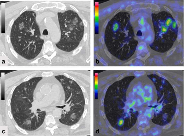
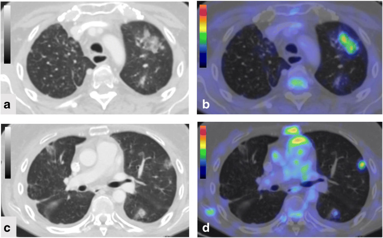
Figure 1 shows an example of a patient with laryngeal carcinoma, proven to be Covid-19 infection free, who was studied during the pandemic and presented a CO-RADS 5. Figure 2 shows an example of a thymoma patient studied for staging in 2019 who presented a CO-RADS 5 due to an infection other than Covid-19.
Fig. 1.
FDG-PET/CT images in a 80-year-old patient, proven to be Covid-19 infection free, in follow-up for laryngeal carcinoma during the pandemic. CT images (a, c) consisted of CO-RADS 5 finding and FDG-PET/CT images (b, d) showed multiple areas of increased tracer activity with a SUVmax of 6.0
Fig. 2.
FDG-PET/CT images in a 41-year-old patient with thymoma studied for staging in 2019 in which CT images (a, c) consisted of CO-RADS 5 finding and FDG-PET/CT images (b, c) showed multiple areas of increased tracer activity with a SUVmax of 3.7
Discussion
The results of our experience in southern Italy show that, despite the restrictive medical measures for Covid-19 emergency, the number of FDG-PET/CT imaging studies was unchanged during the pandemic compared with the previous year. Yet, the presence of abnormal FDG-PET/CT findings, suspicious for interstitial pneumonia, was not associated with Covid-19 infection, as confirmed by negative clinical data and/or laboratory tests. Of note, on the basis of our findings, CT abnormalities, according to CO-RADS classification, seem to be not specific for Covid-19 infection. Taken together, these preliminary results indicate that Covid-19 infection was contained in our southern Italian patient series. Conversely, the initial experience in northern Italy [6–8] describing the potential diagnostic usefulness of FDG-PET/CT imaging for Covid-19 infection reported a significant percentage of positive cases reflecting the higher diffusion of the infection in that region.
In our study, the majority (94%) of patients evaluated during the pandemic consisted of patients with oncological diseases in whom imaging studies could not be postponed, thus explaining the unmodified number of FDG-PET/CT studies compared with the corresponding period of 2019. In this regard, medical measures in Italy were not restrictive for such patients and thus these imaging studies could be performed despite the emergency.
In patients studied during the pandemic, the results of FDG-PET/CT imaging studies did not indicate with certainty the presence of Covid-19 infection in the majority (85%) of cases (n = 22). In particular, in such patients, CT findings consisted of CO-RADS 1, 2, or 3, therefore with no suspicion, low, or indeterminate level of suspicion for Covid-19 infection. On the other hand, in the remaining 4 patients (15%), CT findings consisted of CO-RADS 4 (n = 3) and CO-RADS 5 (n = 1) suggesting a high or very high level of suspicion for Covid-19 infection. However, no cases of Covid-19 infection were proven. Reverse-transcriptase polymerase chain reaction from pharyngeal swabs is considered to be the gold standard for the diagnosis of Covid-19 infection, but a high false-negative rate has been reported [10]. Although CT scan has been showed to be useful in terms of diagnostic sensitivity for Covid-19 infection [11], its accuracy in differentiating such infection from other viral pneumonia was reported to be limited [12]. Our results confirm these observations suggesting a high diagnostic sensitivity, but a low diagnostic specificity.
Similarly, the results of the corresponding period of 2019 showed that the majority (94%) of patients had oncological diseases with no significant difference in terms of initial diagnosis compared with patients enrolled during the pandemic. However, the percentage of abnormal FDG-PET/CT findings (4%) suspicious for interstitial pneumonia corresponding to 2019 period was significantly lower compared with that (9%) of the pandemic. In particular, these findings occurred in 14 patients consisting in the majority (64%) of cases of CT CO-RADS 2 or 3, therefore with low or indeterminate level of suspicion. In the other 36% of cases, CT findings consisted of CO-RADS 4 (n = 3) and CO-RADS 5 (n = 2) suggesting a high or very high level of suspicion. No cases of Covid-19 infection were proven. Hence, the distribution of CO-RADS findings was similar to that observed during the pandemic time confirming the high sensitivity of CT to detect viral pneumonia other than Covid-19 [11, 12].
FDG-PET/CT imaging has been recently reported to be useful to incidentally diagnose Covid-19 infection as demonstrated by initial experiences in Wuhan and in northern Italy [4–8]. In this regard, Zou et al. [4] initially described a patient of Wuhan with suspected lung malignancy on chest CT who underwent FDG-PET/CT for further evaluation. A large FDG-avid mass was detected in the right lung as well as abnormal tracer uptake was observed in loco-regional lymph nodes as well as in bone marrow. CT fusion images demonstrated the presence of multifocal ground-glass opacities with areas of focal consolidation and laboratory tests confirmed Covid-19 infection. Moreover, Qin et al. [5] described similar findings in additional four patients from Wuhan. Albano et al. [6] reported their Italian experience in Brescia on incidental FDG-PET/CT findings suggestive of Covid-19 infection in asymptomatic patients living in a high prevalence region of northern Italy. In 6 (9%) of 65 patients with various malignancies, studied in a short period of 10 days, they observed unexpected CT signs of interstitial pneumonia with increased FDG activity due to Covid-19 infection as confirmed by laboratory tests. Similarly, Kirienko et al. [7] and Setti et al. [8] from Bergamo reported a total of six patients with incidental FDG-PET/CT imaging findings suspicious for Covid-19 infection in asymptomatic patients. Therefore, these data show that incidental FDG-PET/CT findings suggestive of Covid-19 infection may occur and hence it is fundamental that nuclear medicine physicians acquire diagnostic skills to recognize typical findings on the co-registered CT images. In this regard, the potential role of FDG-PET/CT in patients with known or suspicious Covid-19 infection has not been demonstrated [13]. An increased FDG uptake in pulmonary or lymph nodal lesions in patients with Covid-19 infection is not surprising as acute inflammatory and infectious pulmonary lesions are characterized by augmented metabolic activity. These findings are not specific for Covid-19 infection and, therefore, FDG-PET/CT should not be recommended for evaluating patients with known or suspected Covid-19 infection. However, these incidental imaging findings should prompt medical staff to adopt all safety measures to protect the population and personnel from exposure to Covid-19 sources [14, 15].
This study has some potential limitations. First, the retrospective design of the study. We did not exclude patients with known lung pathologies that may present with CT features described in Covid-19. It should be also considered that various clinical conditions (e.g., metastasis, therapeutic effects, and therapeutic complications) can determine a radiological simil-Covid-19 pattern. Finally, although patients under medical therapy had a higher CO-RADS value, alternative etiological causes for lung changes could not be clearly determined.
Conclusion
According to our results and other recently reported experiences, FDG-PET/CT imaging may be useful to confirm or ruling out the presence of Covid-19 infection. Nuclear medicine physicians should be on alert since incidental abnormal findings suspicious for Covid-19 may occur on FDG-PET/CT scans. Our preliminary results from an experience in the south of Italy also show that despite the restrictive medical measures for the emergency, the number of FDG-PET/CT studies was unchanged during the pandemic compared with the previous year. Of note, since no cases positive for Covid-19 infection were observed, these imaging data indicate that Covid-19 infection was contained in our series of patients from southern Italy.
Authors’ contribution
SM, SDV, and AC conceptualized the paper; SM, CGM, CB, AA, and LA evaluated and reported the imaging findings; SM and MP drafted the manuscript; and all the authors revised and commented on the paper and approved the final version of the manuscript.
Compliance with ethical standards
Conflict of interest
The authors declare that they have no conflict of interest.
Ethical approval
All procedures performed in this study involving human participants were in accordance with the ethical standards of the institutional and/or national research committee and with the 1964 Helsinki declaration and its later amendments or comparable ethical standards.
Informed consent
Informed consent was obtained from all individual participants included in the study.
Footnotes
This article is part of the Topical Collection on Infection and inflammation.
Publisher’s note
Springer Nature remains neutral with regard to jurisdictional claims in published maps and institutional affiliations.
References
- 1.Nacoti M, Ciocca A, Giupponi A, Brambillasca P, Lussana F, Pisano M, et al. At the epicenter of the Covid-19 pandemic and humanitarian crises in Italy: changing perspectives on preparation and mitigation. NEJM Catalyst. 2020;21. 10.1056/CAT.20.0080.
- 2.Stempniak M. Mednax sees ‘meaningful’ decline in radiology volume during pandemic, revises revenue forecasts. Radiology Business; https://www.radiologybusiness.com/topics/healthcare-economics/mednax-radiology-volume-coronavirus-covid-19-imaging; Published Mar 25, 2020.
- 3.Cavallo JJ, Forman HP. The economic impact of the COVID-19 pandemic on radiology practices [published online ahead of print, 2020 Apr 15]. Radiology. 2020:201495. 10.1148/radiol.2020201495. [DOI] [PMC free article] [PubMed]
- 4.Zou S, Zhu X. FDG PET/CT of COVID-19 [published online ahead of print, 2020 Mar 6]. Radiology. 2020:200770. 10.1148/radiol.2020200770.
- 5.Qin C, Liu F, Yen TC, Lan X. (18)F-FDG PET/CT findings of COVID-19: a series of four highly suspected cases. Eur J Nucl Med Mol Imaging. 2020;47:1281–1286. doi: 10.1007/s00259-020-04734-w. [DOI] [PMC free article] [PubMed] [Google Scholar]
- 6.Albano D, Bertagna F, Bertoli M, Bosio G, Lucchini S, Motta F, et al. Incidental findings suggestive of COVID-19 in asymptomatic patients undergoing nuclear medicine procedures in a high-prevalence region. J Nucl Med. 2020;61:632–636. doi: 10.2967/jnumed.120.246256. [DOI] [PubMed] [Google Scholar]
- 7.Kirienko M, Kirienko M, Padovano B, Serafini G, Marchianò A, Gronchi A, et al. CT, [18F]FDG-PET/CT and clinical findings before and during early Covid-19 onset in a patient affected by vascular tumour [published online ahead of print, 2020 Apr 25]. Eur J Nucl Med Mol Imaging. 2020:1–2. 10.1007/s00259-020-04822-x. [DOI] [PMC free article] [PubMed]
- 8.Setti L, Kirienko M, Dalto SC, Bonacina M, Bombardieri E. FDG-PET/CT findings highly suspicious for COVID-19 in an Italian case series of asymptomatic patients [published online ahead of print, 2020 Apr 27]. Eur J Nucl Med Mol Imaging. 2020:1–8. 10.1007/s00259-020-04819-6. [DOI] [PMC free article] [PubMed]
- 9.Prokop M, van Everdingen W, van Rees Vellinga T, van Ufford JQ, Stöger L, Beenen L, et al. CO-RADS - a categorical CT assessment scheme for patients with suspected COVID-19: definition and evaluation [published online ahead of print, 2020 Apr 27]. Radiology. 2020:201473. 10.1148/radiol.2020201473. [DOI] [PMC free article] [PubMed]
- 10.Lan L, Xu D, Ye G, Xia C, Wang S, Li Y, et al. Positive RT-PCR test results in patients recovered from COVID-19. JAMA. 2020;323:1502–1503. doi: 10.1001/jama.2020.2783. [DOI] [PMC free article] [PubMed] [Google Scholar]
- 11.Fang Y, Zhang H, Xie J, Lin M, Ying L, Pang P, et al. Sensitivity of chest CT for COVID-19: comparison to RT-PCR [published online ahead of print, 2020 Feb 19]. Radiology. 2020:200432. 10.1148/radiol.2020200432. [DOI] [PMC free article] [PubMed]
- 12.Bai HX, Hsieh B, Xiong Z, Halsey K, Choi JW, Tran TML, et al. Performance of radiologists in differentiating COVID-19 from viral pneumonia on chest CT [published online ahead of print, 2020 Mar 10]. Radiology. 2020:200823. 10.1148/radiol.2020200823. [DOI] [PMC free article] [PubMed]
- 13.Treglia G. The role of 18F-FDG PET for COVID-19 infection: myth versus reality [published online ahead of print, 2020 Apr 30]. Clin Transl Imaging. 2020. 10.1007/s40336-020-00367-z. [DOI] [PMC free article] [PubMed]
- 14.Tulchinsky M, Fotos JS, Slonimsky E. Incidental CT findings suspicious for Covid-19 associated pneumonia on nuclear medicine exams: recognition and management plan [published online ahead of print, 2020 Apr 9]. Clin Nucl Med. 2020. 10.1097/RLU.0000000000003100. [DOI] [PMC free article] [PubMed]
- 15.Lu Y, Yan SX, Lan X, Zhu X, Macapinlac HA. Nuclear medicine in responding to global pandemic COVID-19-American College of Nuclear Medicine member experience [published online ahead of print, 2020 Apr 15]. Eur J Nucl Med Mol Imaging. 2020:1–3. 10.1007/s00259-020-04799-7. [DOI] [PMC free article] [PubMed]


