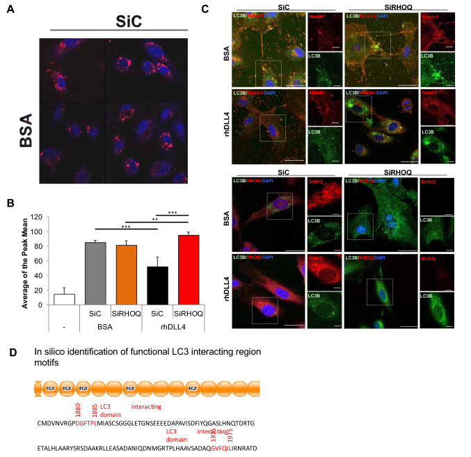
Fig. 10.
Loss of RHOQ increases autophagy and leads to degradation of Notch1 receptor. HUVECs cultured on BSA or rhDLL4 (1 μg/ml)-coated plates and effects on autophagosomes were assessed 16 h later by staining cells with autophagy tracker (red) with changes in tracker levels, a visualised by confocal microscopy (nuclei stained with DAPI (blue)), and assessed by b FACs or c cells were fixed and immuno-fluorescence staining for localisation changes in RHOQ (red) or Notch1 (red) and LC3B (green) proteins and visualised by confocal microscopy (nuclei stained with DAPI (blue)) or d illustrating LC3B binding sites on Notch1 (Error bars = S.D. Key: *p < 0.05, **p < 0.01, ***p < 0.0001 one-way ANOVA or unpaired Student’s t-test between data group and control, Scale bar = 20 nm; representative images and data of n = 3 independent experiments)

