PLOS ONE logoLink to PLOS ONE
. 2020 Jun 24;15(6):e0232111. doi: 10.1371/journal.pone.0232111

Ex-vivo cultured human corneoscleral segment model to study the effects of glaucoma factors on trabecular meshwork

Ramesh B Kasetti 1,#, Pinkal D Patel 1,#, Prabhavathi Maddineni 1, Gulab S Zode 1,*
Editor: Paloma B Liton2
PMCID: PMC7314024  PMID: 32579557

Abstract

Glaucoma is the second leading cause of irreversible blindness worldwide. Primary open angle glaucoma (POAG), the most common form of glaucoma, is often associated with elevation of intraocular pressure (IOP) due to the dysfunction of trabecular meshwork (TM) tissues. Currently, an ex vivo human anterior segment perfusion cultured system is widely used to study the effects of glaucoma factors and disease modifying drugs on physiological parameters like aqueous humor (AH) dynamics and IOP homeostasis. This system requires the use of freshly enucleated intact human eyes, which are sparsely available at very high cost. In this study, we explored the feasibility of using human donor corneoscleral segments for modeling morphological and biochemical changes associated with POAG. Among the number of corneas donated each year, many are deemed ineligible for transplantation due to stringent acceptance criteria. These ineligible corneoscleral segments were obtained from the Lions Eye Bank, Tampa, Florida. Each human donor anterior corneoscleral segment was dissected into four equal quadrants and cultured for 7 days by treating with the glaucoma factors dexamethasone (Dex) or recombinant transforming growth factor (TGF) β2 or transduced with lentiviral expression vectors containing wild type (WT) and mutant myocilin. Hematoxylin and Eosin (H&E) staining analysis revealed that the TM structural integrity is maintained after 7 days in culture. Increased TUNEL positive TM cells were observed in corneoscleral quadrants treated with glaucoma factors compared to their respective controls. However, these TUNEL positive cells were mainly confined to the scleral region adjacent to the TM. Treatment of corneoscleral quadrants with Dex or TGFβ2 resulted in glaucomatous changes at the TM, which included increased extracellular matrix (ECM) proteins and induction of endoplasmic reticulum (ER) stress. Western blot analysis of the conditioned medium showed an increase in ECM (fibronectin and collagen IV) levels in Dex- or TGFβ2-treated samples compared to control. Lentiviral transduction of quadrants resulted in expression of WT and mutant myocilin in TM tissues. Western blot analysis of conditioned medium revealed decreased secretion of mutant myocilin compared to WT myocilin. Moreover, increased ECM deposition and ER stress induction was observed in the TM of mutant myocilin transduced quadrants. Our findings suggest that the ex-vivo cultured human corneoscleral segment model is cost-effective and can be used as a pre-screening tool to study the effects of glaucoma factors and anti-glaucoma therapeutics on the TM.

Introduction

Glaucoma is the second leading cause of irreversible blindness affecting nearly 70 million people worldwide [1]. Primary open angle glaucoma (POAG) is the most common form of glaucoma. The majority of POAG patients experience chronic elevation in intraocular pressure (IOP), which is the only modifiable and treatable risk factor associated with the disease [2]. The balance between aqueous humor secretion from the ciliary body and its outflow through the trabecular meshwork (TM) is critical for maintenance of IOP homeostasis within an acceptable physiological range. In POAG, there is an increased resistance to the aqueous humor outflow through the TM, which leads to IOP elevation [3, 4]. However, the molecular pathology underlying TM dysfunction and IOP elevation are yet to be fully understood. Moreover, the majority of current treatments do not address the underlying pathology of glaucomatous TM damage. Of note, Rho-kinase inhibitors are the newest class of glaucoma drugs that directly target the TM to increase aqueous outflow [57]. Several factors are involved in glaucomatous TM damage, resulting in elevation of IOP. MYOC was the first glaucoma gene identified [8] and is responsible for approximately 4% of POAG [9]. Although the normal role for wild type (WT) myocilin is unknown, mutations in MYOC cause a deleterious gain-of-function leading to inhibition of its secretion [10], intracellular accumulation and protein stress within TM cells [1113]. Another major contributor to glaucomatous TM damage is the increased expression of the pro-fibrotic cytokine, transforming growth factor (TGF) β2 in the aqueous humor and TM of POAG patients [1417]. In addition, glucocorticoid (GC) therapy can cause ocular hypertension (OHT) and development of iatrogenic open angle glaucoma in susceptible individuals [18, 19]. A potent GC, dexamethasone (Dex) is known to cause pathological alterations in the TM such as increased ECM synthesis and deposition, induction of ER stress, reduced TM phagocytic function and cytoskeletal remodeling [2028]. Since clinical presentations of GC-induced glaucoma are similar to POAG in many ways [29], several laboratories have utilized GC-induced OHT to understand glaucomatous TM dysfunction. Treatment with Dex or TGFβ2 elevates IOP in mice in vivo [3032] and in perfusion cultured human anterior segments ex vivo [3335]. Both Dex and TGFβ2-induced OHT are associated with changes to the TM cytoskeleton [33, 36] and ECM deposition [28], which stiffen the TM [35, 37].

There is an unmet need in glaucoma research for a versatile model system that closely recapitulates the disease condition in humans. Although there are remarkable anatomical and physiological similarities between humans and animal models of glaucoma, the results obtained with animal models rarely translate to disease modifying outcomes in human patients. The opportunity to work with gifted human donor tissues is a unique privilege shared by the ocular research community. Perfusion cultured human anterior segments have been frequently used as an ex-vivo model in glaucoma research [34, 35, 3841]. The anterior segments used in perfusion culture are obtained from freshly enucleated human eyes post-mortem for studying physiological parameters like AH dynamics and IOP homeostasis. However, sparse availability, very high cost and high failure rates have impeded the extensive use of the perfusion culture model in glaucoma research.

The cornea is the most commonly donated organ of the human body worldwide. In the United States, 116,990 corneas were donated in 2016, of which only 63,596 were used for transplantation [42]. Due to stringent transplantation eligibility criteria, the majority of corneas are deemed ineligible [43]. These ineligible corneoscleral segments are easily available with lower processing cost compared to intact whole globes. These corneoscleral segments consist of a complete cornea, sclera, and an intact TM rim. Moreover, the patient medical history is readily available. Several labs utilize these tissues to isolate primary TM cells. Since these corneoscleral segments can be cultured in stationary media for several days, it is possible to utilize corneoscleral segments to test the effects of glaucoma factors on intact TM tissues. This study investigates the feasibility of using these corneoscleral segments to detect the morphological and biochemical changes associated with glaucoma pathogenesis. To test this corneoscleral segment model in recapitulating the disease phenotype, we treated these explants with factors known to induce glaucomatous pathology including Dex, TGFβ2, and the mutant form of myocilin protein (Y437H). We examined whether these glaucoma factors induce similar pathological changes in the TM of ex-vivo cultured human corneoscleral segments.

Material and methods

Antibodies

Antibodies were purchased from the following sources; fibronectin (catalog# ab2413, Abcam), Collagen type IV (catalog # SAB4500369, Sigma-Aldrich), ATF4 (catalog# sc-200, Santa Cruz Biotechnology), CHOP (catalog# 13172, Novus Biologicals), GAPDH (catalog# 3683; Cell Signaling Technology), GRP78 (N-20, catalog# sc-1050, Santa Cruz Biotechnology), FLAG (catalog# A8592, Sigma), Myocilin (36–135, catalog# H00004653, Abnova).

Ex-vivo culture of human corneoscleral segments

Human corneoscleral segments with intact TM rim were obtained from the Lions Eye Bank (Tampa, Florida) in accordance with Declaration of Helsinki guidelines. The corneoscleral segments were dissected into 4 equal quadrants and each quadrant was cultured in a 24-well plate using DMEM medium supplemented with 10% FBS, L-glutamine and 1% Pen-strep (Fig 1). Cultured medium was changed every two days. Quadrants were treated either with 100nM Dex or 5 ng/ml recombinant TGFβ2 (in 0.5% FBS containing medium) or transduced with lentiviral vectors expressing FTS (FLAG and S) tagged WT and mutant myocilin for 7 days with appropriate controls.

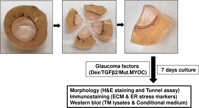
Fig 1. Schematic showing the experimental design.

Fig 1

Human corneoscleral segments with an intact TM rim were dissected into 4 equal quadrants and each quadrant was cultured and treated with glaucoma-inducing factors for 7 days. Each quadrant was then either fixed, or TM tissue and conditioned medium was isolated. Fixed tissues were utilized for H&E and immunohistochemical analysis while TM tissue and conditioned medium was analyzed by Western blot.

H & E staining

Cultured corneoscleral quadrants were fixed overnight in freshly prepared 4% paraformaldehyde in PBS. Fixed tissues were washed 3 times with PBS, dehydrated with ethanol and embedded in paraffin. Paraffin-embedded quadrants were then sectioned at 5 μm thickness, deparaffinized in xylene and rehydrated and stained with hematoxylin and eosin.

TUNEL assay

TUNEL assay was performed on human cultured corneoscleral quadrant sections using a TUNEL Apo-Green Detection Kit (Biotool, Houston, TX, USA) in accordance with the manufacturer’s recommended protocol. The quadrants were cultured at zero or 7 days either with 10% FBS or 0.5% FBS containing DMEM culture medium. Quadrants were then fixed in 4% PFA, processed, and embedded in paraffin. Five-micron sections were cut using a microtome. Sections were dewaxed, rehydrated, and permeabilized by incubating with 20μg/ml proteinase K solution for 20 minutes at room temperature. Sections were then incubated with equilibration buffer for 10 minutes and allowed 60 minutes incubation with the TUNEL reaction mixture (containing Apogreen labeling mix and Recombinant TdT enzyme) at 37°C in the dark. Following 3 washes in PBS, sections were mounted using DAPI mounting solution. TUNEL-positive green fluorescence images were taken using a Keyence microscope (Itasca, IL, USA).

Immunostaining

Cultured corneoscleral segments were fixed overnight in freshly prepared 4% PFA at 4°C, processed and embedded in paraffin. The paraffin embedded quadrants were cut into 5 μm sections. Sections were dewaxed, rehydrated and subjected to antigen retrieval by incubating sections in citrate buffer (pH 6) for 2hrs in a water bath at 60°C. Sections were then allowed to cool to room temperature and blocked with 0.2% Triton-X-100 containing 10% normal goat serum for 2hrs. Slides were incubated overnight with primary antibody (1:500 dilution) in blocking buffer, and then washed 3 times with PBS followed by a 2-hour incubation with the appropriate Alexa Fluor secondary antibodies (1:1000; Life technologies, Grand Island, NY, USA). Sections were mounted with DAPI-mounting solution. Images were captured using a Keyence microscope (Itasca, IL, USA).

Western blot analysis

TM tissue from each cultured corneoscleral quadrant was peeled off and lysed in radioimmunoprecipitation (RIPA) lysis buffer containing protease inhibitors (Life technologies, Grand Island, NY, USA). The insoluble material was separated out by centrifugation and supernatants (lysates) were collected. The protein concentration in the lysates was measured using the DC protein assay kit (Bio-Rad, CA, USA). Equal amounts of tissue lysates were loaded on denaturing 4–12% gradient polyacrylamide ready-made gels (NuPAGE Bis-Tris gels, Life technologies, Grand Island, NY, USA) and transferred onto PVDF membranes. Blots were blocked with 10% non-fat dried milk for 1 hour then incubated overnight with specific primary antibodies at 4°C on a rotating shaker. The membranes were washed three times with PBST and incubated with corresponding HRP-conjugated secondary antibody for 90 minutes. The proteins were then visualized using ECL detection reagents (SuperSignal West Femto Maximum Sensitivity Substrate; Life Technologies, Grand Island, NY, USA). To analyze secreted proteins, conditioned medium (1ml) was mixed with 10 μl resin (StrataClean Resin; Agilent, Inc., Santa Clara, CA, USA) and rotated overnight at 4°C to concentrate protein from the medium. Samples were centrifuged and the pellet was analyzed by Western blot as described above. Coomassie staining of the gel was performed as a loading control to ensure equal protein loading.

Lentivirus preparation

HEK293 cells were co-transfected with lentiviral construct containing FTS (FLAG and S) tagged wild type or mutant myocilin and lentivirus packing plasmids (p9.81 and pMDG) using lipofectamine 3000. Six hours post-transfection, medium containing lipid-DNA complexes was replaced with 10ml of lentivirus packing medium (DMEM with 5% FBS). The first collection of lentivirus supernatant was carried out after 24hrs of transfection and the second collection after 52hrs of transfection. The first and second collections were pooled and centrifuged to remove any cell debris and filtered through a 45μm filter. Supernatants containing virus particles were aliquoted and stored at -80°C. Different concentrations of lentivirus supernatants containing WT or mutant myocilin were tested for transduction efficiency on TM tissue of cultured corneoscleral segments. One ml of supernatant per well of a 24 well plate was determined as an effective titer.

Statistical analysis

Statistical analyses were performed using GraphPad Prism (GraphPad Software, La Jolla, CA). All data are represented as mean ± SEM. The data between two groups were analyzed using the unpaired two-tailed student’s t-test. TUNEL-positive cells in the TM region were counted and analyzed using one-way analysis of variance, followed by Tukey’s multiple comparison test. P less than or equal to 0.05 was considered significant.

Results

TM structural integrity and cellularity is maintained in the cultured human corneoscleral segments

Human corneoscleral segments with an intact TM rim were received from the eye bank and dissected into 4 equal quadrants. Each quadrant was cultured in a 24-well plate and treated with glaucoma-inducing factors for 7 days. Each quadrant was then either fixed, or TM tissue and conditioned medium was isolated. Fixed tissues were utilized for H & E and immunohistochemical analysis while TM tissues and conditioned medium were analyzed by Western blot (Fig 1).

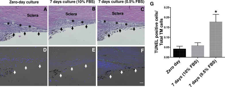
Preservation of tissue structural integrity and cell viability is crucial for developing an ex- vivo tissue culture model. Using H&E staining and the TUNEL assay (Fig 2), we first examined whether TM structural integrity and cellularity is maintained after 7 days in an ex-vivo culture conditions. We cultured the corneoscleral segments with two different serum conditions; i) Standard serum media (10% FBS, suitable for Dex and myocilin treatments) and ii) Low serum media (0.5% FBS, suitable for TGFβ2 treatment). H&E analysis revealed no significant changes in the TM structural morphology among the tested groups (Fig 2, upper panel). A small number of TUNEL-positive cells were observed in the TM at zero-day cultured corneoscleral segments. The numbers of TUNEL-positive TM cells were identical between the zero-day and 7-day cultured quadrants with standard serum conditions. On the contrary, a significant increase in TUNEL-positive cells were observed in the TM region with low serum conditions. The numbers of TUNEL-positive cells were higher in the scleral tissue compared to the TM region (Fig 2, lower panel).

Fig 2. TM structural integrity is maintained in an ex-vivo cultured corneoscleral segment.

Fig 2

H&E staining (upper panel) and TUNEL assay (lower panel) was performed in human corneoscleral quadrants cultured at zero days (A&D), 7 days with 10% FBS (B&E) and 7 days with 0.5% FBS (C&F) containing medium. H&E staining revealed no obvious morphological changes after 7-days of culture compared to zero-days of culture. The total number of TUNEL positive cells over TM cells (DAPI positive) in TM region were shown graphically (G). No difference in TUNEL-positive TM cells was observed between the zero- day and 7-day cultured group with 10% FBS. A significant increase in the TUNEL- positive cells was observed in quadrants cultured with 0.5% FBS. (n = 4 biological replicates per group, one-way ANOVA, *p<0.05), scale bar is 50μm. Arrows indicate the TM region; asterisks represent the Schlemm’s canal.

Dex- or TGFβ2-treatment increases TM cell apoptosis in an ex-vivo cultured human corneoscleral segments

Progressive loss of TM cells is associated with POAG [44, 45]. We next determined whether Dex- or TGFβ2-treatment leads to accelerated TM cell death in cultured human corneoscleral segments. Cultured human corneoscleral segments were treated with Dex or TGFβ2 along with appropriate controls. H&E staining revealed normal iridocorneal angle tissues including intact TM and SC. Interestingly, we observed significantly increased TUNEL-positive TM cells in human corneoscleral quadrants treated with Dex or TGFβ2 compared to their respective controls (Fig 3).

Fig 3. Dex- or TGFβ2-treatment leads to increased apoptosis of TM cells.

Fig 3

H&E staining (left panel) and TUNEL assay (right panel) of cultured corneoscleral segments treated with either Dex (100nM) (B&F) or TGFβ2 (5ng/ml) (D&H) is compared with corresponding quadrants treated with vehicle (A&E or C&G) for 7-days. The total number of TUNEL-positive cells in the TM region were shown graphically (I). Increased TUNEL-positive (green) cells at the TM region was observed in both Dex- and TGFβ2-treated groups. (n = 3 biological replicates per group), scale bar is 50μm. Arrows indicate the TM region; asterisks represent the Schlemm’s canal. N = 3 each; one-way ANOVA; *p<0.05, ***p<0.0001.

Dex treatment increases ECM deposition and ER stress markers in the TM of cultured corneoscleral segments

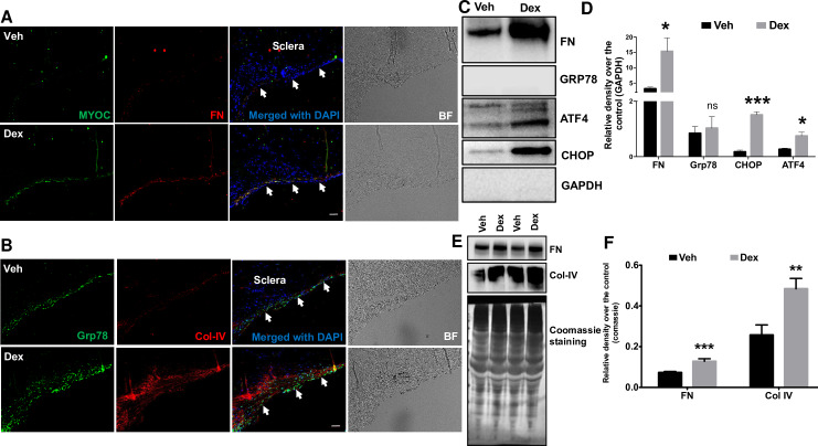
Our previous studies demonstrated that increased ECM deposition and induction of ER stress is associated with human glaucomatous TM tissues and in the TM of mouse models of Dex-induced ocular hypertension [8, 30, 31, 46]. Moreover, Dex is known to increase myocilin expression [8, 47]. Here, we examined whether similar changes can be induced by Dex treatment in the TM of cultured human corneoscleral segments. Immunohistochemical analysis revealed increased expression of myocilin, fibronectin, collagen IV (ECM markers) and GRP78 (ER stress marker) in Dex-treated corneoscleral quadrants compared to the vehicle-treated controls (Fig 4A and 4B). Western blot and densitometric analysis for ECM and ER stress markers in TM tissue lysates and conditioned medium also revealed that Dex treatment increased ECM (fibronectin and Col-IV) and ER stress (ATF4, CHOP and GRP78) proteins (Fig 4C–4F). However, despite the increasing trend, GRP78 expression in the Dex-treated TM was not significant when compared to control. We next examined whether TM cellularity is a contributing factor in Dex-induced changes in the TM of cultured corneoscleral segments. The number of DAPI-stained cells in the TM region were counted and no significant difference between vehicle (121±13.7) and Dex (133.8±8.1) treated groups (n = 5, two-tailed student’s t-test) was found.

Fig 4. Increased ECM accumulation and ER stress induction in the TM of Dex-treated cultured corneoscleral segments.

Fig 4

A) Immunostaining for myocilin and fibronectin, B) GRP78 (ER stress marker) and collagen IV in quadrants cultured for 7 days treated with the vehicle (0.1% ethanol) and Dex (100nM). Dex treatment prominently increased myocilin, fibronectin, collagen IV and GRP78 staining in the TM region. (n = 4 biological replicates, scale bar is 50μm). Western blot and densitometric analysis for FN (ECM marker) and ATF4, CHOP and GRP78 (ER stress markers) in TM tissue lysates (C-D) of vehicle- and Dex- (100nM) treated cultured corneoscleral segments. Dex treatment led to a significant increase in the ECM marker, FN (n = 3 biological replicates) and ER stress markers CHOP and ATF4 but not GRP78 (n = 4 biological replicates). Note that densitometric analysis included only Dex responders. Similarly, conditioned medium (E-F) from Dex-treated corneoscleral segments showed a significant increase in ECM proteins FN and Col IV (n = 8); unpaired t-test, *P<0.05, **P<0.01, ***P<0.001). Arrows indicate the TM region.

TGFβ2 treatment induces ECM deposition in the TM of ex-vivo cultured human corneoscleral segments

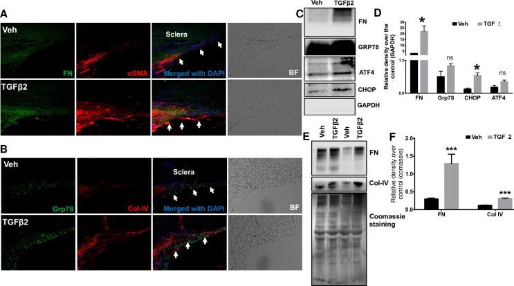
TGFβ2 is known to alter production, degradation and composition of ECM in TM cells [35, 4851]. Several studies have reported increased ECM deposition in human TM cells treated with TGFβ2 [35, 50, 51]. Increased fibronectin levels were observed in ex-vivo human anterior segment perfusates treated with TGFβ2 [35]. Here, we examined whether TGFβ2 treatment also increased ECM deposition in the TM of cultured human corneoscleral segments by analyzing fibronectin and collagen-IV levels, the major ECM components of the TM. Immunohistochemical analysis indicated an increase in fibronectin, collagen-IV (ECM proteins) and GRP78 (ER stress marker) staining in the TM of cultured corneoscleral quadrants treated with TGFβ2 compared to vehicle-treated controls (Fig 5A and 5B). Consistent with previous reports [35, 52], increased α-smooth muscle actin (αSMA) staining was observed in the TM of the TGFβ2-treated group (Fig 5A). Western blot and densitometric analysis on TM tissue lysates (Fig 5C and 5D) obtained from cultured corneoscleral segments revealed a significant increase in the ECM marker, fibronectin and ER stress marker, CHOP. Although ER stress markers GRP78 and ATF4 showed an increasing trend in expression compared to control, the difference was not significant. Furthermore, conditioned medium (Fig 5E and 5F) of cultured corneoscleral quadrants treated with TGFβ2 revealed a significant increase in ECM proteins fibronectin and collagen-IV (ECM proteins). The number of DAPI stained cells in the TM region were not altered significantly between vehicle (119.8±10.5) and TGFβ2 (126.2±10.1) treated samples (n = 5, two-tailed student’s t-test).

Fig 5. Increased ECM accumulation in the TM of TGFβ2-treated cultured corneoscleral segments.

Fig 5

(A and B) Immunostaining for fibronectin (FN), collagen IV (Col-IV), αSMA and GRP78 in vehicle and TGFβ2 (5ng/ml) treated cultured corneoscleral segments. (n = 4 biological replicates, scale bar is 50μm). Western blot and densitometric analysis for FN, Col-IV (ECM markers), ATF4, CHOP, GRP78 in the TM tissue lysates (C-D) and conditioned medium (E-F) of vehicle and TGFβ2-treated cultured corneoscleral quadrants (n = 4 biological replicates for lysates and n = 8 for the conditioned medium), unpaired t-test, *P<0.05, **P<0.01, ***P<0.001. Arrows indicate the TM region.

Lentivirus-mediated expression of mutant myocilin increases ECM deposition and ER stress induction in the TM

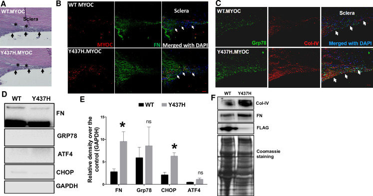
We have previously reported that expression of the mutant form of human myocilin (Y437H) in human TM cells and in the TM of transgenic mice resulted in excessive ECM deposition and ER stress induction [11, 13, 53]. Therefore, we examined whether lentivirus-mediated expression of mutant myocilin protein (Y437H) induces ER stress and ECM deposition in the TM of cultured corneoscleral segments. Lentivirus-mediated transduction of corneoscleral segments showed no adverse effects on TM morphology as determined by H&E analysis (Fig 6A). Transduction of corneoscleral quadrants with WT and mutant myocilin-expressing lentiviral particles resulted in expression of myocilin protein in the TM as determined by immunohistochemistry. However, the inability of the TM cells to secrete the mutant form of myocilin resulted in increased staining of myocilin in the TM of mutant myocilin transduced quadrants compared to WT myocilin-transduced controls (Fig 6B). This finding is consistent with our previous observations and indicates that mutant myocilin is secretory incompetent and accumulates inside the TM cells [11, 13, 53]. Immunohistochemical analysis showed an increase in FN, Col-IV and GRP78 in the TM of corneoscleral quadrants transduced with mutant myocilin compared to WT controls (Fig 6B and 6C). Western blot and densitometric analysis on TM tissue lysates showed a significant increase in the ECM marker FN and the ER stress marker CHOP in mutant myocilin-transduced quadrants compared to WT control (Fig 6D and 6E). Although the difference was not significant, we also observed an increasing trend in the expression of GRP78 and ATF4 in TM tissues of mutant myocilin-transduced quadrants. Western blot analysis of conditioned medium revealed an increase in fibronectin and Col-IV protein levels in the mutant myocilin-transduced corneoscleral segments (Fig 6F). No significant difference in the number of DAPI stained cells in the TM region was observed between WT (139.2±8.1) and mutant myocilin (119±10.1) transduced quadrants (n = 5, two-tailed unpaired student’s t-test).

Fig 6. Lentiviral expression of mutant myocilin induces ER stress and ECM changes in the TM of cultured corneoscleral segments.

Fig 6

The cultured corneoscleral quadrants were transduced with FTS tagged (FLAG & S tag) WT myocilin or mutant myocilin (Y437H) expressing lentiviral particles (1ml of lentivirus supernatant) for 7 days. A) H&E staining and (B&C) immunostaining for myocilin, FN, GRP78 and Col-IV in cultured corneoscleral segments transduced with WT or mutant myocilin. Increased myocilin staining was observed in the TM of mutant myocilin-transduced quadrants compared to WT myocilin. In addition, increased FN and Col-IV staining indicate more ECM accumulation in the TM of mutant myocilin-transduced quadrants (n = 3 biological replicates, scale bar is 50μm). Western blot and densitometric analysis of TM tissue lysates (D-E) and conditioned medium (F) obtained from cultured quadrants transduced with WT and mutant myocilin lentiviral expression vectors. A significant increase in the ECM marker FN (n = 6 biological replicates) and the ER stress marker CHOP (n = 3 biological replicates) was observed in mutant myocilin-transduced TM tissue lysates. Similarly, conditioned medium (F) from mutant myocilin-treated corneoscleral segments showed increases in ECM proteins FN and Col IV. Moreover, WT myocilin was detected in conditioned media of WT myocilin-transduced quadrants while no myocilin was detected in quadrants expressing mutant myocilin indicating that expression of mutant myocilin inhibits its secretion and accumulates in the TM cells. Unpaired t-test, *P<0.05, **P<0.01, ***P<0.001. Arrows indicate the TM region.

Discussion

An ex vivo human anterior segment perfusion culture model has been used to study the effects of drugs on glaucomatous pathophysiology [38, 41, 5456]. This model requires intact human eyes for studying physiological parameters such as IOP homeostasis and aqueous humor dynamics. However, the high processing cost, limited availability of intact human eyes and higher failure rate has restricted the wider use of this model in glaucoma research. In this study, we developed an ex-vivo cultured human corneoscleral segment model to study glaucomatous TM damage. The human corneoscleral segment tissue is a gift that is given post-mortem for corneal transplant. Once excised, the tissue is tested to determine eligibility for transplantation. Due to stringent eligibility criteria, the majority of corneoscleral segments are deemed ineligible for transplant. These ineligible tissues are typically discarded due to low demand for research. The processing fees associated with obtaining corneoscleral segments from the eye bank is considerably lower compared to obtaining whole globes. An excised human corneoscleral segment is comprised of cornea, sclera, and the TM rim. Ciliary body, iris, and lens are promptly removed post-excision. Corneoscleral segments without an intact TM rim were not used for this study.

To determine optimal culture conditions, corneoscleral segments were divided into quadrants and cultured under standard and low serum conditions for 7 days. The structural integrity of the TM was preserved in stationary culture. Histochemical analysis of the quadrants showed no difference in TM morphology between standard (10% FBS) and low serum (0.5% FBS) conditions (Fig 2). TUNEL staining revealed a significant increase in TM cell death at low serum compared to the standard serum conditions. Interestingly, the number of TUNEL-positive cells were higher in adjacent scleral tissue than that of the TM region in quadrants cultured at low serum (Fig 2). Unlike the avascular TM, the vascular sclera appears to be highly serum dependent. Serum starvation can lead to changes in scleral cells, which as a consequence may adversely affect adjacent TM cells. Therefore, we decided to use 10% serum media to avoid any indirect effects of serum starvation on the TM or the adjacent tissues. The role of adjacent tissues should also be taken into consideration when interpreting the effects of drugs on the TM of corneoscleral quadrants. Previous studies with the anterior segment perfusion culture model have used serum-free media for perfusion [34, 38, 39, 41, 57]. Continuous perfusion of fresh media provides constant supply of nutrients to the TM cells eliminating the need for serum in perfusion tissue cultures. The serum dependency of the corneoscleral quadrants in our stationary culture may be due to the lack of continuous perfusion of fresh media. In our study, we observed that serum supplementation was beneficial for the overall health of the tissue. However, we used low serum conditions (0.5% FBS) while treating quadrants with TGFβ2. Considering that serum contains growth factors including TGFβ2, increasing serum concentration might saturate the effect of TGFβ2 on the TM of corneoscleral quadrants. In support of this, we observed that TGFβ2 had minimum effects on ECM proteins when tissues were cultured at high serum compared to low serum (2% and 5% FBS). It is important to note that the prolonged time in culture (4–7 days) before receipt of these corneas may negatively affect tissue viability and cause cell death. However, previous studies have shown that preculture of tissues in stationary media for 7 days prior to perfusion aids in tissue stabilization [39, 55].

Progressive loss of TM cells has been reported with ageing and this loss appears to be accelerated in POAG [44, 45]. Several factors associated with glaucomatous pathology may play a role in accelerating TM cell loss and reduced tissue cellularity. For example, Clark et al. (1995) and Fleenor et al. (2006) observed an increase in TM cell loss in ex vivo perfused human anterior segments with Dex and TGFβ2 treatment respectively [34, 35].

A hallmark feature for a glaucoma model is its ability to mimic the glaucomatous TM pathology in response to glaucoma causing factors. The cultured human corneoscleral quadrants were treated with 3 different glaucoma factors to determine their effect on TM pathology. As expected, Dex treatment of anterior segments resulted in an increased ECM deposition in the TM. Furthermore, Dex treatment also induced ER stress in the TM of cultured human corneoscleral quadrants. A 30% IOP responsive rate was reported in Dex treated ex-vivo perfused human anterior segments [34]. In the present study, we did not evaluate the response rate as corneoscleral segment culture model is unfeasible to IOP and outflow measurements. Instead, we observed Dex-induced ECM changes in the TM only in 50% of cultured corneoscleral segments. Treatment with TGFB2, a glaucoma associated growth factor, resulted in an increased ECM deposition in the TM of corneoscleral segments. Unlike Dex, TGFB2-induced ECM changes in the TM were observed in all cultured quadrants. WT and mutant myocilin were expressed in the TM via lentiviral transduction. Consistent with our previous findings [13, 53], expression of mutant myocilin led to intracellular accumulation, induction of ER stress and increased deposition of ECM proteins in the TM of corneoscleral segments.

We observed that glaucoma causing factors significantly increased chronic ER stress markers, CHOP and ATF4 but not GRP78 in the TM tissues of corneoscleral segments. Although GRP78 expression showed an increasing trend, the difference compared to control was not significant. GRP78 is a key activator of ER stress sensors (IRE1, PERK and ATF6) that activates the UPR pathway. GRP78 is normally bound to these ER stress sensors, thus keeping them inactivated. Under stress conditions or accumulation of misfolded proteins, GRP78 is released from these sensors, resulting into activation of ER stress sensors and the UPR pathway [58]. Since GRP78 is expressed abundantly in TM tissues, a small increase after treatment with glaucoma factors may be sufficient to activate UPR pathway. This is apparent from significantly increased ATF4 and CHOP, which are downstream of GRP78 and are considered classical markers of chronic ER stress.

Although the human ex-vivo corneoscleral segment model is a promising prospect in modeling human glaucoma condition, there are certain limitations to be considered. Since the primary use of these corneoscleral segments is for human transplant, the portion of sclera posterior to the limbus is removed during the excision process. In addition to sclera, ciliary body, iris, and lens are also removed. The removal of scleral tissue reduces the overall diameter of the corneoscleral segments, rendering them unusable for perfusion studies involving aqueous humor dynamics and IOP homeostasis. Furthermore, the removal of ciliary body may affect the TM contractile tone and can lead to biochemical changes in the TM cells. Of note, the ciliary body is also removed even in an ex-vivo human anterior segment perfusion culture to avoid the blockage of outflow with pigment originating from the degenerating pigmented cells from the ciliary body [57]. Unlike intact human eyes used for the perfusion culture (which can be delivered within 24-48hrs of the donor death) the human corneoscleral segments were delivered between 5–7 days. Despite this, we did not observe any notable changes in the TM morphology in these corneoscleral segments. The high variability between different donors is a common issue in research involving post-mortem human tissues. The corneoscleral segments used in this model can be divided into quadrants to perform control and experimental treatments on the same eye, thereby decreasing variability associated with contralateral control and reducing the overall cost of research. However, it is important to note that TM cellularity may differ along the circumferential length of the TM within the same eye. Furthermore, it is now known that the AH outflow through the TM is segmental with some areas along the circumferential TM length experiencing higher flow rate compared to other areas [40, 59, 60]. Therefore, tissues from multiple donors must be tested in order to account for spatial differences in cellularity and segmental flow along the TM. Despite these limitations, the ex-vivo cultured human corneoscleral explant model efficiently simulates glaucomatous pathology, and perhaps provides an attractive cost-efficient model for initial screening to study effects on the TM.

A similar ex-vivo culture method has been reported previously which utilized TM tissues recovered from discarded corneal rims after surgical corneal transplantation [61]. We have refined culture conditions over this previous study. Our current model differs from the previous study in several aspects: 1) We validate TM tissue integrity and cellularity of the cultured corneoscleral segment using H&E staining and TUNEL assays respectively. 2) We show the importance of serum to maintain the tissue integrity of corneoscleral segments in stationary culture. 3) In our study, we treated corneoscleral tissues with biologically relevant doses of Dex (100 nM) and TGFβ2 (5ng/ml) to study their effects on TM tissue in ex vivo cultured human corneoscleral segments while Gonzalez et al, 2012 treated TM rings with higher doses of Dex (250nM) and TGFβ1 (50nM). Of note, increased levels of TGFβ2 but not TGFβ1 in the aqueous humor are associated with ECM remodeling in the TM [14, 6264]. 4) In addition to Dex and TGFβ2, we have shown that effects of mutant myocilin can be studied in intact TM tissues using this model. Considering limited availability of MYOC-associated POAG donor tissues, this approach offers the possibility of understanding the pathology behind MYOC-associated glaucoma. 5) Finally, we also examined the ER stress markers induced by the glaucoma factors.

Supporting information

S1 Data

(PDF)

Acknowledgments

The authors thank the Lions Eye Institute (Tampa, Florida) for supplying the corneoscleral segments, and Sandra Neubauer for expert technical assistance. These studies were supported by the National Institutes of Health; EY028616 (GSZ) and EY026177 (GSZ).

Data Availability

All relevant data are within the manuscript and its Supporting Information files.

Funding Statement

These studies were supported by the National Institutes of Health; EY028616 (GSZ) and EY026177 (GSZ).

References

  • 1.Quigley HA, Broman AT. The number of people with glaucoma worldwide in 2010 and 2020. Br J Ophthalmol. 2006;90(3):262–7. Epub 2006/02/21. 10.1136/bjo.2005.081224 [DOI] [PMC free article] [PubMed] [Google Scholar]
  • 2.Rosenthal J, Leske MC. Open-angle glaucoma risk factors applied to clinical area. J Am Optom Assoc. 1980;51(11):1017–24. Epub 1980/11/01. . [PubMed] [Google Scholar]
  • 3.Rohen JW. Why is intraocular pressure elevated in chronic simple glaucoma? Anatomical considerations. Ophthalmology. 1983;90(7):758–65. Epub 1983/07/01. 10.1016/s0161-6420(83)34492-4 . [DOI] [PubMed] [Google Scholar]
  • 4.Kwon YH, Fingert JH, Kuehn MH, Alward WL. Primary open-angle glaucoma. N Engl J Med. 2009;360(11):1113–24. Epub 2009/03/13. 10.1056/NEJMra0804630 [DOI] [PMC free article] [PubMed] [Google Scholar]
  • 5.Tanna AP, Johnson M. Rho Kinase Inhibitors as a Novel Treatment for Glaucoma and Ocular Hypertension. Ophthalmology. 2018;125(11):1741–56. Epub 2018/07/17. 10.1016/j.ophtha.2018.04.040 [DOI] [PMC free article] [PubMed] [Google Scholar]
  • 6.Kahook MY, Serle JB, Mah FS, Kim T, Raizman MB, Heah T, et al. Long-term Safety and Ocular Hypotensive Efficacy Evaluation of Netarsudil Ophthalmic Solution: Rho Kinase Elevated IOP Treatment Trial (ROCKET-2). Am J Ophthalmol. 2019;200:130–7. Epub 2019/01/18. 10.1016/j.ajo.2019.01.003 . [DOI] [PubMed] [Google Scholar]
  • 7.Rao PV, Pattabiraman PP, Kopczynski C. Role of the Rho GTPase/Rho kinase signaling pathway in pathogenesis and treatment of glaucoma: Bench to bedside research. Exp Eye Res. 2017;158:23–32. Epub 2016/09/07. 10.1016/j.exer.2016.08.023 [DOI] [PMC free article] [PubMed] [Google Scholar]
  • 8.Stone EM, Fingert JH, Alward WL, Nguyen TD, Polansky JR, Sunden SL, et al. Identification of a gene that causes primary open angle glaucoma. Science. 1997;275(5300):668–70. Epub 1997/01/31. 10.1126/science.275.5300.668 . [DOI] [PubMed] [Google Scholar]
  • 9.Alward WL, Fingert JH, Coote MA, Johnson AT, Lerner SF, Junqua D, et al. Clinical features associated with mutations in the chromosome 1 open-angle glaucoma gene (GLC1A). N Engl J Med. 1998;338(15):1022–7. Epub 1998/04/16. 10.1056/NEJM199804093381503 . [DOI] [PubMed] [Google Scholar]
  • 10.Jacobson N, Andrews M, Shepard AR, Nishimura D, Searby C, Fingert JH, et al. Non-secretion of mutant proteins of the glaucoma gene myocilin in cultured trabecular meshwork cells and in aqueous humor. Hum Mol Genet. 2001;10(2):117–25. Epub 2001/01/12. 10.1093/hmg/10.2.117 . [DOI] [PubMed] [Google Scholar]
  • 11.Zode GS, Kuehn MH, Nishimura DY, Searby CC, Mohan K, Grozdanic SD, et al. Reduction of ER stress via a chemical chaperone prevents disease phenotypes in a mouse model of primary open angle glaucoma. J Clin Invest. 2011;121(9):3542–53. 10.1172/JCI58183 [DOI] [PMC free article] [PubMed] [Google Scholar]
  • 12.Joe MK, Sohn S, Hur W, Moon Y, Choi YR, Kee C. Accumulation of mutant myocilins in ER leads to ER stress and potential cytotoxicity in human trabecular meshwork cells. Biochem Biophys Res Commun. 2003;312(3):592–600. Epub 2003/12/19. 10.1016/j.bbrc.2003.10.162 . [DOI] [PubMed] [Google Scholar]
  • 13.Zode GS, Bugge KE, Mohan K, Grozdanic SD, Peters JC, Koehn DR, et al. Topical ocular sodium 4-phenylbutyrate rescues glaucoma in a myocilin mouse model of primary open-angle glaucoma. Invest Ophthalmol Vis Sci. 2012;53(3):1557–65. 10.1167/iovs.11-8837 [DOI] [PMC free article] [PubMed] [Google Scholar]
  • 14.Inatani M, Tanihara H, Katsuta H, Honjo M, Kido N, Honda Y. Transforming growth factor-beta 2 levels in aqueous humor of glaucomatous eyes. Graefes Arch Clin Exp Ophthalmol. 2001;239(2):109–13. Epub 2001/05/25. 10.1007/s004170000241 . [DOI] [PubMed] [Google Scholar]
  • 15.Picht G, Welge-Luessen U, Grehn F, Lutjen-Drecoll E. Transforming growth factor beta 2 levels in the aqueous humor in different types of glaucoma and the relation to filtering bleb development. Graefes Arch Clin Exp Ophthalmol. 2001;239(3):199–207. Epub 2001/06/19. 10.1007/s004170000252 . [DOI] [PubMed] [Google Scholar]
  • 16.Ochiai Y, Ochiai H. Higher concentration of transforming growth factor-beta in aqueous humor of glaucomatous eyes and diabetic eyes. Jpn J Ophthalmol. 2002;46(3):249–53. Epub 2002/06/14. 10.1016/s0021-5155(01)00523-8 . [DOI] [PubMed] [Google Scholar]
  • 17.Tovar-Vidales T, Clark AF, Wordinger RJ. Transforming growth factor-beta2 utilizes the canonical Smad-signaling pathway to regulate tissue transglutaminase expression in human trabecular meshwork cells. Exp Eye Res. 2011;93(4):442–51. Epub 2011/07/05. 10.1016/j.exer.2011.06.011 [DOI] [PMC free article] [PubMed] [Google Scholar]
  • 18.Clark AF. Basic sciences in clinical glaucoma: steroids, ocular hypertension, and glaucoma. J Glaucoma. 1995;4(5):354–69. Epub 1995/10/01. . [PubMed] [Google Scholar]
  • 19.Razeghinejad MR, Katz LJ. Steroid-induced iatrogenic glaucoma. Ophthalmic Res. 2012;47(2):66–80. Epub 2011/07/16. 10.1159/000328630 . [DOI] [PubMed] [Google Scholar]
  • 20.Filla MS, Schwinn MK, Nosie AK, Clark RW, Peters DM. Dexamethasone-associated cross-linked actin network formation in human trabecular meshwork cells involves beta3 integrin signaling. Invest Ophthalmol Vis Sci. 2011;52(6):2952–9. Epub 2011/01/29. 10.1167/iovs.10-6618 [DOI] [PMC free article] [PubMed] [Google Scholar]
  • 21.Johnson D, Gottanka J, Flugel C, Hoffmann F, Futa R, Lutjen-Drecoll E. Ultrastructural changes in the trabecular meshwork of human eyes treated with corticosteroids. Arch Ophthalmol. 1997;115(3):375–83. Epub 1997/03/01. 10.1001/archopht.1997.01100150377011 . [DOI] [PubMed] [Google Scholar]
  • 22.Jones R, 3rd, Rhee DJ. Corticosteroid-induced ocular hypertension and glaucoma: a brief review and update of the literature. Curr Opin Ophthalmol. 2006;17(2):163–7. Epub 2006/03/23. 10.1097/01.icu.0000193079.55240.18 . [DOI] [PubMed] [Google Scholar]
  • 23.Rohen JW. [Fine structural changes in the trabecular meshwork of the human eye in different forms of glaucoma (author's transl)]. Klin Monbl Augenheilkd. 1973;163(4):401–10. Epub 1973/10/01. . [PubMed] [Google Scholar]
  • 24.Shirato S, Murphy CG, Bloom E, Franse-Carman L, Maglio MT, Polansky JR, et al. Kinetics of phagocytosis in trabecular meshwork cells. Flow cytometry and morphometry. Invest Ophthalmol Vis Sci. 1989;30(12):2499–511. . [PubMed] [Google Scholar]
  • 25.Tawara A, Tou N, Kubota T, Harada Y, Yokota K. Immunohistochemical evaluation of the extracellular matrix in trabecular meshwork in steroid-induced glaucoma. Graefes Arch Clin Exp Ophthalmol. 2008;246(7):1021–8. Epub 2008/04/04. 10.1007/s00417-008-0800-0 . [DOI] [PubMed] [Google Scholar]
  • 26.Yun AJ, Murphy CG, Polansky JR, Newsome DA, Alvarado JA. Proteins secreted by human trabecular cells. Glucocorticoid and other effects. Invest Ophthalmol Vis Sci. 1989;30(9):2012–22. Epub 1989/09/01. . [PubMed] [Google Scholar]
  • 27.Zhou L, Li Y, Yue BY. Glucocorticoid effects on extracellular matrix proteins and integrins in bovine trabecular meshwork cells in relation to glaucoma. Int J Mol Med. 1998;1(2):339–46. Epub 1998/12/16. . [PubMed] [Google Scholar]
  • 28.Clark AF, Wordinger RJ. The role of steroids in outflow resistance. Exp Eye Res. 2009;88(4):752–9. Epub 2008/11/04. 10.1016/j.exer.2008.10.004 . [DOI] [PubMed] [Google Scholar]
  • 29.Armaly MF, Becker B. Intraocular pressure response to topical corticosteroids. Fed Proc. 1965;24(6):1274–8. Epub 1965/11/01. . [PubMed] [Google Scholar]
  • 30.Beckel JM, Argall AJ, Lim JC, Xia J, Lu W, Coffey EE, et al. Mechanosensitive release of adenosine 5'-triphosphate through pannexin channels and mechanosensitive upregulation of pannexin channels in optic nerve head astrocytes: a mechanism for purinergic involvement in chronic strain. Glia. 2014;62(9):1486–501. 10.1002/glia.22695 [DOI] [PMC free article] [PubMed] [Google Scholar]
  • 31.Patel GC, Phan TN, Maddineni P, Kasetti RB, Millar JC, Clark AF, et al. Dexamethasone-Induced Ocular Hypertension in Mice: Effects of Myocilin and Route of Administration. Am J Pathol. 2017;187(4):713–23. Epub 2017/02/09. 10.1016/j.ajpath.2016.12.003 [DOI] [PMC free article] [PubMed] [Google Scholar]
  • 32.Shepard AR, Millar JC, Pang IH, Jacobson N, Wang WH, Clark AF. Adenoviral gene transfer of active human transforming growth factor-{beta}2 elevates intraocular pressure and reduces outflow facility in rodent eyes. Invest Ophthalmol Vis Sci. 2010;51(4):2067–76. Epub 2009/12/05. 10.1167/iovs.09-4567 . [DOI] [PubMed] [Google Scholar]
  • 33.Clark AF, Brotchie D, Read AT, Hellberg P, English-Wright S, Pang IH, et al. Dexamethasone alters F-actin architecture and promotes cross-linked actin network formation in human trabecular meshwork tissue. Cell Motil Cytoskeleton. 2005;60(2):83–95. Epub 2004/12/14. 10.1002/cm.20049 . [DOI] [PubMed] [Google Scholar]
  • 34.Clark AF, Wilson K, de Kater AW, Allingham RR, McCartney MD. Dexamethasone-induced ocular hypertension in perfusion-cultured human eyes. Invest Ophthalmol Vis Sci. 1995;36(2):478–89. Epub 1995/02/01. . [PubMed] [Google Scholar]
  • 35.Fleenor DL, Shepard AR, Hellberg PE, Jacobson N, Pang IH, Clark AF. TGFbeta2-induced changes in human trabecular meshwork: implications for intraocular pressure. Invest Ophthalmol Vis Sci. 2006;47(1):226–34. Epub 2005/12/31. 10.1167/iovs.05-1060 . [DOI] [PubMed] [Google Scholar]
  • 36.Clark AF, Wilson K, McCartney MD, Miggans ST, Kunkle M, Howe W. Glucocorticoid-induced formation of cross-linked actin networks in cultured human trabecular meshwork cells. Invest Ophthalmol Vis Sci. 1994;35(1):281–94. Epub 1994/01/01. . [PubMed] [Google Scholar]
  • 37.Raghunathan VK, Morgan JT, Park SA, Weber D, Phinney BS, Murphy CJ, et al. Dexamethasone Stiffens Trabecular Meshwork, Trabecular Meshwork Cells, and Matrix. Invest Ophthalmol Vis Sci. 2015;56(8):4447–59. Epub 2015/07/22. 10.1167/iovs.15-16739 [DOI] [PMC free article] [PubMed] [Google Scholar]
  • 38.Pang IH, McCartney MD, Steely HT, Clark AF. Human ocular perfusion organ culture: a versatile ex vivo model for glaucoma research. J Glaucoma. 2000;9(6):468–79. Epub 2000/12/29. 10.1097/00061198-200012000-00009 . [DOI] [PubMed] [Google Scholar]
  • 39.Acott TS, Kingsley PD, Samples JR, Van Buskirk EM. Human trabecular meshwork organ culture: morphology and glycosaminoglycan synthesis. Invest Ophthalmol Vis Sci. 1988;29(1):90–100. Epub 1988/01/01. . [PubMed] [Google Scholar]
  • 40.Vranka JA, Acott TS. Pressure-induced expression changes in segmental flow regions of the human trabecular meshwork. Exp Eye Res. 2017;158:67–72. Epub 2016/06/24. 10.1016/j.exer.2016.06.009 [DOI] [PMC free article] [PubMed] [Google Scholar]
  • 41.Johnson DH, Tschumper RC. Human trabecular meshwork organ culture. A new method. Invest Ophthalmol Vis Sci. 1987;28(6):945–53. Epub 1987/06/01. . [PubMed] [Google Scholar]
  • 42.Gain P, Jullienne R, He Z, Aldossary M, Acquart S, Cognasse F, et al. Global Survey of Corneal Transplantation and Eye Banking. JAMA Ophthalmol. 2016;134(2):167–73. Epub 2015/12/04. 10.1001/jamaophthalmol.2015.4776 . [DOI] [PubMed] [Google Scholar]
  • 43.Liaboe C VJ, Schmidt GA, Greiner MA. Corneal Transplantation: From Donor to Recipient. 2015. [Google Scholar]
  • 44.Alvarado J, Murphy C, Polansky J, Juster R. Age-related changes in trabecular meshwork cellularity. Invest Ophthalmol Vis Sci. 1981;21(5):714–27. Epub 1981/11/01. . [PubMed] [Google Scholar]
  • 45.Alvarado J, Murphy C, Juster R. Trabecular meshwork cellularity in primary open-angle glaucoma and nonglaucomatous normals. Ophthalmology. 1984;91(6):564–79. Epub 1984/06/01. 10.1016/s0161-6420(84)34248-8 . [DOI] [PubMed] [Google Scholar]
  • 46.Kasetti RB, Maddineni P, Millar JC, Clark AF, Zode GS. Increased synthesis and deposition of extracellular matrix proteins leads to endoplasmic reticulum stress in the trabecular meshwork. Sci Rep. 2017;7(1):14951 Epub 2017/11/04. 10.1038/s41598-017-14938-0 [DOI] [PMC free article] [PubMed] [Google Scholar]
  • 47.Nguyen TD, Chen P, Huang WD, Chen H, Johnson D, Polansky JR. Gene structure and properties of TIGR, an olfactomedin-related glycoprotein cloned from glucocorticoid-induced trabecular meshwork cells. J Biol Chem. 1998;273(11):6341–50. 10.1074/jbc.273.11.6341 . [DOI] [PubMed] [Google Scholar]
  • 48.Tovar-Vidales T, Fitzgerald AM, Clark AF, Wordinger RJ. Transforming growth factor-beta2 induces expression of biologically active bone morphogenetic protein-1 in human trabecular meshwork cells. Invest Ophthalmol Vis Sci. 2013;54(7):4741–8. Epub 2013/06/22. 10.1167/iovs.13-12203 [DOI] [PMC free article] [PubMed] [Google Scholar]
  • 49.Welge-Lussen U, May CA, Lutjen-Drecoll E. Induction of tissue transglutaminase in the trabecular meshwork by TGF-beta1 and TGF-beta2. Invest Ophthalmol Vis Sci. 2000;41(8):2229–38. . [PubMed] [Google Scholar]
  • 50.Sethi A, Jain A, Zode GS, Wordinger RJ, Clark AF. Role of TGFbeta/Smad signaling in gremlin induction of human trabecular meshwork extracellular matrix proteins. Invest Ophthalmol Vis Sci. 2011;52(8):5251–9. Epub 2011/06/07. 10.1167/iovs.11-7587 [DOI] [PMC free article] [PubMed] [Google Scholar]
  • 51.Wordinger RJ, Fleenor DL, Hellberg PE, Pang IH, Tovar TO, Zode GS, et al. Effects of TGF-beta2, BMP-4, and gremlin in the trabecular meshwork: implications for glaucoma. Invest Ophthalmol Vis Sci. 2007;48(3):1191–200. Epub 2007/02/28. 10.1167/iovs.06-0296 . [DOI] [PubMed] [Google Scholar]
  • 52.Torrejon KY, Papke EL, Halman JR, Bergkvist M, Danias J, Sharfstein ST, et al. TGFbeta2-induced outflow alterations in a bioengineered trabecular meshwork are offset by a rho-associated kinase inhibitor. Sci Rep. 2016;6:38319 Epub 2016/12/08. 10.1038/srep38319 [DOI] [PMC free article] [PubMed] [Google Scholar]
  • 53.Kasetti RB, Phan TN, Millar JC, Zode GS. Expression of Mutant Myocilin Induces Abnormal Intracellular Accumulation of Selected Extracellular Matrix Proteins in the Trabecular Meshwork. Invest Ophthalmol Vis Sci. 2016;57(14):6058–69. 10.1167/iovs.16-19610 [DOI] [PMC free article] [PubMed] [Google Scholar]
  • 54.Borras T, Matsumoto Y, Epstein DL, Johnson DH. Gene transfer to the human trabecular meshwork by anterior segment perfusion. Invest Ophthalmol Vis Sci. 1998;39(8):1503–7. Epub 1998/07/11. . [PubMed] [Google Scholar]
  • 55.Bradley JM, Vranka J, Colvis CM, Conger DM, Alexander JP, Fisk AS, et al. Effect of matrix metalloproteinases activity on outflow in perfused human organ culture. Invest Ophthalmol Vis Sci. 1998;39(13):2649–58. Epub 1998/12/18. . [PubMed] [Google Scholar]
  • 56.Johnson DH, Bradley JM, Acott TS. The effect of dexamethasone on glycosaminoglycans of human trabecular meshwork in perfusion organ culture. Invest Ophthalmol Vis Sci. 1990;31(12):2568–71. . [PubMed] [Google Scholar]
  • 57.Gottanka J, Chan D, Eichhorn M, Lutjen-Drecoll E, Ethier CR. Effects of TGF-beta2 in perfused human eyes. Invest Ophthalmol Vis Sci. 2004;45(1):153–8. 10.1167/iovs.03-0796 . [DOI] [PubMed] [Google Scholar]
  • 58.Roussel BD, Kruppa AJ, Miranda E, Crowther DC, Lomas DA, Marciniak SJ. Endoplasmic reticulum dysfunction in neurological disease. Lancet Neurol. 2013;12(1):105–18. Epub 2012/12/15. 10.1016/S1474-4422(12)70238-7 . [DOI] [PubMed] [Google Scholar]
  • 59.Swaminathan SS, Oh DJ, Kang MH, Rhee DJ. Aqueous outflow: segmental and distal flow. J Cataract Refract Surg. 2014;40(8):1263–72. Epub 2014/08/05. 10.1016/j.jcrs.2014.06.020 [DOI] [PMC free article] [PubMed] [Google Scholar]
  • 60.Hann CR, Bahler CK, Johnson DH. Cationic ferritin and segmental flow through the trabecular meshwork. Invest Ophthalmol Vis Sci. 2005;46(1):1–7. Epub 2004/12/30. 10.1167/iovs.04-0800 . [DOI] [PubMed] [Google Scholar]
  • 61.Gonzalez JM Jr., Heur M, Tan JC. Two-photon immunofluorescence characterization of the trabecular meshwork in situ. Invest Ophthalmol Vis Sci. 2012;53(7):3395–404. Epub 2012/04/26. 10.1167/iovs.11-8570 [DOI] [PMC free article] [PubMed] [Google Scholar]
  • 62.Tripathi RC, Li J, Chan WF, Tripathi BJ. Aqueous humor in glaucomatous eyes contains an increased level of TGF-beta 2. Exp Eye Res. 1994;59(6):723–7. Epub 1994/12/01. 10.1006/exer.1994.1158 . [DOI] [PubMed] [Google Scholar]
  • 63.Ozcan AA, Ozdemir N, Canataroglu A. The aqueous levels of TGF-beta2 in patients with glaucoma. Int Ophthalmol. 2004;25(1):19–22. Epub 2004/04/17. 10.1023/b:inte.0000018524.48581.79 . [DOI] [PubMed] [Google Scholar]
  • 64.Kuchtey J, Kunkel J, Burgess LG, Parks MB, Brantley MA Jr., Kuchtey RW. Elevated transforming growth factor beta1 in plasma of primary open-angle glaucoma patients. Invest Ophthalmol Vis Sci. 2014;55(8):5291–7. Epub 2014/07/26. 10.1167/iovs.14-14578 [DOI] [PMC free article] [PubMed] [Google Scholar]

Decision Letter 0

Paloma B Liton

27 Dec 2019

PONE-D-19-32303

Ex-vivo cultured human corneoscleral segment model to study the effects of glaucoma factors on trabecular meshwork

PLOS ONE

Dear Dr. Zode,

Thank you for submitting your manuscript to PLOS ONE. After careful consideration, we feel that it has merit but does not fully meet PLOS ONE’s publication criteria as it currently stands. Therefore, we invite you to submit a revised version of the manuscript that addresses the points raised during the review process.

PLOS ONE now requires that submissions reporting blots or gels include original, uncropped blot/gel image data as a supplement or in a public repository. These requirements apply both to the main figures and to cropped blot/gel images included in Supporting Information. Please provide any missing raw image data for blot/gel results when submiting your revision. 

We would appreciate receiving your revised manuscript by Feb 10 2020 11:59PM. When you are ready to submit your revision, log on to https://www.editorialmanager.com/pone/ and select the 'Submissions Needing Revision' folder to locate your manuscript file.

If you would like to make changes to your financial disclosure, please include your updated statement in your cover letter.

To enhance the reproducibility of your results, we recommend that if applicable you deposit your laboratory protocols in protocols.io, where a protocol can be assigned its own identifier (DOI) such that it can be cited independently in the future. For instructions see: http://journals.plos.org/plosone/s/submission-guidelines#loc-laboratory-protocols

Please include the following items when submitting your revised manuscript:

  • A rebuttal letter that responds to each point raised by the academic editor and reviewer(s). This letter should be uploaded as separate file and labeled 'Response to Reviewers'.

  • A marked-up copy of your manuscript that highlights changes made to the original version. This file should be uploaded as separate file and labeled 'Revised Manuscript with Track Changes'.

  • An unmarked version of your revised paper without tracked changes. This file should be uploaded as separate file and labeled 'Manuscript'.

Please note while forming your response, if your article is accepted, you may have the opportunity to make the peer review history publicly available. The record will include editor decision letters (with reviews) and your responses to reviewer comments. If eligible, we will contact you to opt in or out.

We look forward to receiving your revised manuscript.

Kind regards,

Paloma B. Liton, PhD

Academic Editor

PLOS ONE

Journal Requirements:

1. When submitting your revision, we need you to address these additional requirements.

Please ensure that your manuscript meets PLOS ONE's style requirements, including those for file naming. The PLOS ONE style templates can be found at

http://www.journals.plos.org/plosone/s/file?id=wjVg/PLOSOne_formatting_sample_main_body.pdf and http://www.journals.plos.org/plosone/s/file?id=ba62/PLOSOne_formatting_sample_title_authors_affiliations.pdf

2. We note that your study involved donated tissue/organs. Please provide the following information regarding tissue/organ donors for cases analyzed in your study.

1. Please provide the source(s) of the transplanted tissue/organs used in the study, including the institution name and a non-identifying description of the donor(s).

2. Please state in your response letter and ethics statement whether the transplant cases for this study involved any vulnerable populations; for example, tissue/organs from prisoners, subjects with reduced mental capacity due to illness or age, or minors.

- If a vulnerable population was used, please describe the population, justify the decision to use tissue/organ donations from this group, and clearly describe what measures were taken in the informed consent procedure to assure protection of the vulnerable group and avoid coercion.

- If a vulnerable population was not used, please state in your ethics statement, “None of the transplant donors was from a vulnerable population and all donors or next of kin provided written informed consent that was freely given.”

3. In the Methods, please provide detailed information about the procedure by which informed consent was obtained from organ/tissue donors or their next of kin. In addition, please provide a blank example of the form used to obtain consent from donors, and an English translation if the original is in a different language.

4. Please state whether the donors specifically provided consent for the use of their donated tissue for both transplantation and donation purposes.

5. Please indicate whether the donors were previously registered as organ donors. If tissues/organs were obtained from deceased donors or cadavers, please provide details as to the donors’ cause(s) of death.

6. Please provide the participant recruitment dates and the period during which transplant procedures were done (as month and year).

7. Please discuss whether medical costs were covered or other cash payments were provided to the family of the donor. If so, please specify the value of this support (in local currency and equivalent to U.S. dollars).

8. Please state whether you had access to any identifying information about the donors (e.g. names/addresses) as part of this study."

3.  PLOS ONE now requires that authors provide the original uncropped and unadjusted images underlying all blot or gel results reported in a submission’s figures or Supporting Information files. This policy and the journal’s other requirements for blot/gel reporting and figure preparation are described in detail at https://journals.plos.org/plosone/s/figures#loc-blot-and-gel-reporting-requirements and https://journals.plos.org/plosone/s/figures#loc-preparing-figures-from-image-files. When you submit your revised manuscript, please ensure that your figures adhere fully to these guidelines and provide the original underlying images for all blot or gel data reported in your submission. See the following link for instructions on providing the original image data: https://journals.plos.org/plosone/s/figures#loc-original-images-for-blots-and-gels.

In your cover letter, please note whether your blot/gel image data are in Supporting Information or posted at a public data repository, provide the repository URL if relevant, and provide specific details as to which raw blot/gel images, if any, are not available. Email us at plosone@plos.org if you have any questions.

4. We note that you have included the phrase “data not shown” in your manuscript. Unfortunately, this does not meet our data sharing requirements. PLOS does not permit references to inaccessible data. We require that authors provide all relevant data within the paper, Supporting Information files, or in an acceptable, public repository. Please add a citation to support this phrase or upload the data that corresponds with these findings to a stable repository (such as Figshare or Dryad) and provide and URLs, DOIs, or accession numbers that may be used to access these data. Or, if the data are not a core part of the research being presented in your study, we ask that you remove the phrase that refers to these data.

5. PLOS requires an ORCID iD for the corresponding author in Editorial Manager on papers submitted after December 6th, 2016. Please ensure that you have an ORCID iD and that it is validated in Editorial Manager. To do this, go to ‘Update my Information’ (in the upper left-hand corner of the main menu), and click on the Fetch/Validate link next to the ORCID field. This will take you to the ORCID site and allow you to create a new iD or authenticate a pre-existing iD in Editorial Manager. Please see the following video for instructions on linking an ORCID iD to your Editorial Manager account: https://www.youtube.com/watch?v=_xcclfuvtxQ

[Note: HTML markup is below. Please do not edit.]

Reviewers' comments:

Reviewer's Responses to Questions

Comments to the Author

1. Is the manuscript technically sound, and do the data support the conclusions?

The manuscript must describe a technically sound piece of scientific research with data that supports the conclusions. Experiments must have been conducted rigorously, with appropriate controls, replication, and sample sizes. The conclusions must be drawn appropriately based on the data presented.

Reviewer #1: Partly

Reviewer #2: Partly

**********

2. Has the statistical analysis been performed appropriately and rigorously?

Reviewer #1: I Don't Know

Reviewer #2: No

**********

3. Have the authors made all data underlying the findings in their manuscript fully available?

The PLOS Data policy requires authors to make all data underlying the findings described in their manuscript fully available without restriction, with rare exception (please refer to the Data Availability Statement in the manuscript PDF file). The data should be provided as part of the manuscript or its supporting information, or deposited to a public repository. For example, in addition to summary statistics, the data points behind means, medians and variance measures should be available. If there are restrictions on publicly sharing data—e.g. participant privacy or use of data from a third party—those must be specified.

Reviewer #1: Yes

Reviewer #2: Yes

**********

4. Is the manuscript presented in an intelligible fashion and written in standard English?

PLOS ONE does not copyedit accepted manuscripts, so the language in submitted articles must be clear, correct, and unambiguous. Any typographical or grammatical errors should be corrected at revision, so please note any specific errors here.

Reviewer #1: Yes

Reviewer #2: Yes

**********

5. Review Comments to the Author

Please use the space provided to explain your answers to the questions above. You may also include additional comments for the author, including concerns about dual publication, research ethics, or publication ethics. (Please upload your review as an attachment if it exceeds 20,000 characters)

Reviewer #1: This interesting study discusses the potential of using ineligible human donor corneoscleral segments to model glaucoma. The authors used different factors known to be involved in glaucoma pathogenesis, or known to induce glaucoma and examined the TM tissue. Overall, this study shows a promising model for examining the trabecular meshwork tissue in ex vivo culture.

Comments:

1. In the introduction it states that current glaucoma treatment do not address the pathology of glaucomatous TM damage – Rho Kinase inhibitors are now in clinical use and have been shown to address this.

2. In the methods, a section regarding the statistical analyses conducted needs to be included.

3. There are two copies of figure 2 included – I’m not sure which one is to be published but in the second one, the tissue orientation needs to be the same as the H&E.

4. For the TUNEL assay analysis, I would like to see cell counts conducted by a masked observer as it appears to me that there are significantly more TUNEL positive cells in the TM tissues at 0.5% FBS compared to the 10% FBS. (also, the authors showing delineation of what they are determining is the TM might help readers)

5. Fig 3. Panel labeling is a bit confusing with 2 sets of A and B labels. I think just label the individual panels and not the set of panels. (i.e. take out the labels in black font.)

6. Not sure why there is immunostaining of ATF4 but no Western blot, and Western blot of GRP78 but no immunostaining. For consistency, I think it would be better to see both methods used to assess one or both proteins.

7. Figure 5 shows immunostaining of αSMA but there is not mention of this figure panel/result in the TGFβ2 results section. There is also no mention of GRP78 and it is shown by Western blot.

8. In all figures, labelling of the TM, SC and other visible tissues would be helpful.

9. For all the figures, if the brightfield is to be included in the merge, I would like to see the separate brightfield and immunostaining along with the merged image. Further, I’m not sure why only some figures have the brightfield while others do not? All the images with immunostaining should include the separate brightfield and brightfield merged images.

10. In the discussion, it is mentioned that the serum dependency of the segments may be due to lack of continuous perfusion, did the authors try changing the media of their stationary culture more often to determine if this was the case (every 12 or 24 hours)?

11. Did you observe any morphological changes in the TM with Dex or TGFβ2 treatment? Beam thickening or decreased trabecular spaces as well as the increase in ECM proteins?

12. In the discussion it is mentioned that no changes in TM structural integrity were observed between corneoscleral segments and intact human eyes. Is this data shown?

13. Did the authors compare TM cellularity between the control vs. treated quadrants to determine that it is/is not a factor in these studies?

14. In the discussion, the authors state that compared to the Gonzalez et al. study, the cultured the tissue in its native tissue architecture with minimal surgical intervention. I’m not sure what is meant by this – the previous study followed a similar method of segmenting and culturing tissue with/without treatments. I’d like to see the authors expand on this point.

15. There are some grammatical and spelling errors throughout the paper that need to be corrected. E.g. second line of the abstract “a most common form…” should be either “the most common form…” OR “a common form…”. The third line of the introduction “Majority of POAG patients…” should be “The majority of POAG patients…”

Reviewer #2: Current studies using human anterior segment perfusion organ culture to identify effects of glaucoma factors and disease modifying drugs on aqueous humor dynamics requires freshly enucleated intact human eyes. These are both expensive and have limited availability. This study explores the feasibility of using human donor corneoscleral segments, which are much more widely available and less expensive, for better understanding morphological and biochemical changes associated with POAG. Corneoscleral segments were dissected and cultured prior to treatment with either Dex, TGFβ2, or lentivirus. Then protein expression levels were analyzed to determine whether the TM tissues showed an appropriate response which would indicate the usefulness of the model system as a pre-screening tool for glaucoma therapeutics. The authors show that treatment of the corneoscleral segments with glaucoma factors lead to glaucomatous TM changes, suggesting that this model system which closely resembles the in vivo state of the tissues may be a good system in which to test disease modifying agents.

Overall, this study is of interest and may provide a cost-effective alternative to the need for intact human donor eyes to test potential glaucoma therapeutics. In spite of this, there are several concerns that need to be addressed, including the need for more quantitative methods in assessing cell differences between treatments and tissues, as well as ensuring statistical significance of the data with the appropriate number of biological replicates. Specific comments are provided below:

1) It is not clear whether the authors did any sort of statistical analyses to determine the significance of their data in Figures 2 and 3. Please include cell counts (for example, TUNEL positive cells versus total) to quantitate differences you are observing by eye. This should be included with the appropriate statistical test to determine if this observed difference is statistically significant. This would strengthen the manuscript.

2) In Figures 2 and 3 please provide text and/or arrows to indicate regions of interest. This especially because panels D-E in Figure 2, and panels E-H in Figure 3, are dark and hard to see the blue and green colors.

3) Please include arrows and/or text to indicate anatomical features in Figures 4 and 5, as well as to indicate areas of interest – it is not clear where the TM and SC are in these images and the orientation of the images is different from figure to figure in this manuscript.

4) Did the authors look at any other ER stress markers by Western blot other than GRP78 (Figure 4), such as CHOP or ATF4? It is not clear why the authors chose to show immunostaining for ATF4 but not by western blot. This data would be strengthened and more consistent if other stress markers were shown to be increased in response to Dex treatment by both western blot and immunostaining methods.

5) Please define what lysis buffer was used to lyse the TM tissues prior to running on gels for Western blots. Also, much of the ECM from TM tissues is not soluble in most lysis buffers. It is not clear whether the insoluble material was separated out before loading the gels. Please provide additional details as to whether the material run on gels included insoluble and soluble materials as this may affect the levels of proteins seen by Western blot.

6) Related to the prior point, in Figures 4, 5, and 6 there is Western blot data from TM tissues. The authors state that Coomassie staining of the gels was performed to ensure equal protein loading; however, this is not an appropriate method to normalize samples, especially since the authors are claiming that various proteins are present at different levels. Typically a BCA assay is used to measure total protein in the samples and they are normalized to these concentrations. Please include a more quantitative measure of the samples prior to loading on gels for Western blots.

7) Also for these three figures (4, 5, and 6) the authors state that they had n = 3 for lysates and n = 8 for conditioned medium. Please clarify whether this was three biological replicates or experimental replicates, since this is important information considering biological variability. It is important to show that these experiments have been done in a scientifically rigorous manner using tissues from multiple biological donor eyes.

8) Did the authors observe differences between donor eyes in terms of the observed response to Dex or TGFβ? It is difficult to determine whether all of the tissues tested were from Dex responders and if not, how this would affect the results. Please include additional comments.

**********

6. PLOS authors have the option to publish the peer review history of their article (what does this mean?). If published, this will include your full peer review and any attached files.

If you choose “no”, your identity will remain anonymous but your review may still be made public.

Do you want your identity to be public for this peer review? For information about this choice, including consent withdrawal, please see our Privacy Policy.

Reviewer #1: No

Reviewer #2: No

[NOTE: If reviewer comments were submitted as an attachment file, they will be attached to this email and accessible via the submission site. Please log into your account, locate the manuscript record, and check for the action link "View Attachments". If this link does not appear, there are no attachment files to be viewed.]

While revising your submission, please upload your figure files to the Preflight Analysis and Conversion Engine (PACE) digital diagnostic tool, https://pacev2.apexcovantage.com/. PACE helps ensure that figures meet PLOS requirements. To use PACE, you must first register as a user. Registration is free. Then, login and navigate to the UPLOAD tab, where you will find detailed instructions on how to use the tool. If you encounter any issues or have any questions when using PACE, please email us at figures@plos.org. Please note that Supporting Information files do not need this step.

PLoS One. 2020 Jun 24;15(6):e0232111. doi: 10.1371/journal.pone.0232111.r002

Author response to Decision Letter 0


12 Feb 2020

Authors Response Letter

We very much appreciate the enthusiasm for this manuscript and also thank reviewers for providing valuable insights and suggestions. We feel that we have addressed the reviewers’ concerns as detailed in the response to the reviewers below:

Reviewer #1

1. In the introduction it states that current glaucoma treatment do not address the pathology of glaucomatous TM damage – Rho Kinase inhibitors are now in clinical use and have been shown to address this.

Authors response: We agree with the reviewer’s comment. We have now modified these statements and included a sentence about the newest class of glaucoma treating drugs (Rho kinase inhibitors), which directly act on trabecular meshwork to increase aqueous humor outflow facility (page 3).

2. In the methods, a section regarding the statistical analyses conducted needs to be included.

Authors response: We have now included a separate section for the statistical analysis in the methods (page 9). In addition, we have included statistical analyses of Western blots in all figures.

3. There are two copies of figure 2 included – I’m not sure which one is to be published but in the second one, the tissue orientation needs to be the same as the H&E.

Authors response: We thank the reviewer for pointing out this oversight. We have now removed one copy of Figure 2. We have also now kept the same tissue orientation for both TUNEL and H&E stained images.

4. For the TUNEL assay analysis, I would like to see cell counts conducted by a masked observer as it appears to me that there are significantly more TUNEL positive cells in the TM tissues at 0.5% FBS compared to the 10% FBS. (also, the authors showing delineation of what they are determining is the TM might help readers)

Authors response: We appreciate the reviewer’s suggestion. The number of TUNEL positive cells over the total number of cells (DAPI stained) in the TM region were counted in a masked manner and the data was incorporated in Figure 2 and discussed in the result section (page 11). A significant increase in TUNEL positive cells was observed in 0.5% FBS cultured group compared to zero-day group. Moreover, TUNEL positive cells were increased in 0.5% FBS cultured group compared to 10% FBS group, however this difference was not statistically significant. We have also marked TM region by arrows.

5. Fig 3. Panel labeling is a bit confusing with 2 sets of A and B labels. I think just label the individual panels and not the set of panels. (i.e. take out the labels in black font.)

Authors response: We have now labeled each panel in Figure 3 individually as suggested.

6. Not sure why there is immunostaining of ATF4 but no Western blot, and Western blot of GRP78 but no immunostaining. For consistency, I think it would be better to see both methods used to assess one or both proteins.

Authors response: We appreciate the reviewer’s suggestion. We have now performed additional experiments to maintain consistency both in immunostaining and Western blot data. Immunostaining for FN, Col-IV, Grp78 and Western blot analysis for FN, ATF4, Grp78 and CHOP were uniformly conducted in all 3 groups.

7. Figure 5 shows immunostaining of αSMA but there is not mention of this figure panel/result in the TGFβ2 results section. There is also no mention of GRP78, and it is shown by Western blot.

Authors response: We thank the reviewer for pointing out this oversight. Both αSMA and GRP78 in the Figure 5 are now included in the result section (page #14).

8. In all figures, labelling of the TM, SC and other visible tissues would be helpful.

Authors response: We thank the reviewer for this suggestion. TM, SC and sclera are now clearly labeled in all figures.

9. For all the figures, if the brightfield is to be included in the merge, I would like to see the separate brightfield and immunostaining along with the merged image. Further, I’m not sure why only some figures have the brightfield while others do not? All the images with immunostaining should include the separate brightfield and brightfield merged images.

Authors response: As suggested, we have now included a separate brightfield images for all figures. We did not include the ‘immunostaining merged with brightfield’ since merging the immunostained images with the brightfield obscured the quality of immunostaining.

10. In the discussion, it is mentioned that the serum dependency of the segments may be due to lack of continuous perfusion, did the authors try changing the media of their stationary culture more often to determine if this was the case (every 12 or 24 hours)?

Authors response: We have changed the conditioned medium every 48hrs in a 7-day stationary culture. In ex-vivo perfusion cultured system or in vivo, there is a continuous flow of the culture medium through the conventional outflow path. The similar conditions may not be possible in the stationary culture system and we believe that changing the culture media for every 24hrs may not overcome the problem.

11. Did you observe any morphological changes in the TM with Dex or TGFβ2 treatment? Beam thickening or decreased trabecular spaces as well as the increase in ECM proteins?

Authors response: We have limited the scope of our current study to examine the TM morphology by H&E staining. An ultrastructural (electron microscopic) examination was not conducted to determine the beam thickening or intercellular space in the TM. Based on H&E staining, we did not observe any gross morphological changes in the TM of Dex or TGFβ2 treated tissue compared to their respective controls. Using, immunohistochemical analysis, we further demonstrate that there is increased fibronectin and collagen deposition in Dex or TGFβ2-treated TM tissues compared to vehicle-treated TM (Figures 4-5).

12. In the discussion it is mentioned that no changes in TM structural integrity were observed between corneoscleral segments and intact human eyes. Is this data shown?

Authors response: We appreciate the reviewer pointing this oversight. We did not conduct a side by side comparison between the intact human eyes and cultured corneoscleral segments. We stated these points in the discussion based on our previous observation of H & E stained intact human eye sections. We have now removed this sentence from the discussion.

13. Did the authors compare TM cellularity between the control vs. treated quadrants to determine that it is/is not a factor in these studies?

Authors response: As suggested, we have now compared TM cellularity (DAPI counting) between each treatment and observed no significant difference in TM cellularity between control and treated quadrants. We have stated these findings in the results (page # 13).

14. In the discussion, the authors state that compared to the Gonzalez et al. study, the cultured the tissue in its native tissue architecture with minimal surgical intervention. I’m not sure what is meant by this – the previous study followed a similar method of segmenting and culturing tissue with/without treatments. I’d like to see the authors expand on this point.

Authors response: We have now clarified the differences between ours and a previous study by Gonzalez et al.

15. There are some grammatical and spelling errors throughout the paper that need to be corrected. E.g. second line of the abstract “a most common form…” should be either “the most common form…”OR “a common form…”. The third line of the introduction “Majority of POAG patients…” should be “The majority of POAG patients…”

Authors response: We appreciate the reviewer pointing out these oversights. We have now corrected these errors and also carefully proofread the manuscript for grammatical errors.

Reviewer #2:

1. It is not clear whether the authors did any sort of statistical analyses to determine the significance of their data in Figures 2 and 3. Please include cell counts (for example, TUNEL positive cells versus total) to quantitate differences, you are observing by eye. This should be included with the appropriate statistical test to determine if this observed difference is statistically significant. This would strengthen the manuscript.

Authors response: We appreciate the reviewer’s suggestion. We have now counted total number of TUNEL positive cells in the TM region and data is graphically shown in Figures 2G and 3I with appropriate statistical analyses. In addition, we have included a separate section in the Methods describing these statistical analyses.

2. In Figures 2 and 3 please provide text and/or arrows to indicate regions of interest. This especially because panels D-E in Figure 2, and panels E-H in Figure 3, are dark and hard to see the blue and green colors.

Authors response: As suggested, we have now clearly marked the regions of interests including TM and SC in all figures.

3. Please include arrows and/or text to indicate anatomical features in Figures 4 and 5, as well as to indicate areas of interest – it is not clear where the TM and SC are in these images and the orientation of the images is different from figure to figure in this manuscript.

Authors response: The regions of interest are now clearly marked, and the orientation of images was corrected in all figures.

4. Did the authors look at any other ER stress markers by Western blot other than GRP78 (Figure 4), such as CHOP or ATF4? It is not clear why the authors chose to show immunostaining for ATF4 but not by western blot. This data would be strengthened and more consistent if other stress markers were shown to be increased in response to Dex treatment by both western blot and immunostaining methods.

Authors response: We agree with the reviewer’s suggestion. We have now conducted additional experiments and included Western blot data for GRP78, ATF4 and CHOP for all 3 treatments (Dex/TGFβ2/MYOC). We have also included GRP78 immunostaining data in addition to FN and Col-IV in all the figures. To maintain the consistency throughout the manuscript, we have now removed the ATF4 immunostaining data from the Figure 4.

5. Please define what lysis buffer was used to lyse the TM tissues prior to running on gels for Western blots. Also, much of the ECM from TM tissues is not soluble in most lysis buffers. It is not clear whether the insoluble material was separated out before loading the gels. Please provide additional details as to whether the material run on gels included insoluble and soluble materials as this may affect the levels of proteins seen by Western blot.

Authors response: We have used commercial RIPA lysis buffer containing protease inhibitors to lyse the TM tissues. The insoluble material was separated out by centrifugation and supernatants were subjected to SDS-PAGE and Western blot analysis. Since RIPA buffer contains 0.1% SDS, it is likely that we are able to solubilize most of intracellular and extracellular ECM proteins. We have now included these details in the Methods (Page #8).

6. Related to the prior point, in Figures 4, 5, and 6 there is Western blot data from TM tissues. The authors state that Coomassie staining of the gels was performed to ensure equal protein loading; however, this is not an appropriate method to normalize samples, especially since the authors are claiming that various proteins are present at different levels. Typically a BCA assay is used to measure total protein in the samples and they are normalized to these concentrations. Please include a more quantitative measure of the samples prior to loading on gels for Western blots.

Authors response: We have utilized a Coomassie staining of the gels to examine the equal protein loading in the conditioned medium. We choose this because the phenol red and serum present in the media interferes with the accuracy of BCA assay. It should be noted that a similar approach has been utilized by several previous studies to ensure equal protein loading of the conditioned medium (Wordinger et al., 2007; Kasetti et al, 2016 and 2018; Jain et al., 2017).

7. Also for these three figures (4, 5, and 6) the authors state that they had n = 3 for lysates and n = 8 for conditioned medium. Please clarify whether this was three biological replicates or experimental replicates, since this is important information considering biological variability. It is important to show that these experiments have been done in a scientifically rigorous manner using tissues from multiple biological donor eyes.

Authors response: Experiments in Figures 4, 5 and 6 were conducted in TM tissues of 3 different donors (n=3 biological replicates). Similarly, the conditioned medium was also obtained from 8 different biological donor eyes (3 from tissues used to isolate TM tissue lysates and 5 from tissues that were fixed and used for immunostaining). We have now included this information clearly under each figure in the figure legends.

8. Did the authors observe differences between donor eyes in terms of the observed response to Dex or TGFβ? It is difficult to determine whether all of the tissues tested were from Dex responders and if not, how this would affect the results. Please include additional comments.

Authors response: In the present study, we observed differences between donor eyes in terms of Dex response. Dex treatment induced ECM changes in ~50% of the treated corneoscleral segments. In contrast, TGFB2 mediated induction of ECM was observed in all treated corneoscleral segments. Ideally, it would be better to evaluate Dex response in terms of IOP elevation. However, these corneoscleral segments are unfeasible for use in IOP or outflow measurements. We have included these points in the Discussion (page # 19).

Editorial comments:

We note that your study involved donated tissue/organs. Please provide the following information regarding tissue/organ donors for cases analyzed in your study.

1. Please provide the source(s) of the transplanted tissue/organs used in the study, including the institution name and a non-identifying description of the donor(s).

Authors Response: We have procured the postmortem human donor corneas from the Lions Eye Institute for Transplant and Research, Tampa. The table below describes non-identifying donor information.

Tissue ID Donor Age Male/Female OD/OS Race

8762 59 M OS White

8763 59 M OD White

9436 67 F OS White

9437 67 F OD White

9450 57 M OS Black

9451 57 M OD Black

0658 76 M OD White

0659 76 M OS White

0242 65 M OD White

0243 65 M OS White

0244 62 F OD White

0245 62 F OS White

0286 69 M OD White

0287 69 M OS White

0179 76 M OD Caucasian

0179 76 M OS Caucasian

0252 54 M OD Caucasian

0252 54 M OS Caucasian

0262 69 F OD Caucasian

0262 69 F OS Caucasian

5017 41 M OS Caucasian

5017 41 M OD Caucasian

4983 29 M OS Caucasian

4983 29 M OD Caucasian

12252 73 M OD Other

12252 73 M OS Other

11848 59 NP OD Caucasian

11848 59 NP OS Caucasian

17292 68 M OD Caucasian

17292 68 M OS Caucasian

17331 59 M OD Caucasian

17331 59 M OS Caucasian

0508 60 F OD Hispanic

0508 60 F OS Hispanic

0612 68 F OD Caucasian

0612 68 F OS Caucasian

2. Please state in your response letter and ethics statement whether the transplant cases for this study involved any vulnerable populations; for example, tissue/organs from prisoners, subjects with reduced mental capacity due to illness or age, or minors.

- If a vulnerable population was used, please describe the population, justify the decision to use tissue/organ donations from this group, and clearly describe what measures were taken in the informed consent procedure to assure protection of the vulnerable group and avoid coercion. If a vulnerable population was not used, please state in your ethics statement, “None of the transplant donors was from a vulnerable population and all donors or next of kin provided written informed consent that was freely given.”

Authors response: None of the transplant donors was from a vulnerable population and all donors or next of kin provided written informed consent that was freely given.

3. In the Methods, please provide detailed information about the procedure by which informed consent was obtained from organ/tissue donors or their next of kin. In addition, please provide a blank example of the form used to obtain consent from donors, and an English translation if the original is in a different language.

Authors response: The Lions Eye Institute for Transplant and Research (LEITR) is registered under the Eye Bank Association of America (EBAA), and it follows EBAA regulations to obtain donor tissues. We directly procure the deidentified postmortem tissues from LEITR, which obtains the required informed consent. We have contacted LEITR to provide further information regarding the informed consent obtained and tissue procurement procedure.

4. Please state whether the donors specifically provided consent for the use of their donated tissue for both transplantation and donation purposes.

Authors response: Consent is obtained from donors by Lions Eye Bank.

5. Please indicate whether the donors were previously registered as organ donors. If tissues/organs were obtained from deceased donors or cadavers, please provide details as to the donors’ cause(s) of death.

Authors response: All tissues were obtained from organ donors.

6. Please provide the participant recruitment dates and the period during which transplant procedures were done (as month and year).

Authors response: Not applicable.

7. Please discuss whether medical costs were covered or other cash payments were provided to the family of the donor. If so, please specify the value of this support (in local currency and equivalent to U.S. dollars).

Authors response: Not applicable.

8. Please state whether you had access to any identifying information about the donors (e.g. names/addresses) as part of this study."

Authors response: We do not have access to identifying donor information. We have received only the non-identifying donor information sheets along with rejected donor corneas.

3. PLOS ONE now requires that authors provide the original uncropped and unadjusted images underlying all blot or gel results reported in a submission’s figures or Supporting Information files. This policy and the journal’s other requirements for blot/gel reporting and figure preparation are described in detail at https://journals.plos.org/plosone/s/figures#loc-blot-and-gel-reporting-requirements and https://journals.plos.org/plosone/s/figures#loc-preparing-figures-from-image-files. When you submit your revised manuscript, please ensure that your figures adhere fully to these guidelines and provide the original underlying images for all blot or gel data reported in your submission. See the following link for instructions on providing the original image data: https://journals.plos.org/plosone/s/figures#loc-original-images-for-blots-and-gels.

In your cover letter, please note whether your blot/gel image data are in Supporting Information or posted at a public data repository, provide the repository URL if relevant, and provide specific details as to which raw blot/gel images, if any, are not available. Email us at plosone@plos.org if you have any questions.

Authors response: We have provided original uncropped and unadjusted images of all blots and gels reported in the manuscript figures as per the journal guidelines.

4. We note that you have included the phrase “data not shown” in your manuscript. Unfortunately, this does not meet our data sharing requirements. PLOS does not permit references to inaccessible data. We require that authors provide all relevant data within the paper, Supporting Information files, or in an acceptable, public repository. Please add a citation to support this phrase or upload the data that corresponds with these findings to a stable repository (such as Figshare or Dryad) and provide and URLs, DOIs, or accession numbers that may be used to access these data. Or, if the data are not a core part of the research being presented in your study, we ask that you remove the phrase that refers to these data.

Authors response: We have removed the sentence “data not shown” and also the phrase that refers to this data.

Attachment

Submitted filename: Response to Editor comments.docx

Decision Letter 1

Paloma B Liton

27 Feb 2020

PONE-D-19-32303R1

Ex-vivo cultured human corneoscleral segment model to study the effects of glaucoma factors on trabecular meshwork

PLOS ONE

Dear Dr. Zode,

Thank you for submitting your manuscript to PLOS ONE. After careful consideration, we feel that it has merit but does not fully meet PLOS ONE’s publication criteria as it currently stands. Therefore, we invite you to submit a revised version of the manuscript that addresses the points raised during the review process.

We would appreciate receiving your revised manuscript by Apr 12 2020 11:59PM. When you are ready to submit your revision, log on to https://www.editorialmanager.com/pone/ and select the 'Submissions Needing Revision' folder to locate your manuscript file.

If you would like to make changes to your financial disclosure, please include your updated statement in your cover letter.

To enhance the reproducibility of your results, we recommend that if applicable you deposit your laboratory protocols in protocols.io, where a protocol can be assigned its own identifier (DOI) such that it can be cited independently in the future. For instructions see: http://journals.plos.org/plosone/s/submission-guidelines#loc-laboratory-protocols

Please include the following items when submitting your revised manuscript:

  • A rebuttal letter that responds to each point raised by the academic editor and reviewer(s). This letter should be uploaded as separate file and labeled 'Response to Reviewers'.

  • A marked-up copy of your manuscript that highlights changes made to the original version. This file should be uploaded as separate file and labeled 'Revised Manuscript with Track Changes'.

  • An unmarked version of your revised paper without tracked changes. This file should be uploaded as separate file and labeled 'Manuscript'.

Please note while forming your response, if your article is accepted, you may have the opportunity to make the peer review history publicly available. The record will include editor decision letters (with reviews) and your responses to reviewer comments. If eligible, we will contact you to opt in or out.

We look forward to receiving your revised manuscript.

Kind regards,

Paloma B. Liton, PhD

Academic Editor

PLOS ONE

[Note: HTML markup is below. Please do not edit.]

Reviewers' comments:

Reviewer's Responses to Questions

Comments to the Author

1. If the authors have adequately addressed your comments raised in a previous round of review and you feel that this manuscript is now acceptable for publication, you may indicate that here to bypass the “Comments to the Author” section, enter your conflict of interest statement in the “Confidential to Editor” section, and submit your "Accept" recommendation.

Reviewer #1: (No Response)

Reviewer #2: All comments have been addressed

**********

2. Is the manuscript technically sound, and do the data support the conclusions?

The manuscript must describe a technically sound piece of scientific research with data that supports the conclusions. Experiments must have been conducted rigorously, with appropriate controls, replication, and sample sizes. The conclusions must be drawn appropriately based on the data presented.

Reviewer #1: Partly

Reviewer #2: Yes

**********

3. Has the statistical analysis been performed appropriately and rigorously?

Reviewer #1: Yes

Reviewer #2: Yes

**********

4. Have the authors made all data underlying the findings in their manuscript fully available?

The PLOS Data policy requires authors to make all data underlying the findings described in their manuscript fully available without restriction, with rare exception (please refer to the Data Availability Statement in the manuscript PDF file). The data should be provided as part of the manuscript or its supporting information, or deposited to a public repository. For example, in addition to summary statistics, the data points behind means, medians and variance measures should be available. If there are restrictions on publicly sharing data—e.g. participant privacy or use of data from a third party—those must be specified.

Reviewer #1: Yes

Reviewer #2: Yes

**********

5. Is the manuscript presented in an intelligible fashion and written in standard English?

PLOS ONE does not copyedit accepted manuscripts, so the language in submitted articles must be clear, correct, and unambiguous. Any typographical or grammatical errors should be corrected at revision, so please note any specific errors here.

Reviewer #1: Yes

Reviewer #2: Yes

**********

6. Review Comments to the Author

Please use the space provided to explain your answers to the questions above. You may also include additional comments for the author, including concerns about dual publication, research ethics, or publication ethics. (Please upload your review as an attachment if it exceeds 20,000 characters)

Reviewer #1: The authors have made significant improvements to the study however, in its current form, the data does not support the conclusions. There are some details that need to addressed further.

1. GRP78 is not significant under any of the treatments in the study as measured by Western blot. This needs to be discussed or more experiments need to be conducted to investigate this.

2. Under Dex and mutant MYOC treatments, cellular fibronectin is not significant, even though the secreted form is. This needs to be addressed in the discussion, or more experiments conducted to investigate.

3. Could the authors discuss why they investigated secreted ColIV in conditioned media and not the cellular form by Western blot? I’d like to see cellular ColIV measured by Western blot for comparison.

4. In Fig 6B. there is no visible MYOC present in the indicated TM region under the lentiviral treatment. This does not support the author’s statements regarding accumulated cellular mutant MYOC in TM cells.

5. Finally, the sentence regarding comparison of the TM structural intergrity of these corneoscleral quadrants to intact human eyes is still included the discussion

Reviewer #2: (No Response)

**********

7. PLOS authors have the option to publish the peer review history of their article (what does this mean?). If published, this will include your full peer review and any attached files.

If you choose “no”, your identity will remain anonymous but your review may still be made public.

Do you want your identity to be public for this peer review? For information about this choice, including consent withdrawal, please see our Privacy Policy.

Reviewer #1: No

Reviewer #2: No

[NOTE: If reviewer comments were submitted as an attachment file, they will be attached to this email and accessible via the submission site. Please log into your account, locate the manuscript record, and check for the action link "View Attachments". If this link does not appear, there are no attachment files to be viewed.]

While revising your submission, please upload your figure files to the Preflight Analysis and Conversion Engine (PACE) digital diagnostic tool, https://pacev2.apexcovantage.com/. PACE helps ensure that figures meet PLOS requirements. To use PACE, you must first register as a user. Registration is free. Then, login and navigate to the UPLOAD tab, where you will find detailed instructions on how to use the tool. If you encounter any issues or have any questions when using PACE, please email us at figures@plos.org. Please note that Supporting Information files do not need this step.

PLoS One. 2020 Jun 24;15(6):e0232111. doi: 10.1371/journal.pone.0232111.r004

Author response to Decision Letter 1


3 Apr 2020

Authors Response Letter

We appreciate the reviewers’ valuable insights and suggestions which have substantially improved our manuscript. We have carefully addressed all concerns and performed additional experiments to address these concerns. These changes are highlighted in blue in the revised manuscript. A detailed response to the reviewer’s concerns is provided below:

1. GRP78 is not significant under any of the treatments in the study as measured by Western blot. This needs to be discussed or more experiments need to be conducted to investigate this.

Authors response: We agree with the reviewer’s comment and are thankful for their suggestion. Although there is a trend showing an increase in GRP78 expression in response to treatment with glaucoma factors, this increase is not significant in Western blot analysis. Please note that immunostaining for GRP78 as shown in Figures 4B, 5B and 6C clearly demonstrate that glaucoma factors increase GRP78 staining in the TM. We have added this clarification in the results and further included the following explanation in the discussion:

“We observed that glaucoma causing factors significantly increased chronic ER stress markers, CHOP and ATF4 but not GRP78 in the TM tissues of corneoscleral segments. Although GRP78 expression showed an increasing trend, the difference compared to control was not significant. GRP78 is a key activator of ER stress sensors (IRE1, PERK and ATF6) that activates the UPR pathway. GRP78 is normally bound to these ER stress sensors, thus keeping them inactivated. Under stress conditions or accumulation of misfolded proteins, GRP78 is released from these sensors, resulting into activation of ER stress sensors and UPR pathway [1]. Since GRP78 is expressed abundantly in TM tissues, a small increase after treatment with glaucoma factors may be sufficient to activate the UPR pathway. This is apparent from significantly increased ATF4 and CHOP, which are downstream of GRP78 and are considered classical markers of chronic ER stress.”

2. Under Dex and mutant MYOC treatments, cellular fibronectin is not significant, even though the secreted form is. This needs to be addressed in the discussion, or more experiments conducted to investigate.

Authors response: We thank the reviewer for their suggestion. In the present study, we observed differences in Dex response between donor eyes. Dex treatment-induced ECM changes were observed in ~50% of the treated corneoscleral segments. In our previous version of this manuscript, we included data from the non-responders as well as Dex responders for densitometric analysis. We re-analyzed the data using only the Dex responders and observed a significant increase in FN expression in the lysates (Figure 4D). For the mutant MYOC treatments, 3 additional biological replicates were run and analyzed to show significant change in FN expression (Figure 6E). We have edited the results and figures to include this new data.

3. Could the authors discuss why they investigated secreted ColIV in conditioned media and not the cellular form by Western blot? I’d like to see cellular ColIV measured by Western blot for comparison.

Authors response: We agree with the reviewer’s comment and appreciate their constructive feedback. The TM tissue peeled from each quadrant is only sufficient to run a single Western blot. Furthermore, the ColIV antibody did not work on the stripped blots that were previously probed with FN antibody since the band sizes for both FN and ColIV fall in the same vicinity. This is why we were only able to probe for FN in lysates, whereas FN and ColIV both were analyzed in the conditioned medium, which was available in sufficient quantities for multiple Western blots.

4. In Fig 6B. there is no visible MYOC present in the indicated TM region under the lentiviral treatment. This does not support the author’s statements regarding accumulated cellular mutant MYOC in TM cells.

Authors response: We thank the reviewer for their comment. We have now revised Figure 6B with new images, which clearly show increased mutant myocilin accumulation compared to WT MYOC.

5. Finally, the sentence regarding comparison of the TM structural integrity of these corneoscleral quadrants to intact human eyes is still included the discussion

Authors response: We sincerely regret this oversight and as per the reviewer’s suggestion, we have made the correction by removing the statement from the discussion (Page 21, line 5) and replaced with the following statement. Note that we mentioned TM morphology since we examined TM morphology via H&E staining.

“Despite this, we did not observe any notable changes in the TM morphology in these corneoscleral segments.”

References:

1. Roussel BD, Kruppa AJ, Miranda E, Crowther DC, Lomas DA, Marciniak SJ. Endoplasmic reticulum dysfunction in neurological disease. Lancet Neurol. 2013;12(1):105-18. Epub 2012/12/15. doi: 10.1016/S1474-4422(12)70238-7. PubMed PMID: 23237905.

Attachment

Submitted filename: Authors response Kasetti Plos 040320.docx

Decision Letter 2

Paloma B Liton

8 Apr 2020

Ex-vivo cultured human corneoscleral segment model to study the effects of glaucoma factors on trabecular meshwork

PONE-D-19-32303R2

Dear Dr. Zode,

We are pleased to inform you that your manuscript has been judged scientifically suitable for publication and will be formally accepted for publication once it complies with all outstanding technical requirements.

Within one week, you will receive an e-mail containing information on the amendments required prior to publication. When all required modifications have been addressed, you will receive a formal acceptance letter and your manuscript will proceed to our production department and be scheduled for publication.

Shortly after the formal acceptance letter is sent, an invoice for payment will follow. To ensure an efficient production and billing process, please log into Editorial Manager at https://www.editorialmanager.com/pone/, click the "Update My Information" link at the top of the page, and update your user information. If you have any billing related questions, please contact our Author Billing department directly at authorbilling@plos.org.

If your institution or institutions have a press office, please notify them about your upcoming paper to enable them to help maximize its impact. If they will be preparing press materials for this manuscript, you must inform our press team as soon as possible and no later than 48 hours after receiving the formal acceptance. Your manuscript will remain under strict press embargo until 2 pm Eastern Time on the date of publication. For more information, please contact onepress@plos.org.

With kind regards,

Paloma B. Liton, PhD

Academic Editor

PLOS ONE

Additional Editor Comments (optional):

Reviewers' comments:

Acceptance letter

Paloma B Liton

13 Apr 2020

PONE-D-19-32303R2

Ex-vivo cultured human corneoscleral segment model to study the effects of glaucoma factors on trabecular meshwork

Dear Dr. Zode:

I am pleased to inform you that your manuscript has been deemed suitable for publication in PLOS ONE. Congratulations! Your manuscript is now with our production department.

If your institution or institutions have a press office, please notify them about your upcoming paper at this point, to enable them to help maximize its impact. If they will be preparing press materials for this manuscript, please inform our press team within the next 48 hours. Your manuscript will remain under strict press embargo until 2 pm Eastern Time on the date of publication. For more information please contact onepress@plos.org.

For any other questions or concerns, please email plosone@plos.org.

Thank you for submitting your work to PLOS ONE.

With kind regards,

PLOS ONE Editorial Office Staff

on behalf of

Dr. Paloma B. Liton

Academic Editor

PLOS ONE

Associated Data

    This section collects any data citations, data availability statements, or supplementary materials included in this article.

    Supplementary Materials

    S1 Data

    (PDF)

    Attachment

    Submitted filename: Response to Editor comments.docx

    Attachment

    Submitted filename: Authors response Kasetti Plos 040320.docx

    Data Availability Statement

    All relevant data are within the manuscript and its Supporting Information files.


    Articles from PLoS ONE are provided here courtesy of PLOS

    RESOURCES