Abstract
Cytotoxic CD4 T cells are linked to cardiovascular morbidities and accumulate in both human immunodeficiency virus (HIV) and cytomegalovirus (CMV) infections, both of which are associated with increased risk of cardiovascular disease. In this report we identify CMV coinfection as a major driver of the cytotoxic phenotype – characterized by elevated CD57 expression and reduced CD28 expression – in circulating CD4 T cells from people living with HIV infection (PLWH) and investigate potential mechanisms linking this cell population to cardiovascular disease. We find that human CD57+ CD4 T cells express high levels of the costimulatory receptor CD2 and that CD2/LFA-3 costimulation results in a more robust and polyfunctional effector response to T cell receptor (TCR) signals, compared to CD28-mediated costimulation. CD57+ CD4 T cells also express the vascular endothelium-homing receptor CX3CR1 and migrate toward CX3CL1-expressing endothelial cells in vitro. IL-15 promotes the cytotoxic phenotype, elevates CX3CR1 expression, and enhances the trafficking of CD57+ CD4 T cells to endothelium, and may therefore be important in linking these cells to cardiovascular complications. Finally, we demonstrate the presence of activated CD57+ CD4 T cells and expression of CX3CL1 and LFA-3 in atherosclerotic plaque tissues from HIV-uninfected donors. Our findings are consistent with a model in which cytotoxic CD4 T cells contribute to cardiovascular disease in HIV/CMV coinfection and in atherosclerosis via CX3CR1-mediated trafficking and CD2/LFA-3-mediated costimulation. This study identifies several targets for therapeutic interventions and may help bridge the gap in understanding how CMV infection and immunity are linked to increased cardiovascular risk in PLWH.
INTRODUCTION
While combination antiretroviral therapy (ART) has dramatically prolonged the life expectancy of people living with human immunodeficiency virus (HIV) infection (PLWH), PLWH are still at higher risk for a number of co-morbidities, including cardiovascular disease (CVD) (1, 2). In PLWH, the ubiquitous pathogen cytomegalovirus (CMV) is a risk factor for CVD and other co-morbidities even when HIV viremia is suppressed by ART (2, 3); CMV is also associated with increased CVD risk in the general population (4). It is becoming increasingly evident that immunologic and inflammatory components drive CVD pathogenesis and severity (5, 6). Therefore, understanding how CMV infection alters immune responses may help identify mechanisms by which CMV contributes to HIV-associated co-morbidities, including CVD.
CMV is an immunodominant pathogen associated with elevated inflammation and T cell activation, particularly in PLWH (3, 7–12). A large proportion of circulating CD4 and CD8 T cells are CMV-specific (13–15). Many CMV-specific CD4 T cells have a phenotype characterized by expression of CD57 and/or lack of the costimulatory receptor CD28 (16–18) that is associated with both cytotoxicity and senescence. Most of our understanding of CD57 as a marker of cytotoxicity comes from studying CD57+ CD8 T cells, which have limited proliferation capacities but maintain cytotoxic potential (19, 20). CD57+ CD4 T cells express a number of cytolytic elements, including granzymes A, B, and K, and perforin, and exhibit killing capacity comparable to that of cytotoxic CD8 T cells in human and animal models (21–23). Cytotoxic CD4 T cells have been identified in patients with a range of cardiovascular conditions including acute coronary syndrome and myocardial infarction, where expansions of this population were associated with increased acute mortality and disease recurrence (24–26). Cytotoxic CD57+ CD4 T cells are also observed in PLWH (21, 27). The proportion of circulating CD4 T cells that expresses CD57 is higher in untreated PLWH compared to matched HIV-uninfected controls, and CD57 expression does not normalize even in patients receiving at least 6 months of ART (28). Since nearly all PLWH are co-infected with CMV, we hypothesize that CMV is an important driver of the expansion of these cytotoxic cells.
The mediators of cytotoxic CD4 T cell homing to sites of vascular inflammation in PLWH are not well understood, although several lines of evidence suggest that the vascular-endothelium homing receptor CX3CR1 may be involved. First, genetic polymorphisms in CX3CR1 that limit its surface expression are associated with reduced coronary artery disease in the general population, independent of other risk factors (29, 30). Second, in HIV-uninfected patients with unstable angina, plaque rupture is predicted by numbers of CX3CR1+ lymphocytes in the blood and by plasma levels of fractalkine (CX3CL1) – the ligand for CX3CR1 expressed by endothelial cells (31, 32). Third, lymphopenia following primary percutaneous coronary intervention is predicted by lymphocyte CX3CR1 expression and coincides with serum CX3CL1 levels in HIV-uninfected patients with ST-elevation myocardial infarction (33). Fourth, in PLWH, plasma levels of CX3CL1 are elevated compared to plasma levels in HIV-uninfected controls (34). Fifth, a higher proportion of peripheral blood CD4 T cells express CX3CR1 in PLWH than in HIV-negative individuals (35) and CX3CR1+ CD4 T cells have been linked to increased carotid intima-media thickness (IMT) in PLWH (36). Notably, although CX3CR1 expression on CD8 T cells is associated with cytotoxic potential (20, 37, 38), and CX3CR1+ CD8 T cells are expanded in PLWH (20, 35, 39), it is not clear whether CX3CR1-expressing CD4 T cells are also cytotoxic. Furthermore, the low CD28 expression on cytotoxic CD4 T cells suggests that they may utilize alternate mechanisms of costimulation. Indeed, interaction between CD2 and its ligand LFA-3 is an effective costimulatory pathway for CD28− T cells (40). As all T cells express CD2 and endothelial cells can express LFA-3 (41, 42), the CD2/LFA-3 axis is a strong candidate for providing an alternative method of costimulation for CD28− cytotoxic CD4 T cells, particularly when targeting vascular tissues.
In this paper we aim to clarify the roles of CMV coinfection and CD2/LFA-3 interactions in the accumulation and function of CD57+ cytotoxic CD4 T cells in PLWH. We propose a mechanism by which these cells traffic toward vascular endothelium, setting the stage for further endothelial damage. We hypothesize that cytotoxic CD4 T cells are a key link between immune activation and CVD risk in PLWH as well as in the general population. To explore this further, we utilize atherosclerotic plaque tissue of HIV-uninfected patients undergoing carotid endarterectomy to examine CD57+ CD4 T cell infiltration and expression of CX3CL1 and LFA-3 in plaque. We identify a potential pathway of cytotoxic CD4 T cell contribution to CVD – by CX3CR1-directed migration and LFA-3-enhanced degranulation. Our findings may help bridge the gap in understanding how CMV infection and immunity are linked to increased CVD risk in PLWH.
MATERIALS AND METHODS
Human Donors and Tissues
All human experiments were approved by the Institutional Review Boards of University Hospitals, Cleveland Medical Center and Moscow State University of Medicine and Dentistry. With informed consent, and in accordance with the Declaration of Helsinki, blood was acquired in Vacutainer tubes containing EDTA (BD) from persons living with HIV infection (PLWH) receiving combination ART with plasma HIV RNA <40 copies/mL (CMV-seronegative, n = 12; CMV-seropositive, n = 67) and HIV-uninfected controls (n = 17). Participant characteristics are shown in Table 1. A subset of the HIV-infected CMV-seropositive donors had been previously characterized (43) for whether they had asymptomatic CMV replication in their seminal plasma (shedders, n = 15) or not (non-shedders, n = 14). We also collected peripheral blood and plaque tissues from donors not known to be infected with HIV undergoing clinically indicated carotid endarterectomy (Cleveland cohort, n = 14; Moscow cohort, n = 10). Characteristics of endarterectomy donors are shown in Table 2. Peripheral blood mononuclear cells (PBMCs) were purified by centrifugation over a Ficoll-Paque cushion (GE Healthcare). In some cases, T cells were further enriched using the Pan T cell Isolation kit (Miltenyi Biotec) and AutoMACS Pro Separator (Miltenyi) system. For atherosclerotic plaque analysis, discarded plaque tissue was acquired in sterile saline following clinically indicated carotid endarterectomy procedures, and processed as described previously (44). In short, plaques were washed in PBS, weighed, and cut into 2mm pieces with a scalpel. Pieces were flash frozen in OCT reagent in a cryomould and preserved at −80°C for sectioning and immunostaining. To derive a single-cell suspension, plaque pieces were digested with 0.2mg/ml DNase I (Roche) and either 0.016mg/ml Liberase (Roche) or 1.25mg/ml Collagenase XI (Sigma) by shaking at 250rpm for 1h at 37°C, strained through a 40μM nylon cell filter (BD), and then washed with PBS.
TABLE 1.
Characteristics of peripheral blood donors.
| HIV-uninfected | HIV-infected, CMV seronegative | HIV-infected, CMV seropositive | P-value | |
|---|---|---|---|---|
| N (% female) | 17 (47.1%) | 12 (16.7%) | 67 (12%) | |
| Age1 | 34 (26-49) | 54.5 (48-57) | 43 (35.5-54) | 0.00472 |
| CD4 count/ul1 | NA3 | 635.5 (437-1266) | 708 (539-911) | NS4,5 |
| CD4 nadir count/ul1 | NA | 179 (68-261) | 301 (138-512) | 0.02225 |
| Time on ART6 (y)1 | NA | 13.5 (7.75-15.25) | 4 (1-13) | 0.01105 |
Median (IQR);
Kruskal-Wallis test;
NA, not applicable;
NS, Not Significant;
Mann-Whitney U test;
ART, antiretroviral therapy
TABLE 2.
Characteristics of atherosclerotic plaque donors.
| Cleveland, USA Cohort | Moscow, Russia Cohort | P-value | |
|---|---|---|---|
| N (% female) | 14 (35.7%) | 10 (10%) | ns |
| Age, Median (IQR) | 72 (62.5-80) | 65.5 (58.25-71.5) | ns |
| Risk Factors (N, %): | |||
| Diabetes | 4 (28.57%) | 1 (10%) | ns |
| Hypertension | 10 (71.43%) | 10 (100%) | ns |
| Hypercholesterolemia | 11 (78.57%) | 4 (40%) | ns |
| Active Tobacco | 3 (21.43%) | 2 (20%) | ns |
| Medication Use: | |||
| Aspirin | 14 (100%) | 6 (60%) | 0.0198 |
| Clopidogrel | 4 (28.57%) | 1 (10%) | ns |
| Statin | 11 (78.57%) | 4 (40%) | ns |
Flow Cytometry
Lymphocytes were identified by forward and side scatter, and phenotype was assessed using the following fluorochrome-conjugated antibodies: anti-CD2 (clone RPA-2.10; BD Biosciences), anti-CD3 (UCHT1; BD), anti-CD4 (SK3; BD), anti-CD8 (SK1; BD), anti-CD27 (M-T271; BD), anti-CD28 (CD28.2; BioLegend), anti-CD45RO (UCHL1; BD), anti-CD57 (HNK-1; BioLegend), anti-CD69 (FN50; BD), anti-CD122 (TU27; BioLegend), anti-CCR7 (3D12; BD), anti-PD-1 (EH12.1; BD), anti-TIM-3 (F38-2E2; BioLegend), anti-TIGIT (MBSA43; eBioscience), anti-CD244/2B4 (2-69; BD), anti-LAG-3 (T47-530; BD), anti-CTLA-4 (BNI3; BioLegend), and anti-CX3CR1 (2A9-1; BioLegend). Cells were stained for 20 minutes in the dark at room temperature, washed, and fixed in PBS containing 1% paraformaldehyde. The data were acquired on a LSRFortessa flow cytometer (BD). Viable cells were gated using Live/Dead Aqua viability dye (Invitrogen) per manufacturer’s instructions. For intracellular detection of transcription factors, after surface staining cells were fixed and permeabilized with Cytofix/Cytoperm (BD) for 20 min on ice, then stained for 40 minutes on ice with anti-T-bet (eBio4B10; eBioscience) and anti-Eomes (WD1928; eBioscience). For detection of intracellular cytokines, cells were cultured for 6 hours in the presence of brefeldin A (GolgiPlug, BD), anti-CD107a (H4A3; BD), and medium control, or with 5μg/mL plate-bound anti-CD3 (HIT3a; BD) alone, with 5μg/mL soluble anti-CD28 (CD28.2; BD), or with 5μg/mL plate-bound recombinant human LFA-3 protein (R&D Systems). After Live/Dead and surface staining, cells were fixed and permeabilized with Cytofix/Cytoperm for 20 min on ice and stained for 40 minutes on ice with anti-IFNγ, (B27; BD), anti-TNF (MAb11, BD), anti-MIP-1β (D21-1351; BD), and anti-IL-2 (5344.111; BD). For intracellular accumulation of cytolytic molecules and transcription factors after stimulation, cells were treated with 20ng/ml IL-15 (R&D Systems) or control for 48 hours, then harvested, stained with Live/Dead and surface antibodies, treated with Cytofix/Cytoperm, stained with anti-granzyme B (GB11; BD), anti-perforin (B-D48; BioLegend), anti-c-myc (9E10; R&D Systems) and anti-Bcl-2 (Bcl-2/100; BD). MitoTracker Green (ThermoFisher) labeling was performed following manufacturer’s instructions. For analysis of STAT5 phosphorylation, cells were treated with 20ng/ml IL-15 (R&D Systems) or control for 45 minutes, then fixed in 16% formaldehyde, washed, and treated with 90% methanol for 20 minutes at −20°C. Cells were then stained with an antibody cocktail containing fluorochrome-conjugated anti-STAT5 pY694 (47/STAT5 (pY694); BD) for 40 minutes on ice.
Histology and Immunostaining
Deparaffinized sections were re-hydrated, processed at 95°C for 20 minutes in 10M sodium citrate, 0.05% Tween 20, pH 6.0 for epitope retrieval, followed by blocking with 2% BSA in TBST (TBS + Triton-X100 0.025%) for 1h at room temperature. For immunostaining, cells and sections were incubated overnight at 4°C with affinity-purified primary antibodies to CX3CL1 (goat polyclonal; R&D Systems) and LFA-3 (1C3; BD), or appropriate isotype controls followed by staining with AlexaFluor488- or Cy3-conjugated anti-goat and anti-mouse secondary antibodies (Life Technologies), as per the manufacturer’s recommendations, then mounted in Vectashield with DAPI (Vector Laboratories) for fluorescence microscopy.
Microscopy and Image Analysis
Sections of RM aorta or carotid atherosclerotic plaques were imaged by epi-fluorescence microscopy using 20X, and 100X oil immersion objectives. ImageJ software (NIH) was used to analyze the acquired digital images of aortic endothelium. Briefly, images of each fluorescence channel were converted to 8-bit monochrome images and the background value was subtracted to 0. The representative numerical fluorescence intensity values were measured for the respective target signal and DAPI. We determined the target to nuclear fluorescence ratio (Fc/n) according to the formula: Fc/n = (Fc-Fb)/(Fn-Fb), where Fb is background auto-fluorescence (45). We also applied iterative deconvolution methods (up to 10 iterations) to enhance and study high-resolution images.
Chemoattraction Assay
Human aortic endothelial cells (HAoECs; PromoCell) were cultured in manufacturer-specified endothelial cell growth medium for seven days. Purified T cells were added to the cultures on the seventh day, separated from HAoECs by a 5 μm collagen-coated transwell membrane (product #3496; Corning), and further cultured in RPMI medium (Gibco) supplemented with 10% FBS (Gemini Bio-Products), 1% Penicillin/Streptomycin (Gibco), and 1% L-glutamine (Gibco). The T cells in the upper chamber were pre-stimulated with recombinant human IL-15 (20ng/ml), for 2 days. The HAoEC monolayer lower chamber was activated with human TNF (10μg/ml) (R&D Systems) for 7 days. T cells from the upper and lower chambers were harvested separately at indicated time points, and examined by flow cytometry. Absolute cell counts from the upper and lower chambers were determined by adding Liquid Counting Beads (BD) prior to analysis by flow cytometry.
Cell culture
Primary HAoECs (PromoCell) were treated for 7 days with recombinant human TNF (R&D Systems). Relative expression of CX3CL1 was measured by real-time PCR (Taqman Gene Expression Assays; ThermoFisher) and in supernatant by ELISA (R&D Systems). Magnetic-bead purified memory CD4 T cells (Memory CD4 T cell isolation kit, Miltenyi Biotec) were stimulated with recombinant human IL-15 (247-ILB; R&D Systems) for 2 days. Supernatants were tested for CX3CL1 and TNF protein by ELISA (R&D Systems). Supernatants were then diluted 1:2 and cultured with HAoECs. After 7 days, culture supernatants were harvested and tested by ELISA for CX3CL1.
Statistics
Comparisons between unrelated groups used nonparametric two-tailed Mann Whitney U tests. Comparisons among three or more groups were performed with nonparametric Kruskal-Wall tests with Dunn’s multiple comparison post-tests. Paired group analyses used Wilcoxon matched-pairs signed rank test. Paired comparisons among three or more groups were performed with nonparametric Friedman test. All statistics were performed using Prism 8 software (GraphPad). Boolean-gated cytokine pie charts were compared using SPICE software (NIH) with 10,000 permutations per test. Differences were considered significant if the P-value was less than 0.05.
RESULTS
CD57+ CD4 Tmem have an effector phenotype and are promoted by CMV coinfection
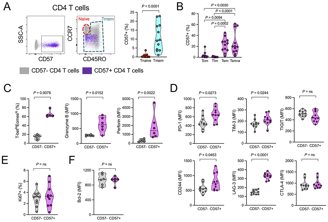
Expression of CD57 on T cells is associated with cytotoxicity, and cytotoxic T cells have been linked to CVD risk (20, 21, 24–26, 36). CD57+ CD4 T cells have been identified in the peripheral blood during HIV and other infections, as well as in elderly (28, 46–48). Here, in a cohort of ART-treated PLWH with CMV coinfection, we found that CD57+ CD4 T cells were confined almost exclusively to the memory T cell (Tmem) compartment (Fig. 1A). Expression of CD57 on CD4 Tmem was highest within the effector memory (Tem; CD45RO+CCR7−CD27−) and terminally-differentiated (Temra; CD45RO−CCR7−) compartments (Fig. 1B). CD57+ CD4 T cells had a robust effector phenotype typified by high T-bet and low eomesodermin (Eomes) expression, and elevated levels of granzyme B and perforin (Fig. 1C). CD57+ CD4 Tmem also had elevated expression of inhibitory receptors, including PD-1, TIM-3, CD244, and LAG-3, compared to CD57− CD4 Tmem (Fig. 1D).
Figure 1.
CD57+ CD4 Tmem have an effector phenotype in PLWH. (A)(Left) Representative dotplots (n = 12) showing CD57 expression on CD4 T cells, and distribution of CD57+ CD4 T cells (purple) within naïve (red) or memory T cell (Tmem, cyan) populations. (Right) Quantification of CD57+ CD4 T cells within naïve or Tmem populations. Significance determined by Mann-Whitney test. (B) Percentage of CD4 Tmem subpopulations that are CD57+. Significance determined by Friedman test. Tcm, CD45RO+CCR7+; Ttm, CD45RO+CCR7−CD27+; Tem, CD45RO+CCR7−CD27−; Temra, CD45RO−CCR7−. (C) Percentage of CD57− or CD57+ CD4 Tmem that are T-bethiEomeslo, and mean fluorescence intensity (MFI) of intracellular granzyme B and perforin expression. Significance determined by Mann-Whitney test. (D) MFI of surface PD-1, TIM-3, TIGIT, CD244 (2B4), LAG-3, and CTLA-4 expression on CD57− and CD57+ CD4 Tmem. Significance determined by Mann-Whitney test. (E) Percentage of CD57− or CD57+ CD4 Tmem that are Ki67+ and (F) MFI of intracellular Bcl-2 expression. Significance determined by Mann-Whitney test.
Expression of CD57 on T cells is associated with shorter lifespan, poor replicative capacity, and immune senescence (19, 28, 49). These observations led us to consider what was driving the accumulation of CD57+ CD4 T cells within the Tmem compartment in HIV infection. Surprisingly, similar proportions of CD57+ and CD57− Tmem were in cell cycle, as evidenced by intracellular expression of Ki67 (Fig. 1E). Notably, we found equivalent expression of the anti-apoptotic molecule Bcl-2 in CD57+ and CD57− CD4 Tmem, suggesting that viability was not reduced in CD57+ CD4 Tmem (Fig. 1F).
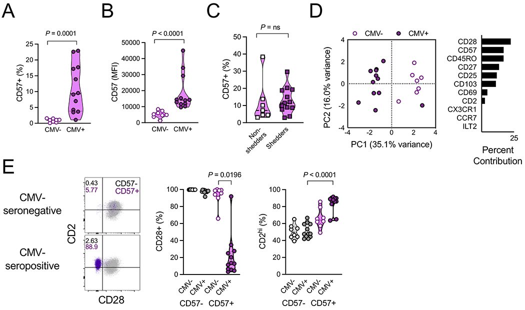
We next asked whether CMV coinfection, which has been shown to drive CD8 T cell expansion, differentiation, and activation in PLWH (12, 50), had a similar effect on CD57+ CD4 Tmem. PLWH who were CMV-seronegative had very few CD57+ CD4 Tmem compared to CMV seropositive donors (Fig. 2A) and the few CD57+ cells that the seronegative donors did maintain exhibited lower surface expression of the CD57 molecule (Fig. 2B). Among a larger cohort of male CMV-seropositive donors, asymptomatic seminal CMV shedding – which is highly correlated with plasma CMV DNA levels (51) – had no discernible effect on the proportion of CD4 Tmem that expressed CD57 (Fig. 2C), suggesting active CMV replication was not necessary for the continued prevalence of CD57+ CD4 Tmem. These observations led us to ask whether CD57+ CD4 Tmem from CMV-seropositive donors were fundamentally different than those from CMV-seronegative donors. Using 11-parameter mean fluorescence intensity (MFI) data acquired by flow cytometry, we compared CD57+ CD4 Tmem in CMV-seronegative and CMV-seropositive donors by principle component analysis (PCA) and found that CD57+ CD4 Tmem were significantly different (P=0.0018, Mann-Whitney test) along PC1, which was driven mainly by the expression of CD28 (Fig. 2D) and CD57 (as evident in Fig. 2B).
Figure 2.
CMV coinfection promotes CD57+CD28−CD2hi CD4 Tmem in PLWH. (A) Percentage of CD4 Tmem expressing CD57 in CMV-seronegative (CMV−; n = 8) and CMV-seropositive (CMV+; n = 12) donors. Significance determined by Mann-Whitney test. (B) Mean fluorescence intensity (MFI) of CD57 expression on CD57+ CD4 Tmem in same donors as in A. Significance determined by Mann-Whitney test. (C) Percentage of CD4 Tmem expressing CD57 in CMV-seropositive PLWH who had previously been characterized for whether they had asymptomatic CMV replication in their seminal plasma (shedders, n = 15) or not (non-shedders, n = 14). Significance determined by Mann-Whitney test. (D) (Left) Principal component analysis of CD57+ CD4 Tmem from donors in A. (Right) Component contributions to principle component 1. (E) (Left) Representative dotplots show CD2 and CD28 expression on CD57− (gray) and CD57+ (purple) CD4 Tmem in same donors as in A. (Right) Percentage of indicated CD4 Tmem populations expressing CD28 or high levels of CD2 (CD2hi). Significance determined by Kruskal-Wallis test with Dunn’s correction for multiple comparisons.
Interactions of CD2 with its ligand LFA-3 (also called CD58) have been shown to be the strongest costimulatory pathway for T cells that lack CD28 (40), so we next examined the expression of the costimulatory receptors CD28 and CD2 on Tmem. CD57+ CD4 Tmem from CMV-seropositive donors were mostly CD28− and had high expression of CD2, whereas in CMV-seronegative donors those few cells that were CD57+ retained CD28 and had relatively lower CD2 expression (Fig. 2E). In both seropositive and seronegative populations, CD57− CD4 Tmem had high CD28 and low CD2 expression.
CD2/LFA-3 costimulation enhances CD57+ CD4 Tmem polyfunctionality
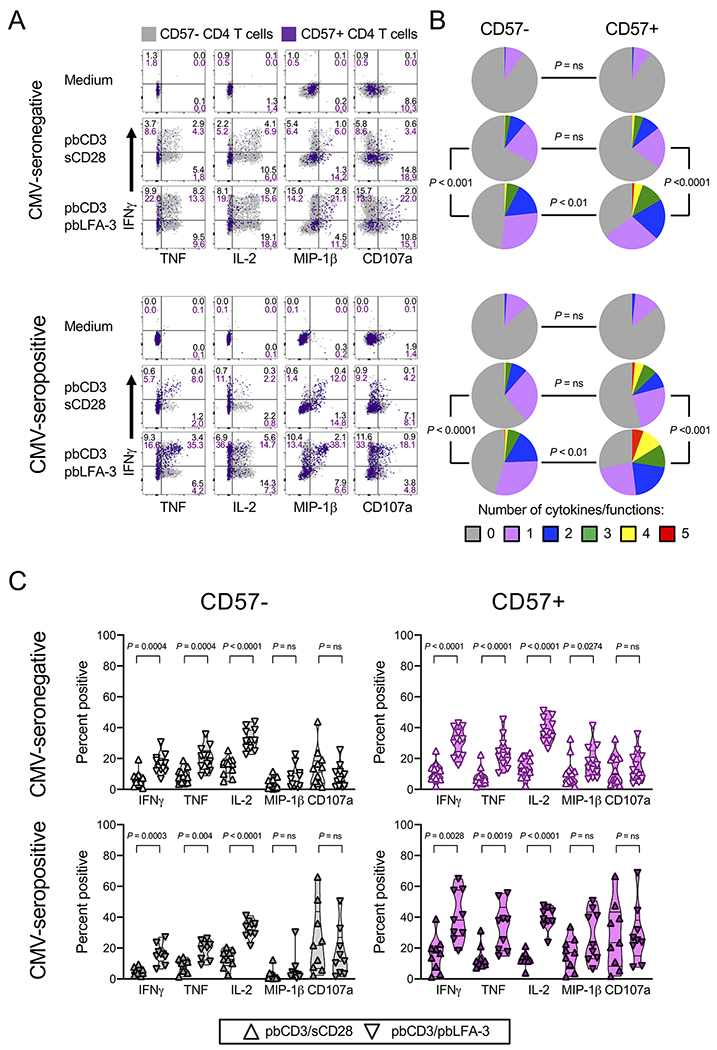
These findings led us to test whether costimulation via the CD2/LFA-3 axis would enhance the function of CD57+ CD4 Tmem, which might be exhausted due to elevated inhibitory receptor expression (Fig. 1D). We stimulated PBMCs from CMV-seronegative and -seropositive PLWH by traditional T cell receptor (TCR) activation (plate-bound anti-CD3 mAb; pbCD3) alone, with CD28 costimulation (soluble anti-CD28 mAb; sCD28), or with LFA-3 costimulation (plate-bound recombinant LFA-3 protein; pbLFA-3), and measured IFNγ, IL-2, MIP-1β, and TNF expression by intracellular flow cytometry, and surface expression of the degranulation marker, CD107a (Fig. 3A, Fig. S1A). Even though CD57− and CD57+ CD4 Tmem from CMV-seropositive donors had very different levels of CD28 expression, the responses of these populations to CD28 costimulation was similar. For all populations, LFA-3 costimulation provided a significantly enhanced response compared to CD28 costimulation (Fig. 3B), or to TCR activation without costimulation (Fig. S1A). Consistent with their elevated expression of CD2, CD57+ cells gave a more robust response than CD57− cells after LFA-3 costimulation in both donor groups, and CD57+ CD4 Tmem from CMV-seropositive donors had significantly more robust response to LFA-3 costimulation than CD57+ cells from CMV-seronegative donors (P=0.03, SPICE pie comparison). For CMV-seropositive donors, the range of responsiveness of CD57+ CD4 Tmem was quite broad, suggesting there might be heterogeneity in this population. Much of the CD2/LFA-3 axis-enhanced polyfunctionality was due to increased IFNγ, IL-2 and TNF production, regardless of CD57 expression (Fig. 3C). Both costimulatory pathways resulted in similar levels of degranulation, as measured by CD107a expression. Interestingly, when compared to anti-CD3 stimulation alone, CD28 costimulation significantly enhanced IL-2 production, even in CD57+ cells from CMV-seropositive donors (Fig. S1B), suggesting that the low, residual CD28 expression on CD57+CD28− CD4 Tmem in CMV-seropositive donors could still provide some costimulatory efficacy. Additionally, our findings suggest that CD57+ CD4 Tmem are not functionally exhausted in terms of ability to express cytokine and CD107 despite elevated inhibitory receptor expression.
Figure 3.
CD2/LFA-3 costimulation enhances CD57+ CD4 Tmem polyfunctionality. (A) Representative dotplots showing IFNγ and TNF, IL-2, MIP-1β, or CD107a expression by CD57− (gray) and CD57+ (purple) CD4 Tmem from CMV-seronegative (n = 12, top) and CMV-seropositive (n = 9, bottom) donors after 6h stimulation with medium control; plate-bound anti-CD3, soluble anti-CD28 (pbCD3/sCD28); or plate-bound anti-CD3, plate-bound LFA-3 (pbCD3/pbLFA-3). (B) Cumulative cytokine/functions in the different groups as in A. Significance determined by pie comparisons using SPICE software, with 10,000 permutations per comparison. (C) Percentage of CD57− and CD57+ CD4 Tmem in each donor group that produce the indicated functions after pbCD3sCD28 or pbCD3pbLFA-3 stimulation. Significance determined by Mann-Whitney test.
IL-15 arms and activates CD57+ CD4 Tmem
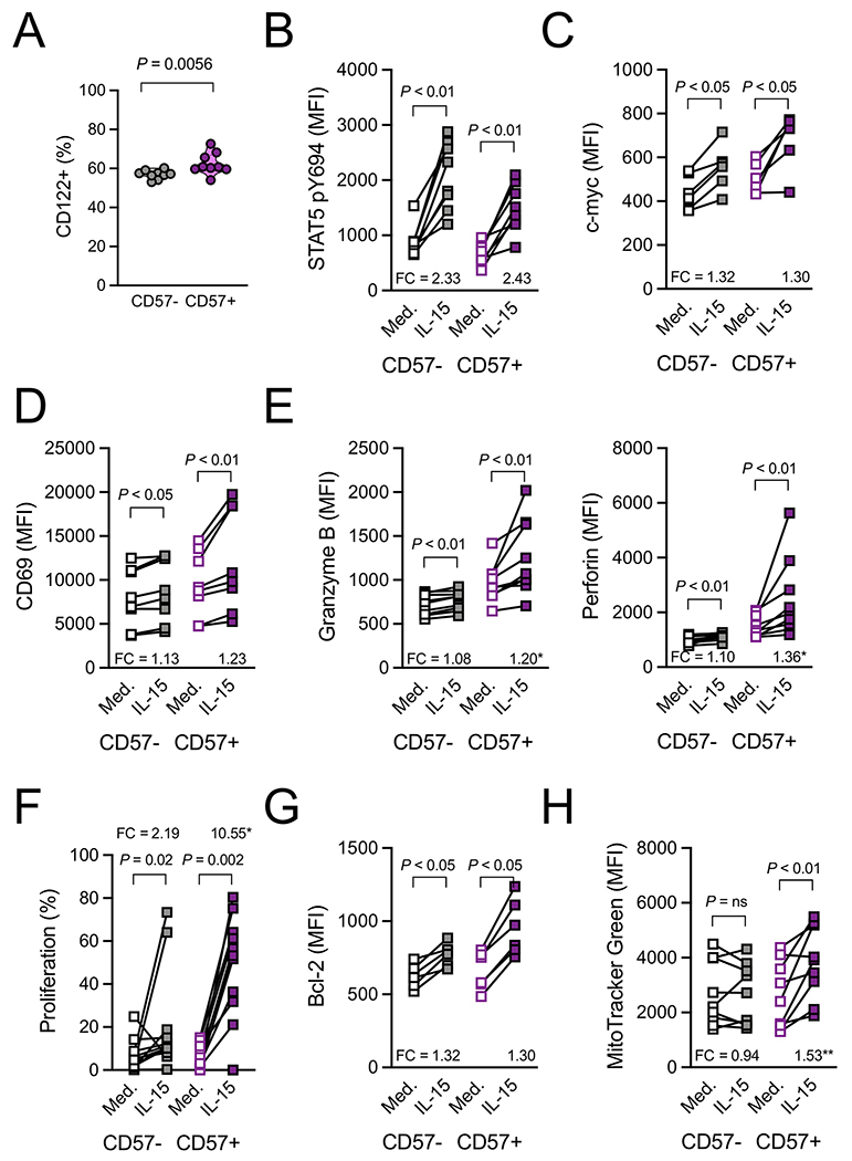
The cytokine IL-15 activates T cells, enhances mitochondrial activity, and promotes the intracellular accumulation of cytolytic molecules such as granzyme B and perforin (52, 53). IL-15 expression in lymph nodes correlates with CD8 T cell numbers in the periphery in chronic (viremic) HIV infection, suggesting that IL-15 may have a role in the immune response to HIV infection (52). In addition, there is evidence that IL-15 expression is increased in the bone marrow of CMV-seropositive compared to CMV-seronegative donors, in HIV-uninfected individuals undergoing hip replacement surgery (54). We therefore wanted to investigate the effects of IL-15 on CD57+ CD4 Tmem. In CMV-seropositive PLWH, CD57+ CD4 Tmem more often express CD122, the β-chain of the IL-2/IL-15 receptor, than do CD57− CD4 Tmem (Fig. 4A) – although both populations have abundant CD122 expression – suggesting potential responsiveness to IL-15 signals. Stimulation with IL-15 promoted robust phosphorylation of STAT5 within 45 minutes (Fig. 4B), and by 2 days had enhanced expression of the master transcriptional regulator c-myc (Fig. 4C), induced surface expression of the early activation and resident memory marker CD69 (Fig. 4D), and promoted intracellular expression of the cytolytic molecules granzyme B and perforin (Fig. 4E) in both CD57− and CD57+ CD4 Tmem populations obtained from HIV-uninfected donors. The enhancement of cytolytic molecule expression by IL-15 was significantly more robust among CD57+ cells than among the CD57− populations. IL-15 also promoted the proliferation of both CD57− and CD57+ CD4 Tmem (Fig. 4F), suggesting that CD57+ CD4 Tmem are not replicatively senescent, but rather can proliferate in response to IL-15. Additionally, IL-15 enhanced expression of the pro-survival factor Bcl-2 in both CD57− and CD57+ CD4 Tmem (Fig. 4G). These results contrast to the effect of IL-15 on mitochondrial biogenesis – mitochondrial mass was increased by IL-15 only in CD57+ CD4 Tmem (Fig. 4H). Thus, CD57+ CD4 Tmem are highly responsive to IL-15, which can be both a homeostatic and an inflammatory stimulus.
Figure 4.
IL-15 activates and arms CD57+ CD4 Tmem. (A) Percentage of CD57− and CD57+ CD4 Tmem from CMV-seropositive donors (n = 9) that express CD122. Significance determined by Mann-Whitney test. (B) Mean fluorescence intensity (MFI) of STAT5 phosphorylated at Y604 on CD57− or CD57+ CD4 Tmem from CMV-seropositive PLWH (n = 8) after 45 minutes of stimulation with medium control or IL-15 (20ng/ml). (C) MFI of intracellular c-myc expression on CD57− or CD57+ CD4 Tmem from CMV-seropositive PLWH (n = 6) after 48h of stimulation with medium control or IL-15 (20ng/ml). (D) MFI of surface CD69 expression and (E) intracellular granzyme B and perforin expression on CD57− or CD57+ CD4 Tmem from HIV-uninfected control donors (n=8-9/group) after 48h of stimulation with medium control or IL-15 (20ng/ml). (F) Percentage of CD57− or CD57+ CD4 Tmem that proliferated (diluted CellTrace Violet dye) from HIV-uninfected control donors (n=12). (G) MFI of intracellular Bcl-2 expression on CD57− or CD57+ CD4 Tmem from CMV-seropositive PLWH donors (n = 6) after 48h of stimulation with medium control or IL-15 (20ng/ml). (H) MFI of MitoTracker Green staining on CD57− or CD57+ CD4 Tmem from HIV-uninfected control donors (n=9) after 48h of stimulation with medium control or IL-15 (20ng/ml). (B-H) Significance determined by Wilcoxon signed rank test. Differences in fold change (FC) determined by Mann-Whitney test. *P ≤ 0.05; **P ≤ 0.01.
CD57+ CD4 Tmem traffic to cytokine-treated vascular endothelium
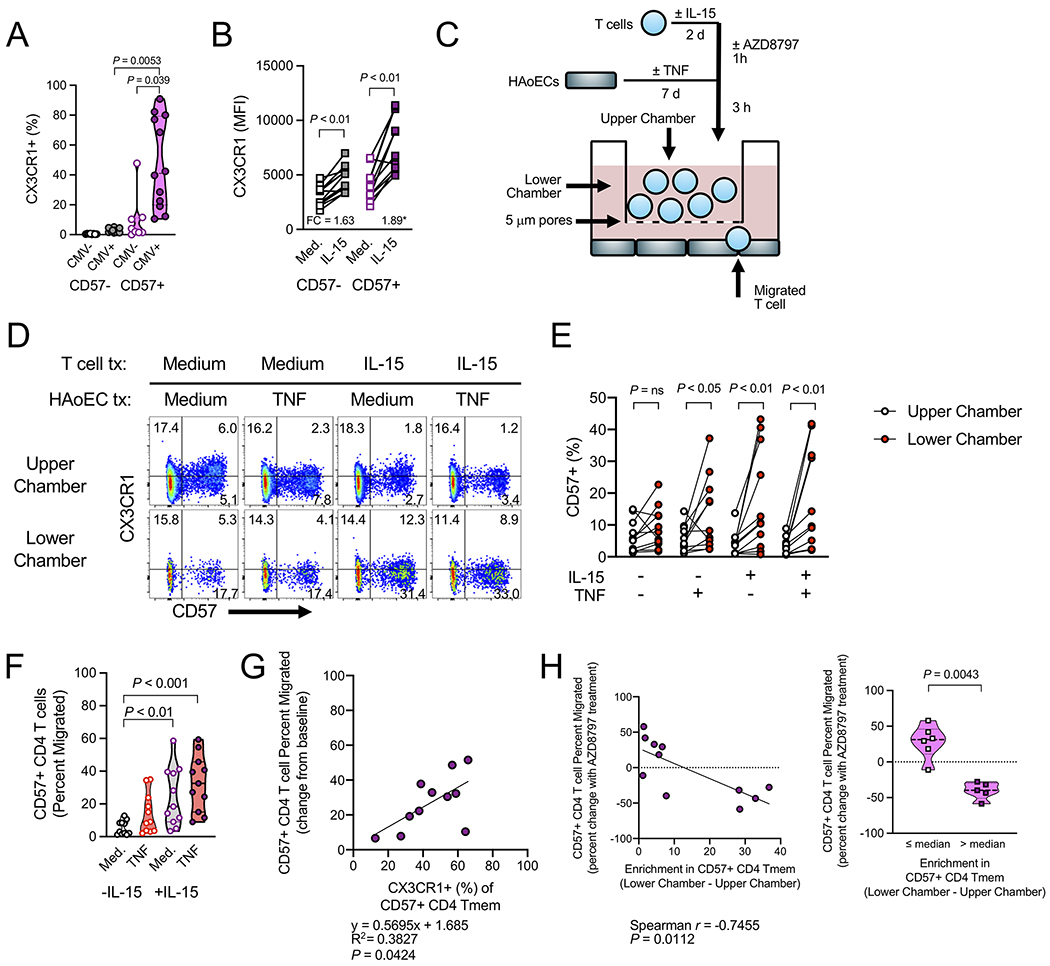
CD4 Tmem that lack CD28 express both CD57 and the endothelium-homing receptor CX3CR1 (46, 55), and CX3CL1 promotes the migration of CX3CR1+ CD4 T cells in vitro (36, 56). In addition, the proportion of CX3CR1+ CD4 T cells positively correlates with carotid IMT and IMT progression over time in PLWH (36). CX3CR1+ CD4 T cells might therefore provide a cellular link from HIV to CVD risk, which is increased in PLWH (2). Only CD57+ CD4 Tmem from CMV-seropositive PLWH donors exhibited CX3CR1 expression (Fig. 5A). While IL-15 treatment upregulated surface expression of CX3CR1 in both CD57− and CD57+ CD4 Tmem from CMV-seropositive PLWH, CD57+ cells showed significantly greater CX3CR1 upregulation in response to IL-15 (Fig. 5B). One of the effector molecules whose TCR stimulation-induced production was enhanced by LFA-3 costimulation was TNF, and TNF is a potent inducer of CX3CL1 gene expression and protein secretion by HAoECs in vitro (Fig. S2A). Notably, while IL-15 induces TNF production from sorted memory CD4 T cells in vitro (Fig. S2B), the amount produced is insufficient to elicit CX3CL1 production from HAoECs in culture (Fig. S2C). In contrast, both CD3/CD28 or CD3/LFA-3 stimulation elicited robust release of TNF (Fig. S2D) and likely other factors sufficient to induce endothelial cell CX3CL1 expression (Fig. S2E). Importantly, none of the stimulations elicited CX3CL1 production by the sorted CD4 Tmem (Fig. S2F). We therefore established a short-term transwell migration assay to see if IL-15 and/or TNF promoted CD57+ CD4 Tmem migration (Fig. 5C). T cells from PLWH that were pre-treated, or not, with IL-15 (at a concentration sufficient to enhance CX3CR1 expression, Fig. 5B) were incubated with HAoECs that had been pre-treated, or not, with TNF (at a concentration sufficient to elicit CX3CL1 secretion, Fig. S2A). Examples of CX3CR1 and CD57 staining prior to the migration assay are shown in Fig. S3A. After 3 hours, CD57+ CD4 Tmem in the upper and lower chambers were recovered and quantified. CD4 T cells that had migrated to the lower chamber were enriched for CD57 (Fig. 5D,E), and the CD57+ cells trended toward lower expression of CX3CR1, consistent with exposure to CX3CL1 (Fig. S3B). We next quantified the numbers of CD57+ CD4 Tmem recovered from the lower chambers and expressed those numbers as a percentage of the total starting numbers of CD57+ CD4 Tmem in each assay well. We found an increase in the number of CD57+ CD4 Tmem that migrated when exposed to IL-15, or both TNF and IL-15, compared to medium only control (Fig. 5F).
Figure 5.
IL-15 and TNF enhance the chemoattraction of CD57+ CD4 Tmem toward CX3CL1-expressing endothelial cells. (A) Percentage of CD57− and CD57+ CD4 Tmem from CMV-seronegative (n = 8) and CMV-seropositive (n = 12) donors that express CX3CR1. Significance determined by Kruskal-Wallis test with Dunn’s correction for multiple comparisons. (B) MFI of CX3CR1 staining on CD57− or CD57+ CD4 Tmem from HIV-uninfected control donors (n = 12) after 48h of stimulation with medium control or IL-15 (20ng/ml). Significance determined by Wilcoxon signed rank test. Differences in fold change (FC) determined by Mann-Whitney test. *P ≤ 0.05. (C) Schematic diagram of the transwell assay system. Confluent monolayers of human aortic endothelial cells (HAoECs) were cultured in the presence or absence of TNF (10ng/ml) for 7 days in wells of a 24-well plate. Purified T cells from CMV-seropositive PLWH (n = 11) were exposed to IL-15 (20ng/ml) or medium control for 2 days prior to placement in the upper chamber of a 5μm transwell situated onto the HAoEC monolayer. In some cases, T cells were treated with AZD8797 (500nM) for 1 hour prior to co-culture. After 3 hours, T cells were separately harvested from the upper and lower chambers and analyzed by flow cytometry. (D) Representative dotplots showing CX3CR1 and CD57 expression on CD4 T cells in transwell assay from upper and lower chambers in indicated conditions. (E) Percentage of CD4 T cells recovered from the upper and lower chambers expressing CD57 after 3 hours. Significance determined by Wilcoxon signed rank test. (F) Absolute number of CD57+ CD4 T cells in the lower chamber expressed as a percent of total (upper chamber + lower chamber) CD57+ CD4 T cells (“Percent migrated”) in indicated conditions. Significance determined by Kruskal-Wallis test with Dunn’s correction for multiple comparisons. (G) The proportion of CD57+ CD4 Tmem that expresses CX3CR1 predicts the percent migrated of CD57+ CD4 T cells in the assay after IL-15 and TNF co-treatment (minus no treatment baseline). Significance determined by simple linear regression. (H) (Left) Enrichment in the percentage of CD57+ CD4 Tmem (lower chamber minus upper chamber) versus the percentage change in percent migrated following AZD8797 treatment of CD57+ CD4 Tmem in the IL-15 and TNF co-treatment condition. Significance determined by Spearman analysis. (Right) The percentage change in percent migrated following AZD8797 treatment of CD57+ CD4 Tmem in the IL-15 and TNF co-treatment condition for donors whose CD57+ CD4 Tmem enrichment was at the median value or below and those who enrichment was greater than the median value. Significance determined by Mann-Whitney test.
Intriguingly, the percentage of CD57+ CD4 Tmem that expressed CX3CR1+ at baseline predicts the amount of induced migration of CD57+ CD4 T cells in the IL-15 and TNF co-treated condition (Fig. 5G), suggesting that the CX3CL1/CX3CR1 axis may be important for the migration of CD57+ CD4 T cells toward activated endothelium. To test this, we treated the T cells with the CX3CR1 antagonist AZD8797 (57–59), prior to the migration assay. While we found no significant reductions in the proportion of CD57+ CD4 T cells that migrated in any of the experimental groups following AZD8797 treatment (Fig. S3C), we did observe an interesting correlation in the IL-15 and TNF co-treated group. For donors with proportional enrichment of CD57+ cells in the lower chamber (values greater than median), AZD8797 treatment resulted in a significant inhibition of migration, but for donors without CD57+ cell enrichment (values less than or equal to the median), AZD8797 treatment paradoxically resulted in more CD57+ CD4 T cell migration (Fig. 5H). In other words, for donors whose CD57+ CD4 T cells were more migratory than the overall CD4 T cell pool, AZD8797 treatment impaired the migration, but for other donors, AZD8797 treatment resulted in increased migration by all CD4 T cells, with no selective effect on CD57+ cells. Thus, our data show that there is heterogeneity in the susceptibility of CD57+ CD4 T cells to CX3CL1 and suggest other factors are likely also important in the migration of CD57+ CD4 T cells toward activated endothelial cells – particularly in the absence of CX3CR1 functionality.
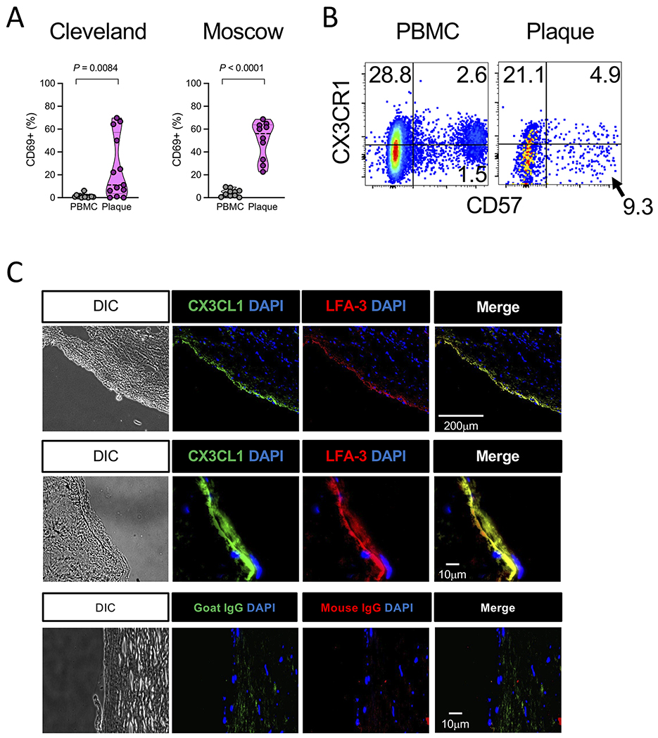
CVD is a leading non-AIDS cause of death in PLWH and in the general population (60–62). Elevated CVD risk in PLWH could be due to an over-abundance of factors that also promote CVD in the general population, such as persistent inflammation, immune cell activation, and CMV infection (1, 2), and we hypothesize that risk is linked to CD57+ CD4 T cells. We therefore examined the CD4 T cell infiltrate in atherosclerotic plaques of HIV-uninfected persons undergoing clinically-indicated carotid endarterectomy to characterize the cells that accumulate at sites of vascular damage in the general population. Endarterectomies were performed at two locations – Cleveland, Ohio, USA and Moscow, Russia – and experiments were run in parallel. In both cohorts we observed a profound upregulation of CD69 expression on CD4 T cells recovered from plaques compared to CD69 expression on peripheral blood cells from the same donors (Fig. 6A). CD69 expression on plaque CD4 T cells is reflective of activation and a tissue resident phenotype (63). Notably, IL-15, which promotes CD69 expression on CD4 Tmem in vitro (Fig. 4D), has been demonstrated within plaque tissues (64, 65). Conceivably, CD69 expression on plaque T cells is the result of recent exposure to inflammatory cytokine or TCR signals. CD57+ CD4 Tmem within plaques also had evidence of CX3CL1 exposure (Fig. 6B), suggesting these cells were capable of trafficking to sites of endothelium damage in vivo. Immunohistologic examination of the plaque tissue showed expression of CX3CL1 and LFA-3 on the vascular endothelium (Fig. 6C). Thus, our data are consistent with a model in which CX3CR1+ CD57+ CD28− CD4 Tmem migrate toward CX3CL1-expressing endothelial cells in the nascent plaque and potentially contribute to vascular damage via cytokine and lytic granule release induced by CD2/LFA-3-mediated costimulation.
Figure 6.
Atherosclerotic plaque tissue contains CD57+ CD4 Tmem and CX3CL1 and LFA-3 proteins. (A) Percentage of CD4 T cells expressing CD69 in PBMCs or donor-matched plaque tissue from Cleveland (n = 14) or Moscow (n = 10) cohorts. Significance determined by Mann-Whitney test. (B) Representative dotplots (n = 14) showing surface CD57 expression on CD4 T cells derived from the PBMCs or plaque tissue. (C) Representative images (n = 4) from cryopreserved carotid endarterectomy tissue sections show DAPI, CX3CL1, and LFA-3, or isotype control staining in the carotid endothelium and sub-endothelial regions at low magnification (top row) or high magnification (middle and bottom rows).
DISCUSSION
Identifying targetable factors that promote atherosclerosis is a major goal of current cardiovascular research. It is becoming increasingly clear that cardiovascular complications like myocardial infarction have immune and inflammatory etiologies, and the contributions of immune cells such as CD4 T cells to these processes are coming into focus. In this report, we show that cytomegalovirus (CMV) coinfection – a major driver of inflammation and T cell activation and expansion (3, 11–14, 50) – is associated with an expanded subpopulation of CD4 memory T cells (Tmem) that express CD57 in the peripheral blood of people living with HIV (PLWH). CD57+ CD4 Tmem are enriched for cytolytic molecules and expression of the vascular endothelium-homing receptor CX3CR1. CD4 Tmem can be isolated from atherosclerotic plaques, suggesting they can traffic to sites of endothelial activation and may contribute to cardiovascular disease (CVD) by damaging endothelial cells via targeted release of cytolytic granules or cytokines that can induce endothelial dysfunction.
CD57+ CD4 Tmem in CMV-seropositive PLWH lack the costimulatory receptor CD28 but have elevated levels of CD2. Costimulation via the CD2/LFA-3 axis elicits a polyfunctional response that is enhanced in CD57+ CD4 Tmem compared with that in CD57− CD4 Tmem, which have lower CD2 expression. We also show that LFA-3 is highly expressed in plaques in close proximity to CX3CL1, providing an environment in which CD57+ CD4 Tmem could utilize CX3CR1/CX3CL1 interactions to migrate into an inflamed vasculature. To explore this, we established a short-term chemoattraction assay and demonstrated that IL-15-treated CD57+ CD4 T cells upregulate CX3CR1 expression and exhibit greater migration toward TNF-treated cultured primary endothelial cells that upregulate CX3CL1. Additionally, the proportion of CD57+ CD4 Tmem that expressed CX3CR1 in the absence of treatment predicted the magnitude of induced migration. Our data are consistent with a model in which pro-inflammatory effector molecules promote CVD by activating CD57+ CD4 Tmem and endothelial cells. Reciprocally, activated CD57+ CD4 Tmem release TNF to further promote endothelial cell activation and CX3CL1 release. CMV infection drives the expansion of CD57+ CD4 Tmem and could be a key component in this model.
The elements that CD57+ CD4 T cells recognize in the vasculature or in plaque tissues are undefined, although it is likely that many CD57+ CD4 Tmem are specific for CMV (7, 48, 66). Latent CMV is harbored within CD34+ hematopoietic stem cells; the virus can reactivate as infected cells differentiate along the myeloid lineage into macrophages (67). If monocytes carrying CMV genomes enter the subendothelium and differentiate into CMV antigen-expressing foam cells, these could provide the source of CMV antigens in the context of MHC class II. We recently showed that activated T cell-derived TNF promotes expression of the pro-coagulant tissue factor on monocytes in vitro (68). Thus, interactions between CMV-specific CD4 T cells and CMV-infected myeloid cells could plausibly contribute mechanistically to clot formation and CVD development. Alternatively, vascular endothelial cells can express MHC class II (42). Since CMV has been shown to infect endothelial cells in vitro (69, 70), and CMV DNA can be found in vascular tissues (71, 72), CMV-infected endothelial cells might be capable of presenting both CMV antigens and LFA-3 costimulatory signals to infiltrating CD57+ CD4 Tmem as well as to CMV-specific CD8 T cells.
Interestingly, the nominally homeostatic cytokine IL-15 could be a key player in our observations. IL-15 activates T cells and NK cells and has been postulated to act as a danger signal to ‘call in the troops’ to sites of infection or tissue damage (73). Consistent with this role, IL-15 has been demonstrated within atherosclerotic plaques (64, 65) and IL-15 promotes the proliferation of CD57+ CD4 T cells from patients with rheumatoid arthritis (74). Our data here suggest that IL-15 in plaques may serve a mechanistic role in atherosclerosis at least in part by activating and enhancing the migration of cytotoxic CD57+ CD4 Tmem, and possibly by directly eliciting release of TNF and/or other proinflammatory factors. Circulating levels of IL-15 are increased in PLWH (75), and we find increased IL-15 protein in aortas of SIV/SHIV-infected macaques compared to uninfected control animals (Panigrahi and Chen, et al., submitted), thus IL-15 may be an important contributor to the increased CVD risk in PLWH.
In conclusion, we have identified a potential mechanism for the contribution of cytotoxic CD57+ CD4 Tmem to the elevated CVD risk in PLWH and the HIV-infected elderly: CD57+ CD4 Tmem expressing CX3CR1 are recruited to the inflamed vasculature where they could further damage endothelium via CD2/LFA-3-mediated degranulation. Future studies are needed to confirm and build upon our findings in vitro and in vivo. FDA-approved drugs that target CMV (e.g. valganciclovir, letermovir) and TNF (e.g. adalimumab, etanercept) provide therapeutic strategies that could be applied in the clinic. Indeed, blocking TNF activity improves endothelial function in patients with rheumatoid arthritis (76). As more therapies to target CX3CL1, IL-15, and CD2/LFA-3 interactions become available, it will be important to examine them in the settings of HIV/CMV coinfection and elderly HIV-uninfected persons with atherosclerosis.
Supplementary Material
KEY POINTS.
CMV coinfection promotes the generation of CD57+ CD4 Tmem in PLWH
CD2/LFA-3 costimulation enhances the functionality of CD57+ CD4 Tmem
IL-15 and TNF enhance chemoattraction of CD57+ CD4 Tmem to CX3CL1+ endothelial cells
ACKNOWLEDGMENTS
We thank Steven Juchnowski and Sadeer Al-Kindi for their help acquiring atherosclerotic plaque tissue specimens, and Daniela Moisi and Dominic Dorazio for their excellent technical assistance.
This work was supported by National Institutes of Health (NIH) grant HL134544 to N.T.F.; The San Diego Primary Infection Resource Consortium (NIH AI106039, S. Little, PI) and the Translational Virology Core at the San Diego Center for AIDS Research (AI036214) to S.G.; a Career Development Award (51K2CX001471-03) from the U.S. Department of Veterans Affairs to C.L.S.; NIH grants (AI076174, AI069501) and the Richard J. Fasenmyer Foundation awarded to M.M.L.; and the CWRU Center for AIDS Research Catalytic Awards (AI036219) to D.A.Z. and M.L.F.
Footnotes
CONFLICTS OF INTEREST
N.T.F serves as a consultant for Gilead. The work of L.M. was funded by the NICHD Intramural Program. All other authors declare no competing interests.
REFERENCES
- 1.Lederman MM, Funderburg NT, Sekaly RP, Klatt NR, and Hunt PW. 2013. Residual immune dysregulation syndrome in treated HIV infection. Adv Immunol 119: 51–83. [DOI] [PMC free article] [PubMed] [Google Scholar]
- 2.Longenecker CT, Sullivan C, and Baker JV. 2016. Immune activation and cardiovascular disease in chronic HIV infection. Curr Opin HIV AIDS 11: 216–225. [DOI] [PMC free article] [PubMed] [Google Scholar]
- 3.Freeman ML, Lederman MM, and Gianella S. 2016. Partners in Crime: The Role of CMV in Immune Dysregulation and Clinical Outcome During HIV Infection. Curr HIV/AIDS Rep 13: 10–19. [DOI] [PMC free article] [PubMed] [Google Scholar]
- 4.Wang H, Peng G, Bai J, He B, Huang K, Hu X, and Liu D. 2017. Cytomegalovirus Infection and Relative Risk of Cardiovascular Disease (Ischemic Heart Disease, Stroke, and Cardiovascular Death): A Meta-Analysis of Prospective Studies Up to 2016. J Am Heart Assoc 6. [DOI] [PMC free article] [PubMed] [Google Scholar]
- 5.Deeks SG 2011. HIV infection, inflammation, immunosenescence, and aging. Annu Rev Med 62: 141–155. [DOI] [PMC free article] [PubMed] [Google Scholar]
- 6.Hansson GK, and Libby P. 2006. The immune response in atherosclerosis: a double-edged sword. Nat Rev Immunol 6: 508–519. [DOI] [PubMed] [Google Scholar]
- 7.Koch S, Larbi A, Ozcelik D, Solana R, Gouttefangeas C, Attig S, Wikby A, Strindhall J, Franceschi C, and Pawelec G. 2007. Cytomegalovirus infection: a driving force in human T cell immunosenescence. Ann N Y Acad Sci 1114: 23–35. [DOI] [PubMed] [Google Scholar]
- 8.Pawelec G 2012. Hallmarks of human “immunosenescence”: adaptation or dysregulation? Immun Ageing 9: 15. [DOI] [PMC free article] [PubMed] [Google Scholar]
- 9.Lichtner M, Cicconi P, Vita S, Cozzi-Lepri A, Galli M, Lo Caputo S, Saracino A, De Luca A, Moioli M, Maggiolo F, Marchetti G, Vullo V, d’Arminio Monforte A, and Study IF. 2015. Cytomegalovirus coinfection is associated with an increased risk of severe non-AIDS-defining events in a large cohort of HIV-infected patients. J Infect Dis 211: 178–186. [DOI] [PubMed] [Google Scholar]
- 10.Gianella S, Massanella M, Wertheim JO, and Smith DM. 2015. The Sordid Affair Between Human Herpesvirus and HIV. J Infect Dis 212: 845–852. [DOI] [PMC free article] [PubMed] [Google Scholar]
- 11.Gianella S, and Letendre S. 2016. Cytomegalovirus and HIV: A Dangerous Pas de Deux. J Infect Dis 214 Suppl 2: S67–74. [DOI] [PMC free article] [PubMed] [Google Scholar]
- 12.Freeman ML, Mudd JC, Shive CL, Younes SA, Panigrahi S, Sieg SF, Lee SA, Hunt PW, Calabrese LH, Gianella S, Rodriguez B, and Lederman MM. 2016. CD8 T-Cell Expansion and Inflammation Linked to CMV Coinfection in ART-treated HIV Infection. Clin Infect Dis 62: 392–396. [DOI] [PMC free article] [PubMed] [Google Scholar]
- 13.Sylwester AW, Mitchell BL, Edgar JB, Taormina C, Pelte C, Ruchti F, Sleath PR, Grabstein KH, Hosken NA, Kern F, Nelson JA, and Picker LJ. 2005. Broadly targeted human cytomegalovirus-specific CD4+ and CD8+ T cells dominate the memory compartments of exposed subjects. J Exp Med 202: 673–685. [DOI] [PMC free article] [PubMed] [Google Scholar]
- 14.Naeger DM, Martin JN, Sinclair E, Hunt PW, Bangsberg DR, Hecht F, Hsue P, McCune JM, and Deeks SG. 2010. Cytomegalovirus-specific T cells persist at very high levels during long-term antiretroviral treatment of HIV disease. PLoS One 5: e8886. [DOI] [PMC free article] [PubMed] [Google Scholar]
- 15.Stone SF, Price P, Khan N, Moss PA, and French MA. 2005. HIV patients on antiretroviral therapy have high frequencies of CD8 T cells specific for Immediate Early protein-1 of cytomegalovirus. AIDS 19: 555–562. [DOI] [PubMed] [Google Scholar]
- 16.Khan N, Shariff N, Cobbold M, Bruton R, Ainsworth JA, Sinclair AJ, Nayak L, and Moss PA. 2002. Cytomegalovirus seropositivity drives the CD8 T cell repertoire toward greater clonality in healthy elderly individuals. J Immunol 169: 1984–1992. [DOI] [PubMed] [Google Scholar]
- 17.Appay V, Dunbar PR, Callan M, Klenerman P, Gillespie GM, Papagno L, Ogg GS, King A, Lechner F, Spina CA, Little S, Havlir DV, Richman DD, Gruener N, Pape G, Waters A, Easterbrook P, Salio M, Cerundolo V, McMichael AJ, and Rowland-Jones SL. 2002. Memory CD8+ T cells vary in differentiation phenotype in different persistent virus infections. Nat Med 8: 379–385. [DOI] [PubMed] [Google Scholar]
- 18.van Leeuwen EM, Remmerswaal EB, Vossen MT, Rowshani AT, Wertheim-van Dillen PM, van Lier RA, and ten Berge IJ. 2004. Emergence of a CD4+CD28− granzyme B+, cytomegalovirus-specific T cell subset after recovery of primary cytomegalovirus infection. J Immunol 173: 1834–1841. [DOI] [PubMed] [Google Scholar]
- 19.Brenchley JM, Karandikar NJ, Betts MR, Ambrozak DR, Hill BJ, Crotty LE, Casazza JP, Kuruppu J, Migueles SA, Connors M, Roederer M, Douek DC, and Koup RA. 2003. Expression of CD57 defines replicative senescence and antigen-induced apoptotic death of CD8+ T cells. Blood 101: 2711–2720. [DOI] [PubMed] [Google Scholar]
- 20.Le Priol Y, Puthier D, Lecureuil C, Combadiere C, Debre P, Nguyen C, and Combadiere B. 2006. High cytotoxic and specific migratory potencies of senescent CD8+ CD57+ cells in HIV-infected and uninfected individuals. J Immunol 177: 5145–5154. [DOI] [PubMed] [Google Scholar]
- 21.Johnson S, Eller M, Teigler JE, Maloveste SM, Schultz BT, Soghoian DZ, Lu R, Oster AF, Chenine AL, Alter G, Dittmer U, Marovich M, Robb ML, Michael NL, Bolton D, and Streeck H. 2015. Cooperativity of HIV-Specific Cytolytic CD4 T Cells and CD8 T Cells in Control of HIV Viremia. J Virol 89: 7494–7505. [DOI] [PMC free article] [PubMed] [Google Scholar]
- 22.Munier CML, van Bockel D, Bailey M, Ip S, Xu Y, Alcantara S, Liu SM, Denyer G, Kaplan W, P. S. group, Suzuki K, Croft N, Purcell A, Tscharke D, Cooper DA, Kent SJ, Zaunders JJ, and Kelleher AD. 2016. The primary immune response to Vaccinia virus vaccination includes cells with a distinct cytotoxic effector CD4 T-cell phenotype. Vaccine 34: 5251–5261. [DOI] [PubMed] [Google Scholar]
- 23.Hildemann SK, Eberlein J, Davenport B, Nguyen TT, Victorino F, and Homann D. 2013. High efficiency of antiviral CD4(+) killer T cells. PLoS One 8: e60420. [DOI] [PMC free article] [PubMed] [Google Scholar]
- 24.Hsue PY, Hunt PW, Sinclair E, Bredt B, Franklin A, Killian M, Hoh R, Martin JN, McCune JM, Waters DD, and Deeks SG. 2006. Increased carotid intima-media thickness in HIV patients is associated with increased cytomegalovirus-specific T-cell responses. AIDS 20: 2275–2283. [DOI] [PubMed] [Google Scholar]
- 25.Liuzzo G, Biasucci LM, Trotta G, Brugaletta S, Pinnelli M, Digianuario G, Rizzello V, Rebuzzi AG, Rumi C, Maseri A, and Crea F. 2007. Unusual CD4+CD28null T lymphocytes and recurrence of acute coronary events. J Am Coll Cardiol 50: 1450–1458. [DOI] [PubMed] [Google Scholar]
- 26.Broadley I, Pera A, Morrow G, Davies KA, and Kern F. 2017. Expansions of Cytotoxic CD4(+)CD28(−) T Cells Drive Excess Cardiovascular Mortality in Rheumatoid Arthritis and Other Chronic Inflammatory Conditions and Are Triggered by CMV Infection. Front Immunol 8: 195. [DOI] [PMC free article] [PubMed] [Google Scholar]
- 27.Juno JA, van Bockel D, Kent SJ, Kelleher AD, Zaunders JJ, and Munier CM. 2017. Cytotoxic CD4 T Cells-Friend or Foe during Viral Infection? Front Immunol 8: 19. [DOI] [PMC free article] [PubMed] [Google Scholar]
- 28.Palmer BE, Blyveis N, Fontenot AP, and Wilson CC. 2005. Functional and phenotypic characterization of CD57+CD4+ T cells and their association with HIV-1-induced T cell dysfunction. J Immunol 175: 8415–8423. [DOI] [PubMed] [Google Scholar]
- 29.Moatti D, Faure S, Fumeron F, Amara Mel W, Seknadji P, McDermott DH, Debre P, Aumont MC, Murphy PM, de Prost D, and Combadiere C. 2001. Polymorphism in the fractalkine receptor CX3CR1 as a genetic risk factor for coronary artery disease. Blood 97: 1925–1928. [DOI] [PubMed] [Google Scholar]
- 30.McDermott DH, Halcox JP, Schenke WH, Waclawiw MA, Merrell MN, Epstein N, Quyyumi AA, and Murphy PM. 2001. Association between polymorphism in the chemokine receptor CX3CR1 and coronary vascular endothelial dysfunction and atherosclerosis. Circ Res 89: 401–407. [DOI] [PubMed] [Google Scholar]
- 31.Ikejima H, Imanishi T, Tsujioka H, Kashiwagi M, Kuroi A, Tanimoto T, Kitabata H, Ishibashi K, Komukai K, Takeshita T, and Akasaka T. 2010. Upregulation of fractalkine and its receptor, CX3CR1, is associated with coronary plaque rupture in patients with unstable angina pectoris. Circ J 74: 337–345. [DOI] [PubMed] [Google Scholar]
- 32.Kasama T, Wakabayashi K, Sato M, Takahashi R, and Isozaki T. 2010. Relevance of the CX3CL1/fractalkine-CX3CR1 pathway in vasculitis and vasculopathy. Transl Res 155: 20–26. [DOI] [PubMed] [Google Scholar]
- 33.Boag SE, Das R, Shmeleva EV, Bagnall A, Egred M, Howard N, Bennaceur K, Zaman A, Keavney B, and Spyridopoulos I. 2015. T lymphocytes and fractalkine contribute to myocardial ischemia/reperfusion injury in patients. J Clin Invest 125: 3063–3076. [DOI] [PMC free article] [PubMed] [Google Scholar]
- 34.Kulkarni M, Bowman E, Gabriel J, Amburgy T, Mayne E, Zidar DA, Maierhofer C, Turner AN, Bazan JA, Koletar SL, Lederman MM, Sieg SF, and Funderburg NT. 2016. Altered Monocyte and Endothelial Cell Adhesion Molecule Expression Is Linked to Vascular Inflammation in Human Immunodeficiency Virus Infection. Open Forum Infect Dis 3: ofw224. [DOI] [PMC free article] [PubMed] [Google Scholar]
- 35.Combadiere B, Faure S, Autran B, Debre P, and Combadiere C. 2003. The chemokine receptor CX3CR1 controls homing and anti-viral potencies of CD8 effector-memory T lymphocytes in HIV-infected patients. AIDS 17: 1279–1290. [DOI] [PubMed] [Google Scholar]
- 36.Sacre K, Hunt PW, Hsue PY, Maidji E, Martin JN, Deeks SG, Autran B, and McCune JM. 2012. A role for cytomegalovirus-specific CD4+CX3CR1+ T cells and cytomegalovirus-induced T-cell immunopathology in HIV-associated atherosclerosis. AIDS 26: 805–814. [DOI] [PMC free article] [PubMed] [Google Scholar]
- 37.Nishimura M, Umehara H, Nakayama T, Yoneda O, Hieshima K, Kakizaki M, Dohmae N, Yoshie O, and Imai T. 2002. Dual functions of fractalkine/CX3C ligand 1 in trafficking of perforin+/granzyme B+ cytotoxic effector lymphocytes that are defined by CX3CR1 expression. J Immunol 168: 6173–6180. [DOI] [PubMed] [Google Scholar]
- 38.Bottcher JP, Beyer M, Meissner F, Abdullah Z, Sander J, Hochst B, Eickhoff S, Rieckmann JC, Russo C, Bauer T, Flecken T, Giesen D, Engel D, Jung S, Busch DH, Protzer U, Thimme R, Mann M, Kurts C, Schultze JL, Kastenmuller W, and Knolle PA. 2015. Functional classification of memory CD8(+) T cells by CX3CR1 expression. Nat Commun 6: 8306. [DOI] [PMC free article] [PubMed] [Google Scholar]
- 39.Mudd JC, Panigrahi S, Kyi B, Moon SH, Manion MM, Younes SA, Sieg SF, Funderburg NT, Zidar DA, Lederman MM, and Freeman ML. 2016. Inflammatory Function of CX3CR1+ CD8+ T Cells in Treated HIV Infection Is Modulated by Platelet Interactions. J Infect Dis 214: 1808–1816. [DOI] [PMC free article] [PubMed] [Google Scholar]
- 40.Leitner J, Herndler-Brandstetter D, Zlabinger GJ, Grubeck-Loebenstein B, and Steinberger P. 2015. CD58/CD2 Is the Primary Costimulatory Pathway in Human CD28−CD8+ T Cells. J Immunol 195: 477–487. [DOI] [PubMed] [Google Scholar]
- 41.Springer TA, Dustin ML, Kishimoto TK, and Marlin SD. 1987. The lymphocyte function-associated LFA-1, CD2, and LFA-3 molecules: cell adhesion receptors of the immune system. Annu Rev Immunol 5: 223–252. [DOI] [PubMed] [Google Scholar]
- 42.Pober JS, Merola J, Liu R, and Manes TD. 2017. Antigen Presentation by Vascular Cells. Front Immunol 8: 1907. [DOI] [PMC free article] [PubMed] [Google Scholar]
- 43.Gianella S, Massanella M, Richman DD, Little SJ, Spina CA, Vargas MV, Lada SM, Daar ES, Dube MP, Haubrich RH, Morris SR, Smith DM, and California T Collaborative Treatment Group. 2014. Cytomegalovirus replication in semen is associated with higher levels of proviral HIV DNA and CD4+ T cell activation during antiretroviral treatment. J Virol 88: 7818–7827. [DOI] [PMC free article] [PubMed] [Google Scholar]
- 44.Grivel JC, Ivanova O, Pinegina N, Blank PS, Shpektor A, Margolis LB, and Vasilieva E. 2011. Activation of T lymphocytes in atherosclerotic plaques. Arterioscler Thromb Vasc Biol 31: 2929–2937. [DOI] [PMC free article] [PubMed] [Google Scholar]
- 45.Nastasie MS, Thissen H, Jans DA, and Wagstaff KM. 2015. Enhanced tumour cell nuclear targeting in a tumour progression model. BMC Cancer 15: 76. [DOI] [PMC free article] [PubMed] [Google Scholar]
- 46.Koch S, Larbi A, Derhovanessian E, Ozcelik D, Naumova E, and Pawelec G. 2008. Multiparameter flow cytometric analysis of CD4 and CD8 T cell subsets in young and old people. Immun Ageing 5: 6. [DOI] [PMC free article] [PubMed] [Google Scholar]
- 47.Fernandez S, French MA, and Price P. 2011. Immunosenescent CD57+CD4+ T-cells accumulate and contribute to interferon-gamma responses in HIV patients responding stably to ART. Dis Markers 31: 337–342. [DOI] [PMC free article] [PubMed] [Google Scholar]
- 48.Pera A, Vasudev A, Tan C, Kared H, Solana R, and Larbi A. 2017. CMV induces expansion of highly polyfunctional CD4+ T cell subset coexpressing CD57 and CD154. J Leukoc Biol 101: 555–566. [DOI] [PubMed] [Google Scholar]
- 49.Focosi D, Bestagno M, Burrone O, and Petrini M. 2010. CD57+ T lymphocytes and functional immune deficiency. J Leukoc Biol 87: 107–116. [DOI] [PubMed] [Google Scholar]
- 50.Barrett L, Stapleton SN, Fudge NJ, and Grant MD. 2014. Immune resilience in HIV-infected individuals seronegative for cytomegalovirus. AIDS 28: 2045–2049. [DOI] [PubMed] [Google Scholar]
- 51.Gianella S, Anderson CM, Vargas MV, Richman DD, Little SJ, Morris SR, and Smith DM. 2013. Cytomegalovirus DNA in semen and blood is associated with higher levels of proviral HIV DNA. J Infect Dis 207: 898–902. [DOI] [PMC free article] [PubMed] [Google Scholar]
- 52.Younes SA, Freeman ML, Mudd JC, Shive CL, Reynaldi A, Panigrahi S, Estes JD, Deleage C, Lucero C, Anderson J, Schacker TW, Davenport MP, McCune JM, Hunt PW, Lee SA, Serrano-Villar S, Debernardo RL, Jacobson JM, Canaday DH, Sekaly RP, Rodriguez B, Sieg SF, and Lederman MM. 2016. IL-15 promotes activation and expansion of CD8+ T cells in HIV-1 infection. J Clin Invest 126: 2745–2756. [DOI] [PMC free article] [PubMed] [Google Scholar]
- 53.Younes SA, Talla A, Pereira Ribeiro S, Saidakova EV, Korolevskaya LB, Shmagel KV, Shive CL, Freeman ML, Panigrahi S, Zweig S, Balderas R, Margolis L, Douek DC, Anthony DD, Pandiyan P, Cameron M, Sieg SF, Calabrese LH, Rodriguez B, and Lederman MM. 2018. Cycling CD4+ T cells in HIV-infected immune nonresponders have mitochondrial dysfunction. J Clin Invest 128: 5083–5094. [DOI] [PMC free article] [PubMed] [Google Scholar]
- 54.Pangrazzi L, Naismith E, Meryk A, Keller M, Jenewein B, Trieb K, and Grubeck-Loebenstein B. 2017. Increased IL-15 Production and Accumulation of Highly Differentiated CD8(+) Effector/Memory T Cells in the Bone Marrow of Persons with Cytomegalovirus. Front Immunol 8: 715. [DOI] [PMC free article] [PubMed] [Google Scholar]
- 55.Suarez-Alvarez B, Rodriguez RM, Schlangen K, Raneros AB, Marquez-Kisinousky L, Fernandez AF, Diaz-Corte C, Aransay AM, and Lopez-Larrea C. 2017. Phenotypic characteristics of aged CD4(+) CD28(null) T lymphocytes are determined by changes in the whole-genome DNA methylation pattern. Aging Cell 16: 293–303. [DOI] [PMC free article] [PubMed] [Google Scholar]
- 56.van de Berg PJ, Yong SL, Remmerswaal EB, van Lier RA, and ten Berge IJ. 2012. Cytomegalovirus-induced effector T cells cause endothelial cell damage. Clin Vaccine Immunol 19: 772–779. [DOI] [PMC free article] [PubMed] [Google Scholar]
- 57.Karlstrom S, Nordvall G, Sohn D, Hettman A, Turek D, Ahlin K, Kers A, Claesson M, Slivo C, Lo-Alfredsson Y, Petersson C, Bessidskaia G, Svensson PH, Rein T, Jerning E, Malmberg A, Ahlgen C, Ray C, Vares L, Ivanov V, and Johansson R. 2013. Substituted 7-amino-5-thio-thiazolo[4,5-d]pyrimidines as potent and selective antagonists of the fractalkine receptor (CX3CR1). J Med Chem 56: 3177–3190. [DOI] [PubMed] [Google Scholar]
- 58.Ridderstad Wollberg A, Ericsson-Dahlstrand A, Jureus A, Ekerot P, Simon S, Nilsson M, Wiklund SJ, Berg AL, Ferm M, Sunnemark D, and Johansson R. 2014. Pharmacological inhibition of the chemokine receptor CX3CR1 attenuates disease in a chronic-relapsing rat model for multiple sclerosis. Proc Natl Acad Sci U S A 111: 5409–5414. [DOI] [PMC free article] [PubMed] [Google Scholar]
- 59.Cederblad L, Rosengren B, Ryberg E, and Hermansson NO. 2016. AZD8797 is an allosteric non-competitive modulator of the human CX3CR1 receptor. Biochem J 473: 641–649. [DOI] [PMC free article] [PubMed] [Google Scholar]
- 60.Antiretroviral Therapy Cohort C 2010. Causes of death in HIV-1-infected patients treated with antiretroviral therapy, 1996-2006: collaborative analysis of 13 HIV cohort studies. Clin Infect Dis 50: 1387–1396. [DOI] [PMC free article] [PubMed] [Google Scholar]
- 61.Croxford S, Kitching A, Desai S, Kall M, Edelstein M, Skingsley A, Burns F, Copas A, Brown AE, Sullivan AK, and Delpech V. 2017. Mortality and causes of death in people diagnosed with HIV in the era of highly active antiretroviral therapy compared with the general population: an analysis of a national observational cohort. Lancet Public Health 2: e35–e46. [DOI] [PubMed] [Google Scholar]
- 62.Xu J, Murphy SL, Kochanek KD, Bastian B, and Arias E. 2018. Deaths: Final Data for 2016. Natl Vital Stat Rep 67: 1–76. [PubMed] [Google Scholar]
- 63.Mueller SN, and Mackay LK. 2016. Tissue-resident memory T cells: local specialists in immune defence. Nat Rev Immunol 16: 79–89. [DOI] [PubMed] [Google Scholar]
- 64.Houtkamp MA, van Der Wal AC, de Boer OJ, van Der Loos CM, de Boer PA, Moorman AF, and Becker AE. 2001. Interleukin-15 expression in atherosclerotic plaques: an alternative pathway for T-cell activation in atherosclerosis? Arterioscler Thromb Vasc Biol 21: 1208–1213. [DOI] [PubMed] [Google Scholar]
- 65.Wuttge DM, Eriksson P, Sirsjo A, Hansson GK, and Stemme S. 2001. Expression of interleukin-15 in mouse and human atherosclerotic lesions. Am J Pathol 159: 417–423. [DOI] [PMC free article] [PubMed] [Google Scholar]
- 66.Pourgheysari B, Khan N, Best D, Bruton R, Nayak L, and Moss PA. 2007. The cytomegalovirus-specific CD4+ T-cell response expands with age and markedly alters the CD4+ T-cell repertoire. J Virol 81: 7759–7765. [DOI] [PMC free article] [PubMed] [Google Scholar]
- 67.Sinclair J, and Sissons P. 2006. Latency and reactivation of human cytomegalovirus. J Gen Virol 87: 1763–1779. [DOI] [PubMed] [Google Scholar]
- 68.Freeman ML, Panigrahi S, Chen B, Juchnowski S, Sieg SF, Lederman MM, Funderburg NT, and Zidar DA. 2019. CD8+ T-Cell-Derived Tumor Necrosis Factor Can Induce Tissue Factor Expression on Monocytes. J Infect Dis 220: 73–77. [DOI] [PMC free article] [PubMed] [Google Scholar]
- 69.Fish KN, Soderberg-Naucler C, Mills LK, Stenglein S, and Nelson JA. 1998. Human cytomegalovirus persistently infects aortic endothelial cells. J Virol 72: 5661–5668. [DOI] [PMC free article] [PubMed] [Google Scholar]
- 70.Kahl M, Siegel-Axel D, Stenglein S, Jahn G, and Sinzger C. 2000. Efficient lytic infection of human arterial endothelial cells by human cytomegalovirus strains. J Virol 74: 7628–7635. [DOI] [PMC free article] [PubMed] [Google Scholar]
- 71.Melnick JL, Hu C, Burek J, Adam E, and DeBakey ME. 1994. Cytomegalovirus DNA in arterial walls of patients with atherosclerosis. J Med Virol 42: 170–174. [DOI] [PubMed] [Google Scholar]
- 72.Nikitskaya E, Lebedeva A, Ivanova O, Maryukhnich E, Shpektor A, Grivel JC, Margolis L, and Vasilieva E. 2016. Cytomegalovirus-Productive Infection Is Associated With Acute Coronary Syndrome. J Am Heart Assoc 5. [DOI] [PMC free article] [PubMed] [Google Scholar]
- 73.Jabri B, and Abadie V. 2015. IL-15 functions as a danger signal to regulate tissue-resident T cells and tissue destruction. Nat Rev Immunol 15: 771–783. [DOI] [PMC free article] [PubMed] [Google Scholar]
- 74.Yamada H, Kaibara N, Okano S, Maeda T, Shuto T, Nakashima Y, Okazaki K, and Iwamoto Y. 2007. Interleukin-15 selectively expands CD57+ CD28− CD4+ T cells, which are increased in active rheumatoid arthritis. Clin Immunol 124: 328–335. [DOI] [PubMed] [Google Scholar]
- 75.Swaminathan S, Qiu J, Rupert AW, Hu Z, Higgins J, Dewar RL, Stevens R, Rehm CA, Metcalf JA, Sherman BT, Baseler MW, Lane HC, and Imamichi T. 2016. Interleukin-15 (IL-15) Strongly Correlates with Increasing HIV-1 Viremia and Markers of Inflammation. PLoS One 11: e0167091. [DOI] [PMC free article] [PubMed] [Google Scholar]
- 76.Ursini F, Leporini C, Bene F, D’Angelo S, Mauro D, Russo E, De Sarro G, Olivieri I, Pitzalis C, Lewis M, and Grembiale RD. 2017. Anti-TNF-alpha agents and endothelial function in rheumatoid arthritis: a systematic review and meta-analysis. Sci Rep 7: 5346. [DOI] [PMC free article] [PubMed] [Google Scholar]
Associated Data
This section collects any data citations, data availability statements, or supplementary materials included in this article.


