Abstract
Balloon expandable stents (BES), which are made of stainless steel, are vulnerable to external compression, leading to deformation or collapse/fracture of the stents. In this report, two cases of complete collapse of BES are presented. In both cases, BES were placed in a heavily calcified aorta and subsequently collapsed without any evidence of external compression. Repeated pulsation of heavily calcified aorta was presumed to be the cause of the stent collapse.
Keywords: balloon expandable stent, stent collapse, aorta
Introduction
Endovascular treatment has been indicated to treat complex lesions such as aortic occlusions due to an advancement of medical devices and increased technical skills. Multiple reports have demonstrated good mid- to long-term results after endovascular treatment comparable to open surgical treatment.1,2) The therapeutic approach may differ depending on the lesion characteristics such as lesion length, occlusion length, location, and degree of calcification. Balloon expandable stents (BES) are commonly used for heavily calcified lesions because of its strong radial force, achieving favorable results.3,4) One drawback of the BES, however, is its vulnerability against external compression. Several reports have shown fracture or collapse of BES at various vessels after external compression of the stents.3,5–8) In this report, two cases of complete collapse of BES in a heavily calcified abdominal aorta are presented without known evidence of external compression.
Case Report
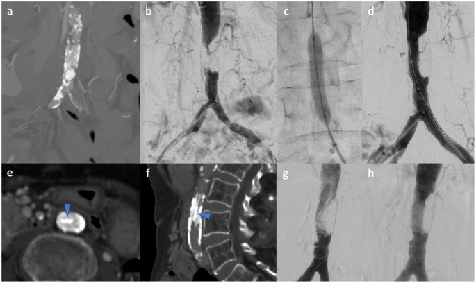
The first case was a 74-year-old woman with severe intermittent claudication of both lower extremities (Rutherford category 3). Preprocedural computed tomography (CT) angiography showed a total occlusion of distal abdominal aorta accompanied by massive calcification occupying the lumen (Fig. 1a). Endovascular treatment was performed to revascularize the occlusion (Fig. 1b). After successful passage of the 0.014-inch guidewire intraluminally, a balloon expandable EXPRESS-LD stent (8×57 mm: Boston Scientific, Marlborough, MA, USA) was implanted to cover the occlusive lesion (Figs. 1c and 1d). After the procedure, her symptom was completely resolved. One year later, she came back to our clinic due to recurrence of the symptom. CT demonstrated a complete collapse of the stent inside the calcified plaque (Figs. 1e and 1f). Although it was very hard to pass through the collapsed stent, a guidewire passage was finally obtained under intravascular ultrasound (IVUS) guidance. Revascularization was achieved with additional placement of a self-expanding stent (S.M.A.R.T. CONTROL, 7×60 mm, Cordis, Hialeah, FL, USA) inside the collapsed BES, resolving her symptom (Figs. 1g and 1h). After the reintervention, stents have been patent for 15 months.
Fig. 1 Collapse of balloon expandable stent in calcified aorta.
(a) Preprocedural computed tomography (CT) angiography shows a total occlusion of distal abdominal aorta accompanied by massive calcification occupying the lumen. (b) Aortogram shows total occlusion of the aorta. (c) A balloon expandable EXPRESS-LD stent (8×57 mm: Boston Scientific, Marlborough, MA, USA) was implanted to cover the occlusive lesion. (d) After the procedure, revascularized aorta was demonstrated. (e, f) CT scan conducted one year later demonstrates a complete collapse of the stent inside the calcified plaque (arrowheads: collapsed stent, (e): axial plane, and (f): sagittal plane). (g) Angiogram shows impaired aortic flow inside the collapsed stent. (h) A self-expanding stent deployed inside the collapsed balloon expandable stent restored the flow
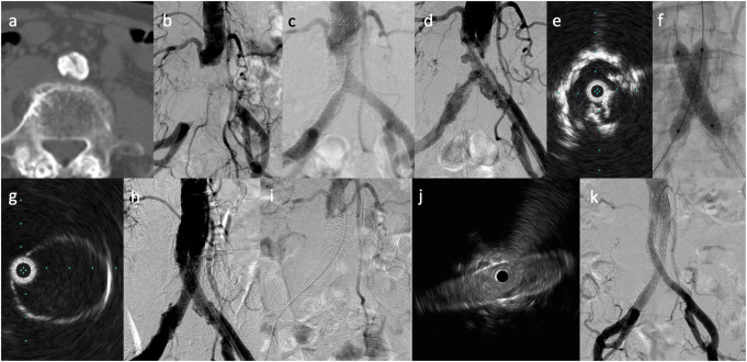
The second case was an 82-year-old woman who had the history of endovascular treatment for heavily calcified occlusions extending from aorta to bilateral common iliac arteries (CIA) (Figs. 2a and 2b). The lesion was treated with implantation of two self-expanding stents (Epic, 10×100 mm, Boston Scientific, Marlborough, MA, USA) from aorta to bilateral CIA in a kissing manner (Fig. 2c). After 30 months of the first endovascular procedure, bilateral claudication recurred. Angiography revealed in-stent occlusive lesions of previously implanted stents (Fig. 2d). IVUS evaluation demonstrated that the lumens of the both stents were occupied by calcified plaques (Fig. 2e). After dilation by noncompliant balloon, two VIABAHN VBX BES (8×40 mm, W. L. Gore & Associates Inc., Flagstaff, AZ, USA) were deployed from aorta to both CIA, achieving good expansion (Figs. 2f–2h). Although the claudication disappeared after endovascular treatment, the symptom recurred one month after the procedure. Angiography showed occlusion of bilateral VIABAHN VBX BES (Fig. 2i). IVUS showed that both VIABAHN VBX were markedly flattened (Fig. 2j). Deployment of another two self-expanding stents (S.M.A.R.T. CONTROL, 8×120 mm) in the “stent in-stent” fashion successfully expanded the collapsed BES and restored the flow (Fig. 2k). After the reintervention, stents have been patent for a year.
Fig. 2 Collapse of balloon expandable stent graft in calcified aorta.
(a) Computed tomography scan demonstrates a massive calcification occupying the aortic lumen. (b) Aortogram demonstrates the occlusion of aorta and bilateral common iliac arteries. (c) The lesion was treated with implantation of two self-expanding stents (Epic, 10×100 mm, Boston Scientific, Marlborough, MA, USA) from aorta to bilateral in a kissing manner. (d) Angiography performed after 30 months of the first endovascular procedure reveals protrusion of calcified plaques inside the previously implanted stents. (e) IVUS evaluation demonstrates that the lumens of the both stents were occupied by calcified plaques. (f) After dilation by noncompliant balloon, two VIABAHN VBX BESs (8×40 mm, W. L. Gore & Associates Inc., Flagstaff, AZ, USA) were deployed from aorta to both CIA. (g) IVUS demonstrates sufficient expansion of the BESs. (h) Aorta was successfully revascularized. (i) Angiography conducted after the recurrence of the claudication shows occlusion of bilateral BESs. (j) IVUS shows that both VIABAHN VBX were markedly flattened. (k) Deployment of another two self-expanding stents in the “stent in-stent” fashion successfully expanded the collapsed BES and restored the flow. BES: balloon expandable stent; CIA: common iliac artery; IVUS: intravascular ultrasound
Discussion
BES placed in superficial vessels such as subclavian vein and carotid artery are subject to the external compression, resulting in deformation/stent fracture.5,6) BES in deep vessels such as iliac arteries have also been reported to cause the stent fracture/deformity. One case report described a complete collapse of BES placed in CIA after Shiatsu (finger pressure) massage treatment.3) A case of fracture of BES placed in CIA was reported in a patient who performed extensive daily stretching and calisthenics.7) Intensive stretching and flexing of his torso and legs were believed to be the most likely cause of the deformation and ultimate fracture. In our cases, BES were deployed in the calcified aortic occlusion in the first case and in the in-stent occlusion in the second case which had been treated for calcified aortic occlusion. The cause of the BES collapse in the cases remains unknown. Both cases had significant calcium occupying the aortic lumen. It was reported that diffuse vessel calcification/calcified plaques were associated with inhomogeneous distribution of mechanical stresses/biomechanical abrasions and subsequent metal fatigues/stent fractures in coronary arteries.9,10) Therefore, it could be assumed that pulsatile pressure of the calcified aortic wall rendered the implanted stent continuous mechanical stress, leading to stent collapse. On the other hand, Kusumoto et al.11) reported a collapse of VIABAHN VBX balloon expandable endoprostheses in an elderly patient with a kyphosis. Plain lumber X-ray showed the balloon expandable stents being compressed by lumber spine while the patient stood up, which they considered to be the cause of the stent collapse. The exact cause of the stent collapse is hard to be proven and further accumulation of cases is needed to clarify its cause. Surprisingly, all of the four cases of the stent collapse which have been reported so far are Japanese, elderly, small, thin patients with heavily calcified aorta.11,12) It is unclear whether the situation could have been avoided by using self-expanding stents rather than BES. In vitro fatigue testing of both BES and self-expanding stents under pulsatile vessel model with calcified plaques may reveal the superiority of one over the other.
Conclusion
In the heavy calcified arterial lesions, particularly in elderly, small Asian patients, the use of BES, despite its high radial strength, may result in complete collapse, and therefore, stent selection should be carefully done in cases of heavily calcified aorta.
Disclosure Statement (COI)
SI received a research grant from W. L. Gore; JS received honoraria from Nipro corporation and Abbott; KK received scholarships from Boston Scientific, Terumo corporation, and Gore Japan. The other authors state that they have no conflicts of interest to disclose.
Author Contributions
Data collection: SI, AK, DF
Writing: TK, SI, AK, FB
Critical review and revision: all authors
Final approval of the article: all authors
Accountability for all aspects of the work: all authors
References
- 1).Nanto K, Iida O, Fujihara M, et al. Five-year patency and its predictors after endovascular therapy for aortoiliac occlusive disease. J Atheroscler Thromb 2019; 26: 989-96. [DOI] [PMC free article] [PubMed] [Google Scholar]
- 2).Schwindt AG, Panuccio G, Donas KP, et al. Endovascular treatment as first line approach for infrarenal aortic occlusive disease. J Vasc Surg 2011; 53: 1550-6.e1. [DOI] [PubMed] [Google Scholar]
- 3).Ichihashi S, Higashiura W, Itoh H, et al. Fracture and collapse of balloon-expandable stents in the bilateral common iliac arteries due to shiatsu massage. Cardiovasc Intervent Radiol 2012; 35: 1500-4. [DOI] [PubMed] [Google Scholar]
- 4).Ichihashi S, Higashiura W, Itoh H, et al. Intravascular ultrasound assessment of acute expansion of the balloon-expandable stent in heavy calcified iliac artery lesions or in lesions resistant to dilation by a self-expanding stent. Ann Vasc Surg 2014; 28: 1449-55. [DOI] [PubMed] [Google Scholar]
- 5).Bjarnason H, Hunter DW, Crain MR, et al. Collapse of a Palmaz stent in the subclavian vein. AJR Am J Roentgenol 1993; 160: 1123-4. [DOI] [PubMed] [Google Scholar]
- 6).Mathur A, Dorros G, Iyer SS, et al. Palmaz stent compression in patients following carotid artery stenting. Cathet Cardiovasc Diagn 1997; 41: 137-40. [DOI] [PubMed] [Google Scholar]
- 7).Sawhney R, Allen D, Nanavati S. Kissing balloon-expandable iliac stents complicated by stent fracture. Journal of Vascular and Interventional Radiology: JVIR 2008; 19: 1519-20. [DOI] [PubMed] [Google Scholar]
- 8).Ihara M, Ueno A, Tsuda Y, et al. A case of repeated occlusion in the common iliac artery due to an unexpected stent deformation. Cardiovasc Interv Ther 2015; 30: 162-7. [DOI] [PubMed] [Google Scholar]
- 9).Morlacchi S, Pennati G, Petrini L, et al. Influence of plaque calcifications on coronary stent fracture: a numerical fatigue life analysis including cardiac wall movement. J Biomech 2014; 47: 899-907. [DOI] [PubMed] [Google Scholar]
- 10).Halwani DO, Anderson PG, Brott BC, et al. The role of vascular calcification in inducing fatigue and fracture of coronary stents. J Biomed Mater Res B Appl Biomater 2012; 100B: 292-304. [DOI] [PubMed] [Google Scholar]
- 11).Kusumoto S, Muroya T, Matsumoto Y, et al. Collapse of VBX balloon-expandable endoprosthesis in bilateral common iliac arteries in a lean, elderly patient with bent back. Ann Vasc Surg 2020; S0890-5096. [DOI] [PubMed] [Google Scholar]
- 12).Mizushima S, Mine T, Fujii M, et al. “Squid-capture” modified in situ stent-graft fenestration technique for recurrent abdominal aortic occlusive disease after collapse of balloon-expandable stent. Ann Vasc Surg 2020; S0890-5096. [DOI] [PubMed] [Google Scholar]


